February 2014

It’s All About the Diet: Obesity and Mitochondrial Dysfunction

7359 views

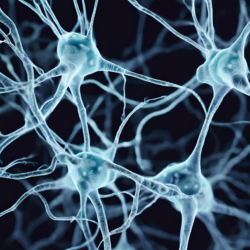
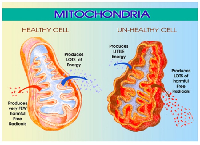
We’ve been learning a lot about mitochondrial dysfunction lately, particularly as it applies to medication and vaccine adverse reactions. Mitochondria are those energy producing powerhouses located within our cells that are critical to every aspect of health. We have over 100,000 trillion mitochondria in our body, each containing 17,000 little assembly lines for making adenosine triphosphate (ATP), the fuel that powers our lives. Mitochondria use ninety percent of the oxygen we breathe, take up 40% percent of the space inside the heart cells and 20% of the space inside the liver cells. Properly functioning mitochondria are critical to human health and survival.

Unfortunately, mitochondria are exquisitely sensitive to their environs and can be damaged easily. Damaged or dysfunctional mitochondria lead to an array of complex and seemingly disparate and untreatable diseases. How mitochondria become damaged is a matter of great interest with research pointing to maternal genetics and epigenetics, environmental exposures, medications and even diet. Mitochondrial dysfunction arguably represents one of the most unrecognized causes of disease in modern medicine and, according to some geneticists, mitochondrial disease represents the next great paradigm in medicine.

The Mitochondrial Cholesterol Transporter

Over the course of my research, I stumbled on a series of papers identifying a cool little transporter channel located on the outer membrane of the mitochondrion called the translocator protein 18kDA or TSPO. For a while, this channel was called the peripheral benzodiazepine receptor because researchers had noticed that the drug diazapam, a benzodiazepine, could bind to a spot on this channel and evoke a reaction. Soon however, researchers learned the function of the TSPO was far more complex than a simple drug binding site and changed the name accordingly.

What is the Mitochondrial Translocator Protein and Why is it Important?

The primary function of the TSPO is to bring cholesterol into the mitochondria. Once inside, another protein, an enzyme called StAR, converts cholesterol into a hormone called pregnenolone and shoots it back out into the cell and beyond. Pregnenolone is the precursor for all steroid hormones and so this TSPO channel is responsible for steroidogenesis in cells, not just in what are considered the typical steroid producing cells like the ovaries, testes, adrenals, but in all cells. TSPO is ubiquitous across mitochondria, meaning steroidogenesis is not limited to the endocrine cells; something many, including myself, have been arguing for decades, but I digress.

In addition to its role in steroidogenesis, the TSPO appears to control many aspects of mitochondrial function. It regulates production of reactive oxygen species (ROS) – those pesky little free radicals that damage mitochondrial DNA. The TSPO also influences outright apoptosis or cell death, and as one might expect, TSPO modulates cellular energy or ATP production. Researchers have found that the functionality of the TSPO channel changes with different disease states and can become dysregulated which then dramatically affects how the mitochondria function. Because of its diverse and seemingly unrelated functions, the TSPO is thought to be part of our host-defense response to disease and injury.

Obesity Impairs Mitochondrial Function via TSPO Downregulation

Here’s where it gets interesting. Once again, we see that diet is critical to mitochondrial functioning via its influence on TSPO activity. In a recent study, Translocator Protein 18 kDa (TSPO) Is Regulated in White and Brown Adipose Tissue by Obesity, researchers demonstrated that a high fat diet downregulates TSPO functioning significantly and the compensatory reaction – obesity – is the survival mechanism.

Study Details. Mice were fed a high fat diet (60% of calories from fat) from 8 weeks of age through 34 weeks of age. Researchers then compared TSPO functioning and other markers of the diet induced obese mice to mice fed a regular diet (13% of calories from fat). They investigated the differences TSPO function and activity in both white fat and brown fat in both groups of mice. Remember white fat stores calories as big fat droplets, while brown fat stores it in smaller droplets and utilizes calories more effectively by burning them for energy. The brown fat is more mitochondrial dense than white fat. So TSPO changes relative to diet and white and brown fat function could be very interesting when understanding obesity.

Results – White Fat. The mice with dietary induced obesity showed a 90% reduction in TSPO mRNA and an 87% reduction in gene expression as measured via a protein called the peroxisome proliferator-activator receptor coactivator (PGC1α). PGC1α is important for mitochondrial biogenesis, making new mitochondria. The researchers also found a 40% reduction in TSPO binding sites, another marker that indicates decreased TSPO gene expression. Interestingly, the shape of the fat cells changed in the diet induced obese mice compared to the regular feed mice. In the obese mice, the white adipocytes were hypertrophic (oversized) and were surrounded by macrophages, the immune cells responsible for ‘eating’ dead or dying cells.

Results – Brown Fat. In the brown fat, the changes in TSPO function between the diet induced obese mice and the regular feed mice were equally dramatic. Visually, the adipocytes were hypertrophic indicating increased fat storage versus energy usage. TSPO gene expression was reduced by 32%, mitochondrial biogenesis was reduced by 31%, while TSPO binding sites decreased by 7%.

A couple more findings. The investigators added a few more conditions to the experiment to determine whether these changes in TSPO could be modified acutely in response to fasting or other stressors. The answer was no. TSPO expression and function were not influenced by acute metabolic changes suggesting a more chronic pattern of metabolic dysfunction.

What This Means: Diet Affects Mitochondrial Function

High fat diet affects mitochondrial function by downregulating a critical receptor – the TSPO channel that brings cholesterol into the mitochondrion. This results in increased cholesterol storage within the fat cells and perhaps decreased conversion from cholesterol to energy (or steroids) inside the mitochondria. The hypertrophic and unhealthy adipocytes then evoke an immune response drawing macrophages and inducing phagocytocis. A dangerous feedback loop ensues.

Though the mechanisms by which a high fat diet induces mitochondrial dysfunction is not clear, one can speculate that a diet high in fat is also low in critical nutrients that are required for proper mitochondrial function and cellular energy production. Deficits in nutrients such as thiamine (vitamin B1) can and do cause severe mitochondrial dysfunction and lead to an array of disease processes from nervous system destabilization to cardiac, GI and reproductive dysfunction. Other nutrients and cofactors are also critical for mitochondrial function. CoQ10, L-Carnitine for example, have been found helpful in mitochondrial induced dysautonomia, and the list goes on.

What’s important to remember is that when the mitochondria are starved of critical nutrients, they don’t function properly and die off. As mitochondria become injured and die, it is possible to see how the hypertrophy and increased cholesterol storage observed in the adipocytes might be a compensatory survival reaction to maintain the requisite demands for mitochondrial cholesterol. The only problem is that as those fat cells become larger and more cholesterol is stored, more mitochondrial dysfunction ensues, reinforcing and continuing the immune response. We get an endless cycle of poorly functioning and dying mitochondria>immune response>larger fat cells with more cholesterol stored>more mitochondrial death>more immune response and so on. Breaking the cycle may be as simple as changing one’s diet and exercising, as both can induce mitochondrial biogenesis.

We Need Your Help

More people than ever are reading Hormones Matter, a testament to the need for independent voices in health and medicine. We are not funded and accept limited advertising. Unlike many health sites, we don’t force you to purchase a subscription. We believe health information should be open to all. If you read Hormones Matter, like it, please help support it. Contribute now.

Yes, I would like to support Hormones Matter.

Photo by Yolanda Djajakesukma on Unsplash.

Another Nail: The Impact of Prescription Painkiller Abuse on Chronic Pain Sufferers

2519 views

Bang, bang, BANG…do you hear that? That’s the sound of nails being hammered into a coffin. No, I’m not trying to be morbid, nor am I in the process of creating a funeral soundtrack. To explain to you what in the world I am talking about, please consider the following current news articles:

  • “Doctor Admits Drug Sales”
  • “Prescription Drug Dealer Allegedly Robbed Over $1 Million from New York City Pharmacy.”
  • “Doctor Pleads Guilty in Drug Trafficking Case”
  • “Sharp Rise in Women’s Death From Overdose of Painkillers”
  • “Fort Worth Officer Accused of DWI on Prescription Drugs Is Fired”
  • “Dentist Pleads Guilty In Prescription Drug Case”

Do you hear the banging now? Each one of these instances of painkiller drug misuse is another nail hammered into the coffin of necessary pain relief for those of us in true need.

Accusations of Addiction

The following is a scene that’s far more common that it should be. A women with endometriosis stumbles into the emergency room screaming in agony. Her endo has flared up and she is in excruciating pain. She waits miserably for at least an hour to be triaged by someone who knows nothing about endo. At triage she is asked what her issue is (as if they can’t tell she’s in excruciating pain!) Due to the fact that she is a confident, strong woman who knows her pain, she immediately advocates for herself: “I have endometriosis and I’m having a flare up. I’m in excruciating pain and I need pain medication!” At the mention of those two words, pain medication, silence abounds. Looks are exchanged between the triage nurses that plainly say ‘oh, she’s one of those’.

Let’s see what he doctor has to say,” she is told condescendingly, and sent back to wait in agony until her name is called. At least an hour passes while she writhes in pain. After finally receiving a coveted bed in the emergency room and struggling to change into an unnecessary hospital gown she is seen by a resident and proceeds to tell him what is wrong. He tells her he has to discuss her situation with his attending and leaves her crying desperately for some pain relief.

Many minutes pass and he comes back with the verdict: “I’m going to tell the nurse to give you some (FILL IN ANY NON-NARCOTIC, UNHELPFUL PAIN MEDICATION).”

She begs, pleads, cries and screams, but he will not give her anything stronger and suggests she go home and see her own doctor.  The nurse comes in with discharge papers and the woman is encouraged to sign them. Right before she does, she finds herself curious to see what they stated her problem was on the discharge papers.  She glances at the papers and is shocked to see what is written: unspecified pelvic pain and drug seeking behavior. The poor woman goes home feeling completely dejected, mortified, and often ready to give up fighting.

An Unfortunate Situation

This horrendous occurrence is far too common for women with endometriosis. Too often we are labeled as ‘drug seeking’ or denied pain medication that we desperately need. But it is not only women with endometriosis who have chronic pain. And we are not the only ones being mistaken for drug addicts or drug seekers. There are far too many stories of people with invisible illnesses being accused of drug addiction when all they are trying to do is ease their pain.

Unfortunately, when we review the statistics on prescription painkiller abuse, it is quite understandable why we are accused of such. While about a third of all Americans (more than 116 million people) deal with some sort of chronic pain that might legitimately warrant prescription pain relief, the non-medical use of prescription pain relievers has been estimated at about 5 percent in people 12 years and older. From 1998 to 2008, the non-medical use of prescription painkillers increased 163 percent and between 1992 and 2003 there was a 90 percent increase in people who admitted abusing them. It is currently estimated that 2.4 million Americans abuse prescription drugs.

The FDA Fights Back

It is statistics like these that cause the FDA to create laws that severely restrict the use of prescription drugs. In October, the FDA recommended that tighter controls be placed on narcotics such as Vicodin, and that the drugs be changed to have a Schedule II classification. This would mean that doctors could no longer call in a prescription to the pharmacy for them or write a prescription for any supply over three months. Additionally, the FDA is recommending that label changes be put in effect for long-acting narcotics such as Oxycontin that describe the risks involved in taking the medication. While this recommendation does not directly restrict the usage of these medications, it is assumed that additional warning labels will create more hesitation on the part of the doctors prescribing them, making it more difficult for chronic pain patients.

What’s a Chronic Pain Patient to Do?

So I ask you, my friends: how are those of us who actually suffer from horrendous, excruciating pain supposed to receive the relief we deserve when others around us are abusing the medication that we need? How are we supposed to be helped if the FDA is forced to restrict prescription drug usage in order to prevent abuse? How do we prove that we are legitimate users, and not abusers? And lastly, how are we supposed to wean off of these medications if there is not enough research being done to discover the cause of our suffering and find better ways to improve our pain? It is a constant, cyclical problem that has one, enormous casualty: those of us who are really in pain.

How Dietary Mayhem Causes Disease: The Choked Engine Syndrome

11399 views

Over the past year, I have written extensively about thiamine deficiency post Gardasil vaccination (here, here, here, here). We now have five cases where thiamine deficiency was identified and clinical symptoms remediated with supplementation. Many more are suspected but recognition and testing have been slow. Thiamine deficiency may not be limited to the post Gardasil population, although that is where we first recognized it. Symptoms of thiamine deficiency and dysfunctional oxidative metabolism have been observed amongst the post fluoroquinolone and post Lupron populations and likely other populations adversely affected by a vaccine or medication, though data are limited. For the current paper, I should like to offer an explanation of the effect of thiamine deficiency in relationship to the stress of the vaccination or medications.

Thiamine Deficiency and Diet

With the widespread ingestion of simple carbohydrates that is almost a hallmark of Western civilization I suggest that the Gardasil vaccination and certain other medications represent “the last straw to break the camel’s back”.  I have included a case report, from my clinical practice, as an example of the effect of a simple nutritional stressor – sugar – imposed on an individual who’s oxidative metabolism was marginal at the time. I have included the references for anybody that wishes to check on how much of this is published.

Cellular Energy and Diet

Present knowledge indicates that cellular energy arises only from oxidation of food sources. The prevalently common form of nutritional mayhem in the U.S. is a high calorie content from simple carbohydrates with insufficient vitamin/mineral content to catalyze efficient oxidation. This form of malnutrition might be compared with functional decline in a choked internal combustion engine. Evidence presented in this case report presented below indicates that simple carbohydrate ingestion can have far-reaching consequences.  A review indicates that a common manifestation of its effect is oxidative stress in the brain, particularly in the limbic system where emotional reflexes originate and where the controls of the autonomic and endocrine systems react automatically to sensory input. Beriberi is the classic example of high calorie carbohydrate malnutrition and is the prototype for dysautonomia (abnormal function of the autonomic nervous system [ANS] ) in its early stages. A later stage results in degeneration of autonomic ganglia and irreversible disease. Symptoms arising from thiamine deficiency or abnormal homeostasis are protean and diverse in nature.

Dysautonomia, Oxidative Stress and Thiamine

Dysautonomia, a common presentation of functional disease and often associated with variable organic diseases caused by loss of oxidative efficiency in the brain, has been reviewed. A hypothesis was presented that there is a combination of genetic risk, different forms of sensory input defined as stress, particularly those imposed by present civilization, and high calorie malnutrition that are collectively responsible. This was presented diagrammatically by the degree of overlap in the “three circles of health, named genetics, stress and nutrition” (1).  It is also known that mitral valve (a heart valve) prolapse (MVP) is widespread in the population and is associated with dysautonomia, although the cause and effect relationship is said to be unknown (2-4). MVP is associated with adrenergic overdrive (the well-known adrenalin rush) in the normally balanced adaptive reactions of the autonomic/endocrine axis (5-8). (The autonomic nervous system and the glands of the endocrine system are under the control of the brain).  Panic disorder, also sometimes associated with MVP, is seen as an example of falsely triggered fight-or-flight reflexes engendered in the limbic brain.  Pasternac and associates (6) showed that symptomatic patients with MVP demonstrated increased resting sympathetic tone and that supine bradycardia (slow heart rate) suggested increased vagal (the vagus is a nerve that runs from the brain to many parts of the body) tone at rest. Davies and associates (7) demonstrated physiologic and pharmacologic hypersensitivity of the sympathetic system in a group of patients with MVP. Sympathoadrenal responses were noted in rats exposed to low oxygen concentration (9) and impaired cerebral autoregulation has been reported in obstructive sleep apnea in human subjects (10). It has also been shown that thiamine deficiency produces traditionally accepted psychosomatic or functional disease (11,12).  A low oxygen concentration results in changes in brain structures similar to those induced in thiamine deficiency (13).

A Case Study of Thiamine Deficiency and Dietary Influence: The Sugar Problem

The Table below shows laboratory results from an 84-year old man who had begun to experience severe insomnia for the first time in his life. He also had painful tenosynovitis (also known as “trigger finger”) in the index finger of the left hand.  He had edited a journal for some 14 years and for several years, had been a member of a bell choir in which he played a heavy base bell in each hand, involving repetitive trauma to the index fingers.  He did not crave sugar, his ingestion of simple carbohydrates being minimal to moderate. The only treatment offered was complete withdrawal from all forms of simple carbohydrates.

Serial laboratory studies revealed a gradual improvement over six months and his weight decreased from 182 to 170 pounds without any other change in diet. Insomnia and tenosynovitis gradually improved. The Table shows that serial laboratory tests over a period of six months, from February to August, showed continued gradual improvement. In September, the day after a minimal ingestion of simple carbohydrate, there was an increase in triglycerides and TPPE.

Understanding the Labs

Notice that the triglycerides dropped from 206 in February to 124 in August, then rose again in September only one day after a minimal amount of sugar.  Triglycerides are part of the routine lipid profile test done by doctors and are well known to be related to the ingestion of simple carbohydrates.  Fibrinogen and HsCRP are both recognized as markers of inflammation.  Notice that both of them decreased between February and August but HsCRP rose again in September like the triglycerides.  The TPPE is the important part of the transketolase test.  The higher the percentage, the greater is the degree of thiamine deficiency.  Notice that it dropped from 35% to zero between February and August, but that it jumped to 8% in September, the day after the ingestion of sweets.  I have provided the normal laboratory values for the discerning reader.

  TABLE 1
Month

Cholesterol

Triglycerides

Fibrinogen

HsCRP

TKA

TPPE

February

169

206

412

7

65

35%

March

155

165

55

25%

May

160

152

312

0.9

85

2%

August

166

124

0.3

59

0%

September*

169

165

220

1

62

8%

Consecutive laboratory blood tests

Cholesterol N <200 mg/dL. Triglycerides N< 150 mg/dL. Fibrinogen N 180-350,g/dL
HsCRP N 0.1-1.0 mg/L. TKA 42-86mU. TPPE 0-18%. *Next day after ingestion of simple carbohydrate.


The abnormal TPPE indicated thiamine deficiency in this patient (14). The increased triglycerides and their steady decrease over time indicated that sugar ingestion was a potent cause of his symptoms. An increase in fibrinogen and hypersensitive CRP are both laboratory markers of inflammation, although the site is not indicated.  Recent studies in mice (15) have shown that high calorie malnutrition activates a normally silent genetically determined mechanism in the hypothalamus, causing either obesity, inflammation or both. The potential association of thiamine with electrogenesis (formation of electrical energy) (16) may have some relationship with brain metabolism and the complex functions of sleep.

Compromised Oxidative Function: Thiamine Deficiency, Beriberi and Diet

It has long been known that beriberi is a classic disease caused by high consumption of simple carbohydrate with insufficient thiamine to process glucose into the citric acid cycle. (This complex chemistry represents the engine of the cell, meaning that it produces the energy for function).  Widespread thiamine deficiency has been reported in many publications(17-20), producing the same brain effects as low oxygen concentration (13,21). In rat studies, this produces an imbalance in the autonomic nervous system (9). Thiamine  deficiency is easily recognized in a clinical laboratory by measuring TKA and TPPE (14).

Thiamine and the Brain

Thiamine triphosphate (TTP) (this is synthesized from thiamine in the brain) is known to be important in energy metabolism. Although its action is still unknown, the work with electric eels has revealed that the electric organ has a high concentration of TTP and may have a part to play in electrogenesis, the transduction of chemical to electrical energy (16,22). The energy for its synthesis from thiamine comes from the respiratory chain. This is also complex chemistry in the formation of energy synthesized within mitochondria, the “engines” of the cell (23), so that any form of disruption of mitochondria would be expected to reduce adequate synthesis of this thiamine ester. Although slowing of the citric acid cycle appears to be the main cause of the biochemical lesion in brain thiamine deficiency (24), the part played by TTP is not yet known. Alzheimer’s disease has been helped by the use of therapeutic doses of thiamin tetrahydrofurfuryl disulfide (TTFD) (25), a more efficient method of administering pharmacologic doses of thiamine (26).

Acetylcholine, the neurotransmitter used by both branches of the autonomic nervous system, is generated from glucose metabolism, requiring  B vitamins, particularly thiamine. Choline is a “conditional nutrient”, meaning that it is derived mainly from diet but is also made in the body. The presence of all these nutrients leads to the synthesis of this neurotransmitter.  It’s depletion would affect both branches of the autonomic nervous system, resulting in dysautonomia.

There is evidence that high-dose thiamin increases the effect of acetylcholine (27). Animal studies have shown that TTFD improves long term memory in mice (28) and it has been shown that it extends the duration of  neonatal seizures in DBA/J2 mice, seizures that normally cease in a few days with normal maturation (29).  These seizures are naturally related to a prolonged effect of this neurotransmitter in this strain of mouse.  The experimental prolongation of the seizures by administration of TTFD indicated that it enhanced the effect of the neurotransmitter. A pilot study in autistic spectrum disorder showed clinical improvement in 8 of the 10 children treated with TTFD (30), a disease that has been shown to have reduced  parasympathetic activity in the heart (31,32). Neural reflexes regulate immunity (33).  Dysautonomia was found in a large number of patients with cancer at Mayo Clinic (34).

Dysautonomia and Thiamine Deficiency         

Evidence has been presented that a common connection exists between dysautonomia, inefficient oxidative metabolism produced mainly by high calorie malnutrition, and organic disease (1). Thiamine enters the equation in terms of its relationship with carbohydrate ingestion and its use by the brain as fuel (35). Decreased transketolase activity in brain cells induced by thiamine deficiency contributes to impaired function of the hippocampus (36) each, part of the limbic system control mechanisms that affect autonomic sympathetic/parasympathetic balance. Erythrocyte (red cells) transketolase indicates abnormal thiamine homeostasis that is commonly achieved by carbohydrate ingestion and deficiency of vitamin B (14).  Beriberi gives rise to functional changes in the autonomic nervous system in its early stages and produces irreversible degeneration in its later stages (37). This, because it represents a largely forgotten aspect of disease, might equate with the wide use of simple carbohydrates in Western civilization. Deficiency of other essential non-caloric nutrients has been associated with dysautonomia (1).

The Role of Nutritional Stress in Post Vaccination and Medication Reactions

Two results of post- Gardasil vaccination have been reported, Postural Orthostatic Tachycardia Syndrome (POTS) and cerebellar ataxia.  POTS, a disease easily confused with beriberi, is one of the many syndromes reported under the general heading of dysautonomia and stress related intermittent episodes of cerebellar ataxia were reported in thiamin dependency (38).  Since the inflammatory reflex has recently been found to be involved with the sympathetic branch of the ANS (39), enhancement of its dysfunction by TD might explain some of the Gardasil affected illnesses.

Conclusion

Thiamine deficiency is now accepted as the major cause of the ancient scourge of beriberi. The underlying mechanisms are still not fully understood for we do not yet know the complete roles of thiamine. The clinical effects are protean and unpredictable. It is, however, clear that thiamine has a vital effect on many aspects of oxidative metabolism and its deficiency can be used as a model for the clinical effects produced by disruption in energy synthesis. It can be summed up under the general heading of dysoxegenosis and thiamine is certainly not the only component that governs this vital life process. The example of beriberi indicates that the brain, peripheral nervous system and the heart are the tissues most affected by the disease, the tissues that rapidly consume oxygen.

The limbic system is a complex computer that organizes all our adaptive survival reflexes and its sensitivity to hypoxia is well known. It is evident that non-caloric nutrient deficiency, especially thiamine, gives rise to the same symptoms and histopathology as mild to moderate hypoxia (oxygen deficiency) and that the leading symptomology is that of dysautonomia. Since the limbic system gives rise to emotional reflexes and mild to moderate hypoxia enhances sympathoadrenal response, it can be expected that an affected individual would be more aggressive and more likely to experience exaggerated fight-or-flight reflexes. A “nursed” emotional grievance might be expected to explode in violence that would otherwise be curtailed or suppressed by normal brain metabolism. It suggests that high calorie malnutrition, particularly that provided by excessive consumption of simple carbohydrates, gives rise to uncontrolled pathophysiological actions that might explain some of the widespread incidence of emotional and psychosomatic disease in contemporary society. It may also explain some of the “hot” juvenile crime and vandalism, much of which is poorly understood in our present civilization. It is also hypothesized that a marginal state of oxidative metabolism, perhaps asymptomatic or with only mild symptoms that are ignored, might be precipitated into clinical expression with a mild degree of stress imposed by a vaccination. The individual in the case reported above appeared to be unusually sensitive to sugar ingestion and this may be an additional genetically determined risk.

We Need Your Help

More people than ever are reading Hormones Matter, a testament to the need for independent voices in health and medicine. We are not funded and accept limited advertising. Unlike many health sites, we don’t force you to purchase a subscription. We believe health information should be open to all. If you read Hormones Matter, like it, please help support it. Contribute now.

Yes, I would like to support Hormones Matter. 

Image created using Canva AI.

References

  1. Lonsdale D. Dysautonomia, a heuristic approach to a revised etiology for disease. eCAM 2009;6(1):3-10.
  2. Orhan A L, Sayar N, Nurkalem Z, Uslu N, Erdem I, Erdem E C, Assessment of autonomic dysfunction and anxiety levels in patients with mitral valve prolapase. Turk Kardiyol Dern Ars 2009;37(4):226-233.
  3. Alpert M A, Murkerji V, Sabeti M, Russell J L, Beitman B D. Mitral valve prolapse, panic disorder, and chest pain. Med Clin North Am 1991;75(5):1119-1133.
  4. Raj A, Sheehan D V. Mitral valve prolapse and panic disorder. Bull Menninger Clin 1990;54(2):199-208.
  5. Di Salvo G, Pergola V, Ratti G, Tedesco M A, Giordano C, Scialdone A, et al. Atrial natriuretic factor and mitral valve prolapse syndrome. Minerva Cardioangiol 2001;49(5):317-325.
  6. Pasternac A, Tubau J F, Puddu P E, Krol R B, de Champlain J. Increased plasma catecholamine levels in patients with symptomatic mitral valve prolapse. Am J Med. 1982;73(6):783-790.
  7. Davies A O. Mares A, Pool J L, Taylor A A. Mitral valve prolapse with symptoms of beta-adrenergic hypersensitivity. Beta 2-adrenergic receptor supercoupling with desensitization on isoproterenol exposure. Am J Med 1987;82(2):193-201.
  8. Boudoulas H, Wooley C F. Mitral valve prolapse syndrome: neuro-endocrinological aspects. Herz 1988;13(4):249-258.
  9. Johnson T S, Young J B, Landsberg L, Dana C A. Sympathoadrenal responses to acute and chronic hypoxia in the rat. J Clin Invest 1983;71:1263-1272.
  10. Urbano F, Roux F, Schindler J, Mohsenin V. Impaired cerebral autoregulation in obstructive sleep apnea. J Appl Physiol 2008;105(6):1852-1857.
  11. Lonsdale D, Shamberger R J. Red cell transketolase as an indicator of nutritional deficiency. Am J Clin Nutr 1980;33:205-211.
  12. Lonsdale D. Three case reports to illustrate clinical applications in the use of erythrocyte transketolase. eCAM 2006;4(2):247-250.
  13. Macey P M, Woo M A, Macey K E, Keens T G, Saeed M M, Alger J R et al. Hypoxia reveals posterior thalamic, cerebellar, midbrain, and limbic deficits in congenital hypoventilation syndrome. J Appl Physiol 2005;98(3):958-969.
  14. Massod M F, McGuire S L, Werner W R. Analysis of blood transketolase activity.Am J ClinPathol 1971;55:465-470.
  15. Zhang X, Zhang G, Zhang H, Karin M, Bai H, Cai D. Hypothalamc 1KKbeta/N-kB and ER stress link overnutrition to energy imbalance and obesity. Cell   2008;135(1):61-73.
  16. Bettendorff L, Michel-Cahay C, Grandfils C, De Rycker C, Schoffeniels E. Thiamine triphosphate and membrane-associated thiamine phosphatases in the electric organ of Electrophorus electricus. J Neurochem 1987;49(2):495-502.
  17. O’Keefe S T, Tormey W P, Glasgow R, Lavan J N. Thiamine deficiency in hospitalized elderly patients. Gerontology 1994;40(1):18-24.
  18. Macias-Matos C, Rodriguez-Ojea A, Chi N, .Zulueta D, Bates C J. Biochemical evidence of thiamine depletion during the Cuban neuropathy epidemic, 1992-1993. Am J Clin Nutr 1996;64(3):347-353.
  19. Mazavet D. Vassilev K, Perrigot M. Neuropathy with non-alcoholic thiamine deficiency: two cases of bladder disorders [article in French]. Ann Readapt Med Phys 2005;48(1):43-47.
  20. Hazell A S, Butterworth R F. Update of cell damage mechanisms in thiamine deficiency: focus on oxidative stress, excitotoxicity and inflammation. Alcohol Alcohol 2009;44(2):141-147.
  21. Vortmeyer A O, Hagel C, Laas R. Hypoxia-ischemia and thiamine deficiency. Clin Neuropathol 1993;12(4):184-190.
  22. Nghiem H O, Bettendorff  L,Changeux J P. Specific phosphorylation of Torpedo 43K raspsyn by endogenous kinase(s) with thiamine triphosphate as the phosphate donor. FASEB J 2000;14(3):543-554.
  23. Gangolf M, Wins P, Thiry M. Thiamine triphosphate synthesis in the rat brain is mitochondrial and coupled to the respiratory chain. J Biol Chem 2010;285(1):583-594.
  24. Bettendorff  L, Sluse F, Goessens G,  Wins P, Grisar T. Thiamine deficiency-induced partial necrosis and mitochondrial uncoupling in neuroblastoma cells are rapidly reversed by addition of thiamine. J Neurochem 1995;65(5):2178-2184.
  25. Mimori Y, Katsuoka H, Nakamura S. Thiamine therapy in Alzheimer’s disease. Matab Brain Dis 1996;11(1):89-94.
  26. Lonsdale D. Thiamine tetrahydrofurfuryl disulfide: a little known therapeutic agent. Med Sci Monit 2004;10(9):RA199-203.
  27. Meador K J. Nichols M E, Franke P, Durbin M W. Evidence for a central cholinergic effect of high-dose thiamine. Ann Neurol 1993;34:724-726.
  28. Micheau J, Durkin D P, Destrade D C, Rolland Y, Jaffard R. Chronic administration of sulbutiamine improves long term memory formation in mice: possible cholinergic mediation.  Pharacol Biochem Behav 1985;23(2):1
  29. Lonsdale D. Effect of thiamine tetrahydrofurfuryl disulfide on audiogenic seizures in DBA/2J mice. Dev Pharmacol Ther 1982;4(1):28-36.
  30. Lonsdale D, Shamberger R J, Audhya T. Treatment of autism spectrum children with thiamine tetrahyhdrofurfuryl disulfide: a pilot study. Neuro Endocrinol Lett 2002;23(4):303-308.
  31. Ming X,  Julu P O O, Brimacombe M, Connor S, Daniels M L. Reduced cardiac parasympathetic activity in children with autism. Brain Dev 2005;27:509-516.
  32. Palmieri L Persico A M. Mitochondrial dysfunction in autism spectrum disorders: cause or effect? Biochem Biopys Acta 2010; May 1 [Epub ahead of print].
  33. Rosas-Ballina M, Tracey K J. The neurology of the immune system: neural reflexes regulate immunity. Neuron 2009;64(1):28-32.
  34. McKeon A, Lennon V A, Lachance D H, Fealey R D, Pittock S J. Ganglionic acetylcholine receptor autoantibody: oncological, neurological and serological accompaniments. Arch Neurol 2009;66(6)(:735-741.
  35. Elmadfa I Majchrzak D, Rust P Genser D. The thiamine status of adult humans depends on carbohydrate intake. Int J Vitam Nutr Res 2001;71(4):217-221.
  36. Zhao Y, Pan X, Zhao J, Wang Y, Peng Y, Zhong C. Decreased transketolase activity contributes to impaired hippocampal neurogenesis induced by thiamine deficiency. J Neurochem 2009;111(2):537-546.
  37. Inouye K, Katsura E. Etiology and pathology of beriberi. In: Thiamine and
  38. Beriberi. Igaku Shoin Ltd. Tokyo;1965:1-28.
  39. Lonsdale D. Faulkner W R, Price J W, Smeby R R. Intermittent cerebellar ataxia associated with hyperpyruvic academia,hyperalaninemia and hyperalaninuria. Pediatrics 1969;43:1025-34.
  40. Martelli D, Yao S T,McKinley M J,McAllen R M. Reflex control of inflammation by sympathetic  nerves, not the vagus. J Physiol 2014; Jan 13 [Epub ahead of print].


Surfing the Sugar Wave: PCOS and Insulin Resistance

4536 views

After a wonderful Valentine’s day filled with candy, chocolate, and carbs, I was riding the sugar wave and loving every minute. On Friday evening, after two cups of coffee, the combination of sugar and coffee was keeping me buzzed and happy. But by Saturday I woke up with a headache, feeling moody and miserable. I felt like a grey cloud was hanging over my head. Why was I in such a funk? By the time Sunday rolled around – after more cake and more carbs, and a barbeque lunch with friends – I came home and crashed…literally.

All day I had felt on the verge of tears and when I walked in the door, they all came tumbling out. My husband watched helplessly as I wept and sobbed uncontrollably, and when I was done he put his arm around me and handed me a cup of soup. But where were these emotions coming from? And why did they come on so suddenly?

What is PCOS?

To fully understand, we must travel back to 2008, the year I was diagnosed with Polycystic Ovarian Syndrome, or PCOS. The syndrome, which affects nearly five million women in the United States, often goes undiagnosed and if left untreated can lead to high cholesterol, diabetes, and heart disease. The name Polycystic Ovarian Syndrome refers to small cysts that form around the ovaries, but it is really an endocrine disorder in which the sex hormones estradial and progesterone are out of balance. Women with PCOS typically have an increased production of androgens (the male sex hormone) causing acne, increased hair growth, irregular menstrual cycles, and male pattern baldness. This can also lead to weight gain and infertility.

Coming away from my doctor’s office I was left confused. What was Polycystic Ovarian Syndrome? My OBGYN had actually read me the disorder from her textbook and then handed me a prescription for birth control pills. Because PCOS has no documented cure and because doctors still know very little about the cause of the disease, patients are often left helpless, searching for answers on the Internet or among their peers. My doctor never once told me there might be a link between what I eat and my disorder. She never once mentioned that diet and exercise were important factors in controlling my PCOS symptoms. And because I was “thin.” she assumed I was healthy.

The truth was, I was far from healthy. I had come from a country where most food was prepared from scratch, ingredients were natural and from the earth, and dinners were bought from the butcher or farmer, not a box. When I moved from Kenya to the US in 1999, my diet drastically changed without me giving it a second thought. I went from rice and beans to TV dinners, from curry and vegetables to Ramen noodles. In college I existed on pasta, fried chicken, alcohol, caffeine, frozen meals, and anything I could lug across campus from the food store. Although outwardly I appeared fit, I was eating the kind of diet that leads to heart disease and high cholesterol.

After college, my diet consisted of pizza, pasta, canned beans, canned soup, and anything else I could fix up really quick on the stove. Because I was a terrible cook I turned to things that were high in sugar, salt, and saturated fat, all the while not understanding the link between these foods and my PCOS. I was at the top of my sugar wave and the more sugar and carbs I consumed, the more I was addicted to them. It was a vicious cycle that I was unwilling to break. I started gaining weight, losing hair, and developing acne, all the while working out as hard as I could and getting nowhere.

It was not until I began to read more about the disease and learn to cook for myself that I saw what I was really doing to myself.

PCOS and Insulin Resistance

In 2011, I began seeing a specialist at local fertility clinic. This was the first time that someone addressed my PCOS as a real syndrome that needed to be attended to. The doctor gave me a food chart and asked that I wrote down all that I ate for a week. She then sat me down and showed me on a diagram just how much sugar I was taking in and how little protein and fiber I was getting. She suggested I try a low carb, high protein diet similar to the Atkins diet. “Why?” I asked. She explained that PCOS has been linked to insulin resistance. “Let me explain it this way,” she said, “For example, if I eat a cupcake and you eat a cupcake, I will burn off that cupcake in a day or so. Whereas women with PCOS may eat the cupcake and it could take a week to burn off that cupcake.”

I was shocked. Why had nobody told me this before?

According to the PCOS Foundation, “Insulin resistance (IR) is a physiological condition where the natural hormone, insulin, becomes less effective at lowering blood sugars. The resulting increase in blood glucose may raise levels outside the normal range and cause adverse health effects.” If left untreated these high insulin levels can lead to a diagnosis of type 2 diabetes. Additionally, increased insulin levels causes the increase of androgen production, leading to excess hair on the body, loss of hair on the head, and acne. It also may lead to infertility.

Sugar Crash  versus Hormone Roller-Coaster

So what caused my crash off the sugar wave?  I believe that my sugar filled weekend, coupled with a sudden stop of food (I hadn’t eaten in 5 hours) lead to a dramatic drop in glucose. Although I am no doctor, I also think I was emotional because my hormones had been up and down all weekend.

Listening to my doctor, I knew right then and there that my sweet tooth had to be curbed, but it took a while to follow through on her advice.

Sugar Addicts Anonymous

It’s not easy being a sugar addict. Every day consists of riding the sugar wave:  I wake up and have tea that is filled with sugar, for breakfast I have cereal or yogurt that is filled with sugar, at lunch I have something with carbs, in the afternoon I have some chocolate, at dinner I have carbs again and some protein, and then just before midnight I raid the fridge looking for something sweet. If I go out to dinner, I must order a dessert. I am, unfortunately, a card carrying member of the Sugar Addicts Anonymous.

How Much Sugar Per Day?

  • Men should have no more than 37.5 grams or 9 teaspoons
  • Women should have no more than 25 grams or 6 teaspoons

…according to the American Heart Association.

It’s not a real club but it really should exist. There are probably hundreds of thousands (if not millions) of us living in the United States. When we live in a country where even our bread has sugar in it – yes, even our bread, just read the labels for High Fructose Corn syrup – it feels like we have been set up to fail. But there is a way out!

The truth is, just like any other addiction, you have to work hard at it. My changes began with watching movies like Food Nation and Forks Over Knives and reading books like “The Omnivore’s Dilemma” and “Women Code.” I came to realize that I needed to nurture my body and think about the things I was putting into it. At first I found sugar alternatives like agave nectar or honey, but then I realized I was using just as much (if not more) honey than I was sugar, so was I making that much of a difference?

Over the course of a year, I cut back on my sugar in small steps like instead of three spoons I had one spoon in my tea. Then I dumped out my yogurts with 18g of sugar and opted for one with 9 grams, and then one with no sugar because it was just natural yogurt (I just added fruit). I learned to read labels, I cut down on my alcohol intake (which was a huge factor), and I increased dark, leafy vegetables and grains.

I can’t say its been an easy road and weekends like this remind me what riding the sugar wave is all about. But I like to think I am riding the sugar lake now with an occasional wave coming up on the horizon. The PCOS symptoms have been under control for the past year and I know that my diet and a good exercise regime has helped to keep them at bay.

And although that chocolate cake might taste oh-so-wonderful as I scarf it down, perhaps this time I’ll give it a pass.

Read more about my sugar addiction on my blog.

Photo credit: Paul Patton

Endometriosis and Risk of Suicide

10509 views

The alarming text came to my phone on a Friday afternoon: “I want to die.” It was from a friend with endometriosis who was suffering with intense pain again, and feeling like the continual suffering was unbearable. That text led to a visit to the ER, which ended up resulting in a three day hospital stay in a short stay mental health unit. Unfortunately, not much has changed: the cycle of pain continues, and my friend remains uncertain of how to cope with the severe pain which is sure to come again.

Sadly, this was not the first incident of severe depression and suicidal thoughts that I have been aware of associated with endometriosis. In the past month alone, throughout our support network I am aware of four other instances where people expressed suicidal thoughts and wanting to die because of the despair and hopelessness of dealing with pain that most people do not understand.  And many people with endometriosis continue to suffer in severe pain despite medical treatment, so it can be particularly difficult to be hopeful for a better future.

Chronic pain from any cause has been shown to be associated with depression. This is not a surprising finding, as anyone who has lived with pain for any significant amount of time will know that the social isolation, inability to participate in normal activities of daily life, and sheer exhaustion, can lead to feelings of depression and hopelessness. Patients with chronic pain have a two to five fold increased risk for developing depression, and each condition affects the other: depression can worsen the perception of pain, and pain can worsen depression. Furthermore, studies have shown that when pain is moderate to severe, impairs daily functioning, and is difficult to treat, it is associated with worse depressive symptoms and outcomes.

Although the association between endometriosis in particular with depression has not been studied, it stands to reason that endometriosis may be even more highly associated with depression than other chronic pain conditions. In general, pain associated with endometriosis tends to be dismissed by medical professionals and thus undertreated (for example, see this story). And people with endometriosis may not get the social and emotional support they need from those around them, because of a general lack of understanding of the disease and the effects it can have on a person’s body and spirit.

One study has examined the association between chronic pain conditions and risk of suicide. Most clinical pain diagnoses were associated with an increased risk of suicide, and the highest risks were for psychogenic pain (pain without a known physical cause, usually attributed to psychological factors), migraines, and back pain. It is particularly interesting, but maybe not surprising, that psychogenic pain posed the greatest risk for suicide in this study. Because psychogenic pain is defined as pain without known physical cause, it is impossible to treat the underlying cause, since it is unknown, and the pain symptom itself is probably undertreated by doctors who claim that the cause is all in the patient’s head.

This type of thinking will sadly be very familiar to endometriosis patients. Many of us have been told that our pain is all or mostly in our heads, or is psychological rather than physical (for example, see this story). Imagine the despair that can be caused by dealing with severe pain, worse than childbirth, month in and month out, or sometimes even every day, and being told by doctors that the pain was just in your head? Knowing it is not, and that without treatment you will have to live with this pain for the rest of your life? And imagine losing your job, or having your partner leave, because of your disease. It is easy to see why depression turns to hopelessness, turns to thinking you would rather die than live.

However, with compassionate and appropriate medical care, the pain of endometriosis can be treated. Pain can be treated with appropriate medical pain management, with diet, acupuncture, and physical therapy. Endometriosis itself can be treated with surgical excision, often with long-lasting relief from pain. Nancy Petersen, who founded the first surgical excision center for endometriosis in the U.S., with David Redwine, MD, has stated that although approximately 75 percent of their patients had previously been told their pain was in their head, most of their patients had biopsy-proven endometriosis, and had pain relief after endometriosis was surgically removed.

Nancy Petersen has stated that “endometriosis may not be fatal, but despair can be.” We need to continue to move towards recognition of endometriosis as the life-altering disease that it is. We need to continue to strive to make effective treatment accessible for all patients with endometriosis. We need to make all care providers understand the severity of the pain that endometriosis can cause, so that the pain itself can be treated appropriately. And we need to recognize the potential for depression and even suicide, and bring these difficult discussions of the emotional consequences of living with endometriosis out into the open.

The History and Ethics of Valentine’s Day

3717 views

On this day, 54 percent of Americans will celebrate with their loved ones this year, at least with their pocketbooks.  According to a poll conducted by the National Retail Foundation, “the average person plans to spend $133.91 on candy, cards, gifts, dinner and more.

Interestingly enough, and contrary to the feminist’s movements call for equality, “Men will spend $108.38 on gifts for their significant others – twice as much as women who will spend $49.41 on their special someone.” And we wonder why men hate Valentine’s Day. Maybe if we increased the sale of beer, bacon, and beef jerky on February 14th, they’d be happier to open their wallet for their special lady.

Without a doubt this is one of the most commercialized holidays, but before I dive into the likelihood that the flowers, chocolates, and jewelry are most likely produced using slave and child labor, I’d like to look at the history of Valentine’s Day.

Lupercalia

The original Valentine’s Day celebration was a Pagan fertility celebration called Lupercalia. In pre and ancient Roman culture the Lupercalia festival ran from February 13-15. The ceremony started with the sacrifice of two goats and a dog. After the sacrifice, two noble, and naked, lads were anointed with the blood of the sacrifice. Next the naked lads would cut off the skin of the animals and wear it on their skin. After feasts and other traditions, these Luperci would run around with whips and strike the woman of the town to ensure fertility and ease the pain of child birth. By the 5th century, public performance of pagan rites were outlawed, and new traditions were born for the new religion of the state.

Saint Valentine’s Day

Saint Valentine wasn’t one particular Saint, but a few saints that were martyred by the church. The one that is probably has the most romantic ties was Valentine of Rome, who performed marriage ceremonies to Roman soldiers after the Emperor Claudius II banned them from marrying  because he thought single men made better soldiers. The story goes that while just before his execution, Saint Valentine wrote a letter and signed it “Your Valentine” supposedly starting the tradition of Valentine’s Day cards. As romantic as a pre-execution greeting card might be, it wasn’t until much later that romance and cards were associated with this holiday.

The Commercialization of Romance

It wasn’t until 1382 that romance was associated with the February 14 holiday. The earliest association of romance and Valentine’s Day is in Chaucer’s poem Parlement of Foules:

For this was on seynt Volantynys day
Whan euery bryd comyth there to chese his make.

[“For this was on St. Valentine’s Day, when every bird cometh there to choose his mate.”]

However, historians now believe that this was just referencing bird mating season, which at that time they believed to start on February 14th.

The first surviving written Valentine’s greeting was written by Charles, Duke of Orleans in 1415. He wrote a rondeau to his wife when he was held in the Tower of London after being captured during the Battle of Agincourt. During the sixteenth century, it became more commonplace to send a greeting to a Valentine and by the seventeenth century it was a common tradition. The advancement of printing technologies of the time likely had an impact on this tradition. This tradition caught on to the US and by 1840 entrepreneurs were taking advantage of people’s hormones and mass producing cards for Saint Valentine’s Day.

2014 – The Age of Unethical Gifts

This year it’s anticipated that American’s will spend $17.3 billion on Valentine’s Day. My boyfriend and I have been together for five years, but have never celebrated this holiday. It is a mutual agreement that we both eagerly shook hands on during our first year together, not because we are unromantic, pertinacious cynics, but because we really don’t need a special day to declare our love (ok I’ll confess – it’s really just because we don’t like to wait in line at restaurants, and I hate the color pink). When I was single, I didn’t celebrate the holiday either, but I also didn’t mope because society deemed me unlovable and unworthy because I was alone on one day of the year – that’s just silly. This year, I’d like to take a moment of your time to inform you that the 17.3 billion dollars you are spending on last minute flowers, cheap heart candy, jewelry, etc. is promoting child and slave labor, and violence against women. Is that love?! I’d also like to introduce you to a new celebration on Feb 14th.

Roses are Red

Americans will spend approximately 1.7 billion dollars on flowers this week. Most of these flowers are imported from Colombia and Ecuador. Both countries are known to have poor labor laws and ethics. Due to the increase in demand for this week and the quick expiration date of flowers, workers will be required to work up to 20 hour days and new employees, often children, will have to be brought on to support the demand.

The Atlantic reports that “At least 8.3% of flowers in the U.S. were cut by child laborers in Ecuador, or about one in 12 stems. During the school year, 80% of the workers in Ecuador’s enormous flower industry are children.”

The good news is that in Colombia child labor in the flower industry has nearly been eradicated thanks to free trade initiatives by the Bush administration in 2006. Unfortunately, this did not improve the overall working conditions of the fields and there are many health related problems due to the pesticides and herbicides, 20% of which are actually banned in the US because of the toxicity, and generally poor conditions reported by the workers.

Furthermore, if you truly believe in women’s rights, you’ll pass on the flowers this year. Women make up about 65% of Colombian and 50% of Ecuadorian flower workers. According to a study conducted by International Labor Rights Forum, “55% of Ecuadorian flower workers have been the victims of sexual harassment. Many women said that they had been asked out by their bosses or supervisors, who offered to improve their jobs in exchange. Alarmingly, we also learned that 19% of flower workers had been forced to have sex with a coworker or superior and 10% had been sexually attacked.” In order to even get and keep the job, these women are forced to take pregnancy tests or show proof of sterilization so employers can avoid paying maternity leave. Violets are definitely blue for these women. An easy, ethical alternative is to find a florist that uses locally or US grown flowers.

Chocolate

According to the World Cocoa Foundation and the International Cocoa Initiative an estimated 70-75% of the world’s cocoa beans are grown on small farms in West Africa. CNN reports “in the Ivory Coast alone, there are an estimated 200,000 children working the fields, many against their will, to create the chocolate delicacies enjoyed around the world.” What does that mean? Unless you are consuming free trade, ethically harvested chocolate, you are supporting human trafficking and slavery. In 2002, legislation was introduced that would mandate a labeling system for chocolate. The initiative decided to take things to the root of the problem and started the “Cocoa Protocol Initiative”. Instead of labeling chocolate products as “child-free labor,” the initiative called for public reporting by African governments as well as an establishment of an audit system and poverty remediation by 2005. That deadline was extended time and time again and the initiative hasn’t been able to make much progress due to civil wars in the area. For an in-depth report of the Cocoa Protocol Initiative, check out Tulane’s oversight and reports of the initiative as they have had representatives in country monitoring the program since 2006.

What is a chocoholic to do? Look for fair-trade chocolate (shop online if you can’t find any at your local store). What’s great about companies selling fair-trade chocolate, they usually donate a percentage of their proceeds to organizations fighting for better labor conditions on cocoa farms and they are almost always certified organic. Win-win

Diamonds are a Girl’s Best Friend

Well as long as you’re not a girl in one of the countries where diamonds are mined. The movie “Blood Diamond” exposed some of the gemstone industries corruption and horrible working conditions for men and women, yet we will still spend $3.9 billion this week on jewelry. Here are some facts from World Vision, a nonprofit organization dedicated to educating consumers on the poor ethics of the gem industry and fighting for better mining practices:

• An estimated 1 million children work in the mining industry worldwide.
• 80-100 million people are estimated to depend on small-scale mining for their livelihood.
• Gold is one of the goods most widely produced with forced or child labor.
• Rubies are the most expensive gem per carat. More than 90 percent of the global ruby trade comes from Myanmar where forced and child labor has been reported.

There are numerous organizations fighting for fair trade and labor laws, but as a consumer we can help reach a solution faster by educating ourselves and asking retailers:

• Where did the precious metals or gemstones come from?
• Where were the materials processed?
• What processes do they have in place to ensure that the highest labor standards are upheld throughout the supply chain of the product?

Not satisfied with the answer? Don’t buy out of obligation to the calendar and take the time to find a fair trade, ethical jeweler.

It gets worst.

Again, if you say you support women’s rights it’s very important that you look into where your jewelry is mined. Countries like the Dominican Republic of the Congo have been plagued with violence as local militias and international companies fight for the natural resources.

International jewelry companies as well as electronics manufacturers finance local militias to control local populations and force labor, as well as control the mines, trade routes, and other strategic lands. One of the tactics used is ‘mass rape’ to intimidate and control local populations. Unfortunately, there’s no way of knowing if your computer, tablet, cell phone, etc. uses ethically mined minerals (essential to the electronic world), so instead of jumping on the latest, greatest device, think about the true cost of that product. Trust me, having an extremely outdated and recycled cell phone isn’t the end of the world!

Vagina Monologues and V-Day

One of my favorite traditions on February 14 is watching a local production of The Vagina Monologues. Playwright Eve Ensler started V-Day in 1998. The website describes it as:

V-Day is a global activist movement to end violence against women and girls. V-Day is a catalyst that promotes creative events to increase awareness, raise money, and revitalize the spirit of existing anti-violence organizations. V-Day generates broader attention for the fight to stop violence against women and girls, including rape, battery, incest, female genital mutilation (FGM), and sex slavery.

Since V-Day started, volunteers around the globe have raised over $100 million, the play has been performed in over 200 countries in 48 languages, benefiting 13,000 anti-violence programs and spread its message to over 1 billion people worldwide.

Happy Ethical Valentine’s Day

If you do celebrate this year, or any other special day throughout the year, do the world a favor and spend some time researching the ethics behind the product you are purchasing. We take a stand each time we purchase items – make sure you are standing on the side of change and fair labor!

Hysterectomy: Impact on Pelvic Floor and Organ Function

60312 views

My life and health were turned upside down after my unwarranted hysterectomy. I touched on the internal and external anatomy changes in a previous article. I am going to go into more detail here on the effects of hysterectomy on the internal anatomy.

Female Anatomy

The uterus sits in the center of the pelvis held in place by four sets of ligaments. The uterus separates the bladder and the bowel and holds those organs in their rightful positions. Once the ligaments are severed and the uterus removed, the bladder and bowel drop down and, without the uterus to separate them, are now adjacent to each other. The nerves and blood vessels that are severed during hysterectomy may also alter the functions of pelvic organs. This female anatomy video explains the anatomical (and other) effects of hysterectomy.

What Every Woman Wants to Know about Hysterectomy

Pelvic Floor Disorders after Hysterectomy

What do medical studies say about the effects of these anatomical changes on the pelvic floor and organ function?

This 2014 U.S. study concluded that hysterectomy is one risk factor for developing pelvic floor disorders. The others are higher Body Mass Index (BMI) and greater parity. There are a number of studies that came to this same conclusion.

This large 31 year Swedish study concluded that hysterectomy, particularly vaginal hysterectomy, even in women with no vaginal births is associated with pelvic organ prolapse surgery. The number of vaginal births further increases this risk.

According to this large Swedish study, vaginal hysterectomy had a higher risk of surgery for pelvic organ prolapse or stress urinary incontinence than other modes of hysterectomy.

Of course, women who undergo pelvic organ prolapse surgery represent only a subset of those who suffer symptoms of bladder and/or bowel dysfunction.

Bladder Function after Hysterectomy

A number of studies have shown no short-term urinary adverse effects of hysterectomy. However, longer-term follow-up shows an increased risk. This large Swedish study over a 31 year period (1973 to 2003) showed a 2.4-fold risk of urinary stress incontinence surgery in women who had hysterectomies for benign conditions. This Danish study of women aged 40 to 60 years also showed a 2.4-fold risk of stress incontinence in women who had a hysterectomy. A small China study showed a 7.6% rate of pelvic organ prolapse and 67.4% rate of urinary incontinence 6 years post total hysterectomy.

A systematic review of 12 MEDLINE articles that used original data published over a 32 year period (January 1966 to December 1997) “was consistent with increased odds for incontinence in women with hysterectomy….Among women who were 60 years or older, summary odds ratio for urinary incontinence was increased by 60% but odds were not increased for women younger than 60 years.” Another review of this same data consistently found an increased risk of incontinence many years after hysterectomy.  However, this study also concluded that “Oral estrogen replacement therapy seems to have little short-term clinical benefit in regard to incontinence and is associated consistently with increased risk of incontinence in women aged 60 years and older in epidemiologic studies.”

The latter statement begs the question “Is the association of oral estrogen and incontinence solely from the oral estrogen or could it be that it’s caused by hysterectomy that prompted the use of estrogen?”

Hysterectomized women of ALL ages were at increased odds for urge (1.9) and bothersome urge (2.6) urinary incontinence (but not stress incontinence) according to this Netherlands study of 1,626 women. This French study of 1,700 women also concluded that hysterectomy increases risk of urge, as well as stress, incontinence regardless of age.

In contrast, this analysis of studies done on urodynamics before and after hysterectomy concluded that “Hysterectomy for benign gynecological conditions does not adversely impact urodynamic outcomes nor does it increase the risk of adverse urinary symptoms and may even improve some urinary function.”

This small study compared incontinence / continence at 1 to 3 years post-hysterectomy and again at 4 to 6 years post-op.  Interestingly, some women went from being continent to incontinent while others went from being incontinent to continent.

Why the conflicting results? There are a few things that come into play, the more obvious ones being study design and size as well as the follow-up period. Mostly, the results depend on the reason for the hysterectomy and whether a bladder suspension was done at the same time. Two common reasons for hysterectomy are fibroids and uterine prolapse. Both conditions can cause urinary symptoms such as frequent urination and incontinence. So symptoms may improve after hysterectomy and if the bladder was suspended at the time of hysterectomy (in the case of prolapse), that would also explain improvement.

Bowel Function after Hysterectomy

Some studies show that hysterectomy negatively affects bowel function. While this small and short-term 2004 study (comparing pre-operative to 6 and 12 months post-operative) concluded that vaginal hysterectomy does not increase incontinence or constipation, abdominal hysterectomy may increase risk “for developing mild to moderate anal incontinence postoperatively and this risk is increased by simultaneous bilateral salpingo-oopherectomy.” In contrast, this small 2007 study found that vaginal hysterectomy significantly increased anal incontinence at the three-year point and at one and three years for abdominal hysterectomy. However, there was “no significant rise in constipation symptoms or rectal emptying difficulties in either cohort through the follow-up.”

This contradicts this small case control study that showed significant short-term decreased bowel frequency and increased urinary frequency after hysterectomy. It also contradicts this larger Netherlands retrospective study in which 31% of women reported severe bowel function deterioration and 11% reported moderate bowel changes after hysterectomy. In the control group which consisted of women who underwent laparoscopic cholecystectomy, 9% reported “disturbed bowel function.”

Constipation, straining, lumpier stools, bloating, and feelings of incomplete evacuation were reported by women who had undergone hysterectomy in this small study.

Abdominal hysterectomy is associated with a significant risk of fecal incontinence and rectoanal intussusception according to this small retrospective study.

Post Hysterectomy Fistula

Hysterectomy increases risk of fistula as documented in the below excerpt from this article:

The uterus precludes fistula formation from the sigmoid colon to the urinary bladder.

As well as this excerpt from this article:

The most common types of fistula are colovesical and colovaginal, against which the uterus can act as an important protective factor.

Diverticulitis is a known risk factor for fistula formation. This large study looked at the risk of fistula formation in hysterectomized women with and without diverticulitis using data from women hysterectomized between 1973 and 2003. Women who had a hysterectomy but no diverticulitis had a 4-fold risk of fistula surgery compared to women who did not have a hysterectomy or diverticulitis. Women who had a hysterectomy and diverticulitis had a 25-fold risk of fistula surgery whereas non-hysterectomized women with diverticulitis had a 7-fold risk.

Vaginal Vault Prolapse

The International Continence Society defines vaginal vault prolapse as “descent of the vaginal cuff below a point that is 2 cm less than the total vaginal length above the plane of the hymen.” This Obstetrics and Gynecology International article states that “it is a common complication of vaginal hysterectomy with negative impact on women’s quality of life due to associated urinary, anorectal and sexual dysfunction.”  The article cited above explains the mechanism for this common complication in section 2 titled “Anatomic Background.”

Table 3 in section 12 compares vaginal and abdominal corrective surgery outcomes using a 5 year follow-up.  Vaginal had significantly higher post-operative incontinence and recurrence rates. The re-operation rate due to recurrence was 33% in the vaginal group versus 16% in the abdominal group.

Surgical mesh is used for many pelvic organ prolapse surgeries. And as shown by the TV ads, surgical mesh has high complication rates. It can cause infection and the mesh can protrude into the vagina leaving sharp edges having obvious negative effects on male partners. And removal of all traces of mesh may be impossible because tissue grows around the mesh.

Women who have not had a hysterectomy and have pelvic organ prolapse may choose to use a pessary instead of undergoing surgery to suspend the uterus (and bladder) or undergo hysterectomy. But a pessary may be difficult to hold in place in women who have had a hysterectomy since the walls of the vagina are no longer supported by the uterus and cervix.

Hysterectomy Consequences

Hysterectomy can have serious consequences on bladder and bowel function and increase risk for future surgeries, but the research is mixed, primarily due to differences in methodology.  Pelvic organ prolapse is also a possibility. Important variables that increase or decrease the risk for future problems include the reason for the hysterectomy and pre-operative bladder and bowel function. If endometriosis, fibroid or other conditions compromise or affect bladder and bowel function pre-surgery, then odds are they will be affected post-surgery and whether there is improvement or further damage depends upon a number of factors, including the surgeon’s skill. In contrast, and I think where most women are interested, is whether these problems can arise post-hysterectomy when no such problems existed pre-surgery. The answer is yes, there is an increased risk for both urinary and bowel incontinence post hysterectomy.

Additional Resources

This RadioGraphics article details the pelvic organ sequelae that can be caused by obstetric and gynecologic surgeries and the imaging techniques for diagnosing them.

This Medscape article details the Long-term Effects of Hysterectomy.


A Call for Improved Thyroid Treatment Options

3473 views

Did you know that there is a controversy regarding the treatment of thyroid disease? Harvard Health estimates that more than 12 million Americans have thyroid disease, many of whom don’t even realize it. Thyroid patients insist that current treatment standards leave too many patients suffering from a lack of diagnosis or ineffective treatment. Both patient populations are left with a diminished quality of life, yet there are few doctors willing to step outside of the current guidelines. In an effort to change this situation a team of patients created ThyroidChange, a nonprofit organization dedicated to improving the treatment of thyroid disease.

What is Thyroid Dysfunction?

The thyroid is a gland in your body which is located at the base of your neck. Go ahead, touch it! Just above your collar bone and just below your “Adam’s apple.” This gland is shaped like a butterfly with two lobes which stretch around your trachea – hence its nickname the butterfly gland. This gland is key to your body’s metabolism. The hormones it secretes kick-start the energy in every cell of your body like a spark plug in your engine or flint to a match. Without appropriate amounts of thyroid hormones, whether too high or too low, the function of your body is greatly impacted. There are over 300 symptoms of insufficient thyroid hormone and they represent dysfunction in every major organ. Thyroid dysfunction is so common that you or someone you know is probably impacted or will be in the future.

Thyroid hormone is a broad term. The thyroid secretes many hormones, but the two most important for this conversation are thyroxine (T4) and triiodothyronine (T3). The pituitary gland located in your brain senses the level of hormones in that area of your brain, and sends out thyroid stimulating hormone (TSH) which excites your thyroid and causes it to release hormones. T3 is used immediately by various cells and T4 is called the storage hormone because it cannot be used by the cells. It is stored to later be converted to the useable and valuable T3. When a patient does not have sufficient levels of T3 in their blood stream, physicians expect a patient’s TSH level to increase, demonstrating the demand for more hormone by the pituitary gland. If a patient’s thyroid is producing too many hormones, the TSH lab value should decrease to tell it to “decelerate” its production.

An individual whose thyroid gland makes too much thyroid hormone is hyperthyroid. Some patients are rendered hyperthyroid due to an autoimmune disease called Graves’. One can also have cancer of the thyroid gland. Treatments for these can vary, but inevitably it leads to a lifetime of hypothyroidism.

One who has insufficient T3 available to their cells is called hypothyroid. The leading cause of insufficient thyroid hormone is the autoimmune disease, Hashimoto’s Thyroiditis. This disease causes one’s own body attacks their thyroid gland making it inefficient.

What is the Current Treatment standard? Why is it Flawed?

The flawed TSH test is used to diagnose and treat individuals. A physician will run the TSH and if the value is above a particular threshold, the physician will declare a patient hypothyroid. The physician will usually prescribe levothyroxine which is synthetic T4. The dosage will be determined by the TSH lab value. Once it hits a number determined by the physician, the patient will be determined as “euthyroid” or “good as new.”

How many disorders are you aware of that have one lab test and one prescribed treatment? Even though there is research demonstrating that the TSH lab test is flawed and that there are alternative treatments to effectively treat thyroid disease, thyroid patients have difficulty accessing these research-proven methods. We have not even begun to discuss that a majority of hypothyroidism is caused by an autoimmune disease that is seen as insignificant and is not necessarily tested for when a patient is being treated for the resulting hormone imbalance.

What is the Change Being Sought by Thyroid Patients?

One can plainly see that current treatments are solely based on T4 substitution and the ability of an individual’s body to convert the prescribed hormone to the active hormone T3. Patient experience shows that if a patient continues to complain about lingering symptoms, physicians focus on current guidelines which promote the flawed TSH lab value instead of drawing additional labs. This further testing may well demonstrate that a patient is lacking the necessary T3 to function properly, but as so few physicians are willing to perform this, many patients continue to suffer.

In a person with low Free T3, raising T4 doses will not alleviate symptoms. These patients are often prescribed drugs such as antidepressants and cholesterol-lowering drugs to mask the symptoms of insufficient T3. (If you are on one of these medications, have you had your thyroid tested? If you are on thyroid medication, have you had your Free T3 tested?). Adding medications increases side effects and does not restore balance to the patient’s hormones. Simple blood tests can demonstrate to a physician why symptoms persist after the employment of current treatment guidelines. Tests such as Free T4 and Free T3 can indicate how much Free and useable T4 and T3 are in a person’s bloodstream. These tests in combination with antibody testing can help to shed light on a patient’s persistent symptoms.

As per the current guidelines in the treatment of hypothyroidism, based on TSH testing most patients are sufficiently relieved of their symptoms by T4-only treatment. Those who are not in the majority are left to struggle for a doctor willing to listen and run extended lab tests. Complicating this further, many doctors and patients are not only unaware of additional lab work, but also of the many treatment options that one can use to treat thyroid disease.

Levothyroxine or synthetic T4 will restore health in some patients. There are also various T3-containing treatment options such as natural thyroid extract and synthetic T3 which can be used to supplement or substitute T4 treatment. Each patient is unique. We need to make sure that each patient has access to the treatment approach to best restore his or her health. ThyroidChange is about patients advocating for other patients.

Who is ThyroidChange?

ThyroidChange is a nonprofit organization advocating for the use of extended lab testing (Free T4, Free T3, TSH, Reverse T3 and thyroid antibodies) and access to various treatment options to suit the individual patient. We host a petition to appeal for such changes and this has garnered more than 16,000 signatures worldwide, demonstrating that there is indeed a genuine need for change in thyroid treatment. Our community unites patient advocacy groups in an effort to gain better care for thyroid patients and to raise awareness of this widespread problem. ThyroidChange also aims to work with medical oversight agencies in order to create a unified standard of care for thyroid patients to increase patient access to effective, modern treatment.

Please help this effort by visiting www.ThyroidChange.org to sign the petition and unite for better thyroid care, or contact ThyroidChange to discuss how you can support this initiative.