estradiol

How Hormones Rise and Fall Throughout the Menstrual Cycle

1623 views

Fertility Awareness Method For Contraception

Back in 2012, I was really sick and while we were trying to figure out what was going on, my doctor recommended I discontinue hormonal birth control for a while. For about 6 months, I used conductivity monitoring to avoid pregnancy. Each morning, I’d record the conductivity of my salivary and vaginal secretions looking for a change to indicate I was approaching ovulation and another change to indicate ovulation had occurred.

Back then, it felt confusing to me and a little black box”ish”, so when I was cleared to go back on hormonal birth control, I went back on it and didn’t give another thought to Fertility Awareness Methods (FAMs), until I decided to ditch hormonal birth control again.

This time, I did a deep dive and discovered new methods alongside familiar methods of FAM, and I went head-over-heels into the science of it.

In the decade since I relied on FAMs last, at-home urinary monitors are now available, and being a data driven girl, this is the method I opted for. Qualitative devices such as the ClearBlue Fertility Monitor (CBFM) didn’t quite offer the numbers I craved, so I went with the Mira Fertility Monitor even though, to date, no FAM endorses the use of this monitor for contraception (though Marquette University is actively testing the Mira against the CBFM with its protocols).

This ability to monitor your hormones at home also revolutionizes maintaining healthy hormonal balance and body literacy. Indeed, body literacy and the natural rise and fall of hormones throughout a healthy cycle is the topic of this post.

Hormones of the Menstrual Cycle

In this article, we will discuss:

  • follicular phase and ovulation
    • follicular development, how follicles are recruited and begin maturing throughout a woman’s reproductive life span
    • how testosterone and estradiol are produced in the developing follicles
    • the role of the hypothalamus and pituitary glands in follicular development and ovulation
    • the role of progesterone in ovulation
  • luteal phase
    • key changes in hormone production during the luteal phase (second half of the cycle)
  • finally, the entire menstrual cycle will be summarized in a single graph showing the rise and fall of hormones throughout the cycle

Why does all of this matter? When you understand how the menstrual cycle works, it becomes much easier to determine hormonal imbalances and much easier to navigate fertility. Women are only fertile for around a maximum of 5 days during any given menstrual cycle and when you have a condition like PCOS (polycystic ovarian syndrome) or experience delayed ovulation (or anovulation) for any reason during a cycle, menstrual cycle literacy makes it possible to pinpoint your fertile days when trying to conceive and naturally improve your chances of conception in each cycle.

For women who are not trying to conceive, cycle awareness is profoundly beneficial to overall health because you are better able to determine which part of your cycle is unhealthy and better able to address the underlying imbalance simply by knowing how your cycle works. Maintaining a healthy cycle throughout your reproductive years is of utmost importance even when your intention is to avoid pregnancy because the reproductive hormones impact every system within your body and are critical for everything from maintaining a healthy weight to a healthy heart.

This particular article (while containing lots of information) is an overview of the topics bulleted above. You will find a more in-depth discussion of these topics in this post.

An Overview of Follicular Development

Non-cyclical follicular development: Early follicular development of pre-antral follicles (follicles that don’t respond to follicular stimulating hormone) happens in a way that is not well understood by modern science and this part of follicular development is not governed by the menstrual cycle but instead occurs throughout a woman’s reproductive years beginning at the onset of puberty and ending with menopause.

Cyclical follicular development: A follicle is a structure within the ovary and it contains an ovum (immature egg). Each ovary houses several hundred thousand follicles at birth and throughout a woman’s reproductive life, these follicles mature and are responsible for releasing the reproductive hormones, estradiol and progesterone, which control release of these hormones:

  • GnRH (gonadotropin releasing hormone) released by the hypothalamus in a pulsed pattern
  • FSH (follicular stimulating hormone) released by the pituitary gland
  • LH (luteinizing hormone) released by the pituitary gland

The brain’s role in follicular development and ovulation: The tempo at which GnRH releases from the hypothalamus controls the secretions of FSH and LH by the pituitary, and these two hormones influence ovarian hormone patterns and those ovarian hormones affect the tempo of GnRH pulses by the hypothalamus. This feedback loop is what the term, hypothalamic-pituitary-ovary (HPO) axis refers to. It is important to know about the brain’s involvement in follicular development and ovulation because when there is a problem with the menstrual cycle, practitioners generally look at where in this axis the misfire is occurring. Conditions like hypothalamic amenorrhea (HA) arise due to an issue with the release of GnRH from the hypothalamus and we will revisit this condition along with others caused by a dysregulation of hormonal release in the brain rather than the ovaries in future articles.

Selection of one follicle for ovulation: Once follicles have matured into antral follicles, further development is governed by FSH and the follicles need FSH to not only continue growing but also to prevent atresia (follicular death). More than one follicle matures during each menstrual cycle and because of the well-designed negative feedback between estradiol concentrations and FSH, the fastest growing follicle generally outcompetes all other follicles by releasing more estradiol, which then suppresses FSH production and starves out the remaining developing follicles. The dominant follicle survives this period of FSH famine because it has more FSH receptors. The additional FSH receptors make it better able to sequester the small amounts of FSH released at this time. It is also larger and has more energy reserves than smaller and slower growing follicles. This is why women typically release only one egg (mature ovum) at ovulation.

Testosterone and estradiol in follicular development: During follicular development, follicles produce both testosterone (and several other androgens [male hormones]) and estradiol (plus small amounts of estrone). The androgens are produced in the theca cell layers. The theca cell layers are not able to convert these androgens into estradiol or estrone because they lack the necessary enzymes. Instead, through diffusion, these androgens enter the granulosa cell layer of the follicle where the necessary enzymes are found (aromatase) to convert testosterone to estradiol and androstenedione to estrone. A separate enzyme converts the estrone into estradiol within the granulosa cells. In conditions like polycystic ovarian syndrome (PCOS), there is an imbalance in the androgen and estradiol ratio with higher levels of androgens suggesting a problem with conversion of these hormones in that condition. We will revisit this in future articles on PCOS.

Ovulation

Progesterone prompts ovulation. Historically, it was thought that the LH surge caused the follicle to release the mature ovum (egg) in a reversal of the negative feedback loop between estradiol and the pulse of GnRH which suppresses release of both FSH and LH from the pituitary. New research suggests that the adrenals release a small surge of progesterone that stimulates ovulation and prompts a rise in LH. This pathway explains why women who are under stress experience delayed ovulation.

Based on my own at-home hormone monitoring of urinary metabolites of estradiol and progesterone plus LH and FSH, I can confirm this pre-ovulatory temporal rise in progesterone. In fact, if this new theory proves correct, it may help explain the sudden shift in the electrolyte composition of vaginal secretions at ovulation.

Progesterone concentrations just prior to ovulation are much lower than concentrations mid-luteal phase, and so it is likely that the adrenal cortex, rather than the developing follicles, are producing the progesterone necessary to prompt the surge in luteinizing hormone (LH). It is also of note that high concentrations of progesterone (like those produced during the luteal phase and during pregnancy) inhibit ovulation. In in-vitro fertilization, when progesterone is given at doses to simulate the blood concentration seen during the luteal phase, this prompts the “vanishing follicle” phenomenon suggesting that a low progesterone concentration is vitally important to successful ovulation.

This theory may also explain why women under stress do not ovulate. It is common for women who develop a cold or illness during the peri-ovulatory phase to have either delayed ovulation or an anovulatory cycle. Other forms of stress (mental, over-exercise, disturbances to the circadian rhythm) are also known to delay ovulation. Considering that pregnenolone is the precursor to both cortisol and progesterone, this progesterone rise theory as the key event leading to ovulation evolutionarily fits the concept of conserving eggs or preventing reproduction when conditions aren’t favorable to pregnancy. Elevated demands for cortisol during times of high stress would deplete the body’s ability to create progesterone.

Role of LH: LH (luteinizing hormone) transforms the follicle into the corpus luteum. While the follicle primarily generated the hormones testosterone and estradiol throughout follicular development and leading up to ovulation, the corpus luteum releases progesterone and estradiol to maintain the uterine lining after ovulation.

Key Takeaways From the Luteal Phase and Menstruation

Progesterone released by the corpus luteum throughout the luteal phase is vitally important for pregnancy because it sustains the uterine lining providing nourishment to the developing embryo until the placenta fully forms around 12 weeks gestational age. It is especially important that concentrations of progesterone be maintained until implantation of the fertilized egg occurs. Luteal phase deficiencies, which we will talk about more in future posts, is one of the common causes of implantation failure.

In the absence of pregnancy, the corpus luteum atrophies between 10 and 16 days after ovulation. As the corpus luteum atrophies, levels of progesterone and estradiol both fall, resulting in atrophy of the uterine lining resulting in onset of menses.

An Overview of a Healthy Menstrual Cycle

In summary, a slowdown in the rate of release of GnRH from the hypothalamus prompts an increase in FSH secretion from the pituitary and this awakens further development in antral follicles within the ovaries. As these follicles mature, both testosterone and estradiol are made by the developing follicles increasing the amount of both these hormones within the body. Estradiol quickens the release rate of GnRH by the hypothalamus which reduces FSH secretions by the pituitary gland.

Historically, it was believed that once estradiol achieved a critical threshold, this negative feedback loop reverses, and FSH spikes along with an LH surge to cause ovulation. New research shows a transient rise in progesterone ahead of the LH surge. This rise in progesterone is about one-tenth the maximum rise in progesterone seen during the luteal phase of the cycle and is presumably produced by the adrenal cortex. If this theory (that a transient concentration-dependent rise in progesterone) prompts ovulation, then this better connects the dots between why stress and undereating cause anovulatory cycles.

Luteinizing hormone, which spikes around the time of ovulation, elicits key changes within the follicle allowing for rupture of the mature egg from the follicle and conversion of the follicle into the corpus luteum. The corpus luteum produces both progesterone and estradiol and in the absence of pregnancy naturally atrophies resulting in falling levels of progesterone and estradiol. As circulating blood concentrations of these two hormones, which are necessary for maintaining the uterine lining fall when the corpus luteum atrophies, the uterine lining itself also atrophies and sloughs off the walls of the uterus leading to the onset of menses between 10 and 18 days after ovulation in a healthy cycle.
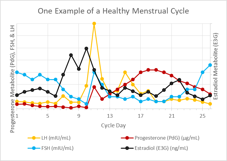
hormones across menstrual cycle
Figure 1. Hormone concentration throughout the menstrual cycle.

In Summary

This very quick overview of the menstrual cycle (aka ovulation cycle) forms the basis of every single fertility awareness method (FAM) today. Whether the method involves monitoring changes in cervical mucus, cervical position, basal body temperature, electrolyte composition of salivary/vaginal secretions, and/or at-home urinary hormone monitoring, these methods are highly reliable for predicting ovulation and are so reliable that their efficacy for avoiding unplanned pregnancy vies that of hormonal birth control.

These methods are also invaluable for shining light on a woman’s reproductive health and elucidating where hormonal imbalance lies within her cycle when things are a bit off. FAMs also provide real time data for women who are tracking their cycles so that you are able to adjust diet and lifestyle to support hormonal balance.

I will refer back to this article often in future posts on FAMs and hormonal health.

We Need Your Help

More people than ever are reading Hormones Matter, a testament to the need for independent voices in health and medicine. We are not funded and accept limited advertising. Unlike many health sites, we don’t force you to purchase a subscription. We believe health information should be open to all. If you read Hormones Matter, and like it, please help support it. Contribute now.

Yes, I would like to support Hormones Matter.   

Photo by Towfiqu barbhuiya on Unsplash.

Fluoroquinolones, Beta Glucuronidase and Oxalosis: New Connections

3660 views

Have you ever taken antibiotics and felt they had possibly both beneficial and detrimental effects on your health? There are plenty of scientific journals that debate just this (here, here). In the functional medicine world, we hear many stories from people about moderate to severe adverse reactions to fluoroquinolones and other antibiotics. This website has many such stories about the fluoroquinolone reactions and has published several articles on the mechanisms. One way fluoroquinolones wreak havoc on health that has not be discussed here is their role in oxalosis. There are several straightforward ways that antibiotics may increase oxalate, but I would like to share some new ideas about what changes they may make that might impact oxalate and inflammation.

What Are Fluoroquinolones?

Fluoroquinolones are a family of antibiotics that have a powerful bactericidal effect by inhibiting bacterial type II DNA topoisomerase. Due to their potent antibacterial effect, fluoroquinolones are most commonly used to assist with a wide variety of infectious conditions. This class of drug contains a black box warning from the FDA, indicating that it can cause serious issues like tendinitis, tendon rupture, peripheral neuropathy, myasthenia gravis and central nervous system effects. The negative effects of these drugs have become so widespread that a nickname has been coined for those who have endured fluoroquinolone injuries, called Floxies.

In our functional medicine circle we have heard many of their stories. In some instances, the original problem for which the antibiotic was prescribed becomes worse after treatment. In other cases, Floxies seem to experience major hormonal shifts with changes to their skin, collagen, hair, and connective tissues. In the worst cases, patients experience tendon ruptures, often of the Achilles, which is one of the more likely areas to be affected. Additionally, some patients develop sudden onset of allergic reactions to things in their environment or diet that they were not previously allergic too. Angioedema, skin rashes, peripheral neuropathy, and air hunger are some of the many other common symptoms that develop after using fluoroquinolone antibiotics. Many of these may be related to newly developing oxalate issues driven by these antibiotics.

What Is Oxalate?

Oxalic acid is an organic acid that is highly corrosive and elevated levels in the body correlate to having high inflammation. Oxalic acid binds to minerals in the body forming insoluble oxalate crystals that can deposit in organs, tissues, joints and the vascular system.

We can ingest exogenous sources of oxalate in the foods we eat, with certain plant foods like spinach having the highest level of oxalate. The liver can produce oxalate endogenously if one has certain SNPs in oxalate metabolism or nutrient deficiencies. There are many other ways to increase oxalate, including certain antibiotics, like fluoroquinolones.

The Antibiotic-Oxalate Connection

Research shows that antibiotics are not selective for only the bad bacteria. They kill off good bacteria as well. Humans cannot metabolize oxalate. We need help from our microbiota, particularly the oxalate degraders. Antibiotics will kill off many beneficial oxalate degrading flora, like oxalobacter formigenes, allowing oxalate levels to increase.

The bacteria in the gut also absorb and produce many B vitamins. Most of the vitamins synthesized endogenously are used by the bacteria themselves, but a small percentage is released into circulation. Both poor absorption and reduced synthesis of B vitamins will have many deleterious effects on health. Among them, increased oxalate synthesis and decreased metabolism and elimination.

Thiamine (B1) and pyridoxine (B6), for example, in addition to all of their other functions in human health, are instrumental in preventing endogenous production of oxalate. The bacteria that produce these vitamins are affected negatively by antibiotics.

Menaquinone or vitamin K producing bacteria also take a hit when antibiotics are used. Reduced vitamin K impacts calcium handling. Calcium homeostasis is critical for cell function in general and oxalate management in particular. It is also important for something called matrix Gla protein expression. Matrix Gla is a protein produced in the bone that inhibits vascular calcification. In other words, matrix Gla prevents the accumulation of oxalate, when expressed. When it is not expressed sufficiently, because vitamin K concentrations are diminished either by diet, antibiotic use, or both, oxalate will accumulate (here, here). Vitamin K is also critical in balancing hormones by multiple mechanisms. It is important to androgen/estrogen balance as seen in this study about PCOS. Hormonal balance plays a significant role in oxalate metabolism, which will be discussed later. These are just a few of the ways in which antibiotics contribute to an increase in oxalate and oxalate-driven inflammation

Beta Glucuronidase: A New Player in Oxalosis

In addition to the antibiotic-induced changes to oxalate-degrading and vitamin-synthesizing flora, antibiotics also alter carbohydrate metabolism. Antibiotics inhibit an enzyme called beta-glucuronidase (BG). BG is a lysosomal enzyme needed for the breakdown of complex carbohydrates and for the proper degradation of other small molecules like glycosaminoglycans (GAGs) such as hyaluronic acid, heparin/heparin sulfate, chondroitin sulfate/dermatan sulfate, and keratin sulfate and glucuronides such as bilirubin.

We have both human (encoded by the GUSB gene) and bacterially produced BG enzymes. Some main bacterial producers of BG are Escherichia coli, Clostridium species, Bacteroides species and Staphylococcus species. As men age, BG will increase. For women though, BG declines over time.

In disease states, certain enzymes become elevated and can therefore reliably indicate various pathological conditions. Beta glucuronidase is one of those enzymes. With metabolic disease and diabetes, for example, BG is elevated and the mix of gut bacteria shifts considerably.

Elevated BG also interrupts an important part of phase II liver detoxification called glucuronidation. This is where a glucuronic acid molecule is added onto toxic substances to inactivate and make them water soluble and easier to excrete. Beta glucuronidase can break the glycosylic bonds freeing the carcinogen and allowing its reabsorption and enterohepatic recirculation, rather than the proper clearance. Elevated BG, because of its impact on glucuronidation/phase II liver detoxification, will decrease the clearance of sex steroid hormones, xenobiotics, pesticides, herbicides, and insecticides, BPA, mycotoxins, pharmaceutical medicines, and more.

Elevated BG can be a marker for, and can contribute to, various issues. The recirculation of these carcinogens can contribute to colon cancer. The poor clearance of estrogens is associated with hormonal cancers like breast and ovarian cancer and endometriosis. In prostate cancer, upregulated BG is related to disease progression. It is not just cancers where BG is problematic, in chronic periodontitis, an 8-fold increase in BG activity has been recorded along with 33-fold increase in BG in bacterial peritonitis. This study details some of these observations.

This is where the fluoroquinolones and other antibiotics enter. A key job that they perform is to inhibit BG enzyme activity and the BG producing bacteria, pushing these levels back down into range. BG inhibition helps limit the recirculation of toxins that contribute to a vast number of diseases. A quick look at studies shows that fluoroquinolones like ciprofloxacin significantly inhibit BG. This study reveals that a combination of other antibiotics such as penicillin and metronidazole can have an inhibitory effect of up to 50% on enzyme activity.

What Happens When Beta Glucuronidase Is Too Low?

While it is clear that elevated levels of BG contribute to serious disease states, there are many substrates in the body that have to be at just the right concentration; where either too much or too little can be harmful to health. BG is one of these substrates. If fluoroquinolones and other antibiotics inhibit BG, and BG manages oxalate accumulation, could repeated use of these drugs over-inhibit BG such that oxalosis develops? I believe so. Below are a few of the pathways that may lead to high oxalate.

Beta-Glucuronidase Effect on Flavonoids

There is a strong relationship between inflammation and deconjugation. Conjugation and deconjugation are terms used to describe the addition or separation of molecules during liver detoxification.

When tissues in the body are experiencing necrosis, inflammation and mitochondrial dysfunction one of the most helpful antioxidants is the flavonoid. Once ingested, flavonoids like quercetin, which is the most abundant flavonoid in our diet, are conjugated via glucuronidation in the liver to their inactive metabolites making them easier to transport through the blood to these problem areas. Once they arrive, they must be deconjugated back into their active aglycone form so that they can be used to suppress the expression of pro-inflammatory genes such as COX-2 and scavenger receptors.

When the inactive flavonoids arrive at the inflamed tissues, phagocytic cells surround the problem area and secrete large amounts of both lactic acid and BG. The lactate is required to create the acidic environment needed for the most optimal BG function. One study shows a 13-fold increase of BG activity from pH 7.4 to pH 5.4. Then, BG secretion is needed for the bioactivation of the inactive flavonoid metabolites back to their aglycone form. From here, the flavonoids can go to work quenching the inflammation. If BG has been inhibited by fluoroquinolones or other antibiotics, flavonoids like quercetin will not be re-activated and will be unable to temper the inflammation. Here are three fascinating studies that explain this in detail (here, here, here). It appears that elevations in BG might follow inflammation, in an attempt to help resolve it

In addition to suppressing pro-inflammatory genes, the diuretic, antioxidant, anti-inflammatory, and antibacterial properties of flavonoids help inhibit the formation of calcium oxalate stones. If BG has been over-inhibited, such that these flavonoids cannot be reactivated, do we not then lose the oxalate preventative benefit that they provide? Possibly, which means oxalate levels would increase.

Ironically, flavonoids are potent inhibitors of BG. Is it possible they are used to resolve the inflammation then inactivate the BG so that its continued accumulation is not able to progress hormonal and other issues? Perhaps BG is involved in a natural feedback cycle of sorts.

Beta-Glucuronidase Effect on Glycosaminoglycans

As mentioned previously, BG is a lysosomal enzyme, which catalyzes the degradation of glycosaminoglycans (GAGs), such as chondroitin sulfate, heparin sulfate, dermatan sulfate and hyaluronan. When there is a mutation in the GUSB gene resulting in a deficiency of BG the GAGs are improperly degraded with consequent intraliposomal and tissue accumulation, creating lysosomal storage issues in a disease called mucopolysaccharidoses.

Now, there is some major interplay between oxalate and GAGs. GAGs are protective against the growth and formation of calcium oxalate crystals by having a strong binding affinity to them (here, here). There appears to be a low excretion rate of sulfated GAGs in stone formers, possibly due to improper degradation. And like GAGs, oxalate also needs to be phagocytized or engulfed into the lysosome where it can be dissolved. If they tend to bind together, it would appear that the increase in BG level by phagocytes may also be important for proper handling of oxalate.

In tendon rupture, a well-recognized side-effect of fluoroquinolone antibiotics, there is an accumulation of non-degraded GAGs (here, here). We know, through observations of people with hyperoxaluria, that oxalate crystals can accumulate around the joints and within tendon sheaths. Could the over suppression of BG by fluoroquinolones and other antibiotics contribute to their accumulation? Possibly.

Beta-Glucuronidase, Oxalate, and Estradiol

We know a consequence of elevated BG is estrogen dominance (here, here), so low BG activity could translate to lower levels of estradiol. Could repeated use of fluoroquinolones diminish estradiol concentrations such that it too influences oxalate build up? Once again, possibly.

Low estradiol (E2) might contribute to elevated oxalate in a couple of ways. Estradiol inhibits the activity of glycolic acid oxidase (GAO), thereby decreasing the amount of glyoxylate converted to oxalate within the peroxisome. Studies show that E2 is significantly lower in kidney stone patients with a significantly higher frequency of calcium oxalate stones in the lowest E2 groups.

Estradiol is the body’s natural anti-androgen via its suppression of 5 alpha reductase, which converts testosterone to the substantially more potent androgen receptor ligand, dihydrotestosterone (DHT). So, it can also help to manage oxalate level by keeping DHT levels in check. A quick look at studies will show that androgens increase oxalate level and stone formation by increasing GAO activity, and by suppressing osteopontin levels while estrogens elevate it, respectively.

However, like BG, E2 is also one of those substrates that has to be kept in balance. There can be harmful effects if E2 becomes either elevated or deficient. This study shows that E2 might attenuate oxalate transport activity via the downregulation of SLC26A6 and therefore enhance cancer cell migration, creating a potential risk for nephrolithiasis and cancer.

Fluoroquinolones, Beta-Glucuronidase, Estradiol and Tendinopathies

Estradiol is very important to maintaining connective tissue in women by improving the tendon collagen synthesis rate and increasing the number of smaller versus larger fibrils. Conversely, this study shows that the natural ageing process, whereby estradiol declines, increases a woman’s risk of tendon rupture to a similar level as seen in men. They postulate that a reduction in tensile strength, decrease in collagen synthesis, fiber diameter, density, and increase degradation in tendon tissue, all play a part. This means that both elevated and diminished estradiol influence connective tissue. Consider, for example, pregnancy in which E2 is quite elevated. It is the E2 induced changes to various collagen synthesizing genes that allow the laxity and the stretching of tendons, ligaments and skin. But, going to the other extreme could low estradiol induce excessive rigidity, also contribute to injury? When fluoroquinolones are used, they inhibit beta glucuronidase. Since BG inhibits estradiol, it is possible that this is one of the mechanisms contributing to the rigidity seen in tendinopathies and ruptures after fluoroquinolone use.

It All Comes Back to the Gut

It is in the gut where the oxalate degrading flora live. It is in the gut where the microbes that absorb and synthesize vitamins and minerals live. It is in the gut where the microbes that produce short chain fatty acids and beta glucuronidase that help balance the estrobolome and other hormones live. All of these factors intersect to manage and determine our inflammation level. When we kill these microbes with antibiotics and other ‘kill tools’ we inevitably impact oxalate metabolism. Fluoroquinolone induced inhibition of beta glucuronidase may be a key player in the cascade leading to oxalosis. While the inhibition of BG may be useful when concentrations are too high, repeated or over-inhibition may be deleterious to health.

The good news is, the body wants to regain homeostasis when pushed out of balance. The cessation of any antibiotics that are depressing BG will allow it to recover over time. Perhaps by taking these other factors and associated biomarkers into consideration we might be able to gently push into some level of homeostasis more quickly to regain health.

We Need Your Help

More people than ever are reading Hormones Matter, a testament to the need for independent voices in health and medicine. We are not funded and accept limited advertising. Unlike many health sites, we don’t force you to purchase a subscription. We believe health information should be open to all. If you read Hormones Matter, like it, please help support it. Contribute now.

Yes, I would like to support Hormones Matter.

Photo by Vardan Papikyan on Unsplash.

Could Altered Vitamin A Metabolism Be Responsible for Endometriosis and Fibroid Growth?

16074 views

Yes, and increased use of environmental toxicants may be partially to blame. Over the last decade researchers have uncovered connections between tissue level vitamin A activity – the retinoic acid pathway – hormone metabolism, and the cell cycle overgrowth noted in fibroid tumor development, breast and ovarian cancer, and endometriotic tissue growth. Moreover, researchers from the environmental side have found that the popular glyphosate-based herbicides alter vitamin A or retinoic acid metabolism which in turn alters androgen and estrogen metabolism. Connecting the dots, we may have a first step to reducing cell growth in these conditions; remove the toxicant exposure and increase nutritional resources. A second step may be to develop locally absorbed vitamin A, applied directly to the aberrant tissues.

What is Vitamin A?

Vitamin A, (retinol, carotene) is a fat-soluble nutrient that we derive solely from dietary sources. It is responsible for a myriad of functions in a vast number of tissues from the eye, to the ovary, to the heart. Historically, nutrition from diet, coupled with the old wives’ tales of good health, carrots for eyesight, and cod liver oil for all that ails you, were all that were needed to maintain healthy levels of Vitamin A in most individuals. However, with the increase in processed foods, modern farming, intense use of herbicides and pesticides, and the general replacement of the old wives’ nutritional wisdom with pharmaceuticals, many men, women, and children are vitamin A deficient and likely do not even know it. The WHO estimates vitamin A deficiency in 19 million pregnant women and 150 million children worldwide. When Vitamin A deficiency reaches its nadir night blindness, maternal mortality, and difficulty fighting infections are common. In women, the first signs of vitamin A deficiency may be unrecognized and include fibroids or endometriosis. Earlier signs of vitamin A deficiency in women could also be menorrhagia (heavy menstrual bleeding) that often precedes fibroid or endometriosis diagnosis, but research is lacking here, or even genital warts of the common HPV strains.

Why Retinoic Acid, Hormones, and Cell Growth

Retinoic acid (RA), is the form of vitamin A stored in the body. RA is what is called a paracrine, perhaps even an intracrine hormone regulator. That means it turns hormone metabolism on or off in the cells within its immediate vicinity (paracrine) or within its own cell (intracrine). This is compared to endocrine control of hormone metabolism – where hormones and the factors that regulate hormone synthesis and metabolism travel vast distances through the blood to reach their targets tissues (the hypothalamus-pituitary – ovarian system is an example of endocrine regulation) or autocrine where the hormone leaves its own cell only to turn around and bind to a receptor on that cell. In contrast, retinoic acid stays close to home and regulates local cell behavior, both internally and proximally. The vitamin A deficiency leading to fibroids or endometriosis represents a cell and tissue level disruption of the retinoic acid pathway that in turn interrupts the normal cell cycle (differentiation, proliferation, and apoptosis -cell death) and elicits all sorts of problems from decreased estrogen metabolism (too much estradiol at the cells), to cell overgrowth, or more specifically, not enough cell death where needed. The results include aberrant cell growth as in fibroids, tumors, and endometriosis.

Retinoic Acid, Progesterone and Estrogen Metabolism

With many women’s health conditions too much estradiol at the tissue level is at the root. Estradiol is an excitatory hormone that tells our cells to go forth and prosper. Progesterone, depending upon the tissue and the relative values of each circulating hormone can work synergistically to enhance estradiol’s actions or it can shut it down entirely via the upregulation of a specific estradiol metabolizing enzyme called 17 beta-hydroxysteroid dehydrogenase type 2  (17B -HSD2).  When these enzyme levels are high, more estradiol is converted to estrone. Since estrone is a less potent estrogen than estradiol, metabolism of estradiol to estrone somewhat inactivates the estrogen and slows cell proliferation. When the enzyme levels are low, more estradiol remains, and cell growth is enhanced.  Vitamin A or retinoic acid mediates the progesterone-dependent activation of this enzyme, effectively regulating estradiol concentrations locally. Too little retinoic acid or a disrupted retinoic acid pathway and estradiol is not converted to estrone – e.g. it is not inactivated. Cell proliferation dominates, while normal cell death or apoptosis is reduced. Fibroids, tumors, or endometriosis ensue.

What Causes Low Retinoic Acid or Reduced Functioning?

Vitamin A is derived entirely from diet. Foods high in vitamin A include brightly colored vegetables, dark leafy greens, carrots, pumpkin, sweet potatoes, bell peppers, and fatty fish oils, like cod liver oil and organ tissues like the liver. Meat and dairy also have high concentrations of vitamin A. Diets high in processed food do not contain sufficient vitamin A to maintain the proper cell cycle balance and so we get too much proliferation and too little apoptosis. Tissues grow and grow and do not die.

Alcohol intake reduces the body’s ability to metabolize retinoic acid because alcohol and the retinoic acid pathway use the same enzymes – alcohol dehydrogenase (ADH1) and aldehyde dehydrogenase (ALDH1) for metabolism. Alcohol competes for the enzyme and so vitamin A from diet cannot be converted to the usable retinoic acid.

Can Toxins Disrupt the Vitamin A Pathway?

Yes, but here is where it gets complicated. Environmental toxins like glyphosate used in common weed killers such as Round-up have a complex relationship with the vitamin A pathway and hormone metabolism. These herbicides and many pesticides are endocrine disruptors, meaning they disrupt ‘normal’ hormone metabolism, often towards a hyper-estrogenic state. Similarly, plastics like BPA and a host of industrial chemicals are also endocrine disruptors that move us towards hyper-estrogenism – a key component of fibroid and endometriosis.

Glysophate activates an enzyme called retinaldehyde dehydrogenase which increases retinoic acid synthesis. This is argued to be the mechanism by which environmental exposures during pregnancy cause birth defects. However, glyphosate also inhibits vitamin A metabolism by a similar mechanism as alcohol, by competing for ADH1 availability, thereby having the ability to reduce vitamin A synthesis. Glyphosate also increases aromatase activity (the enzyme that converts testosterone to estradiol), creating a hyper-estrogenic state and depending upon the time course and the exposure concentration, completely wipes out aromatase activity. So like any true hormone system, that uses a complex chain of compensatory reactions to maintain homeostasis, the reactions to environmental toxins are complicated and non-linear. Nevertheless, they warrant attention, particularly when one is suffering from a condition affected by the environmental toxin in question.

Managing Vitamin A Levels

To determine if you are vitamin A deficient, seek out a lab that specializes in micronutrient testing. The recommended daily values of vitamin A can be found in the Dietary Supplement Fact Sheet.

Vitamin A is a fat-soluble vitamin, meaning that it will be stored in fat, and toxicity from too much vitamin A is possible. It is rare, but nevertheless, if supplementing, vitamin A levels should be monitored by micronutrient testing.

My Two Cents

Much of the research presented here linking local vitamin A deficiencies with endometriotic, fibroid, and cancer growth has not crossed over into clinical care. Moreover, it is complex and far from settled. Except for cancer trials, mostly in males and mostly with oral supplementation, the research regarding dietary vitamin A is limited and mixed. However, I think a local application of an absorbable form of vitamin A or retinoic acid should be investigated for the treatment of endometriotic and fibroid growth in women. Similarly, dietary supplementation within acceptable levels and changes combined with environmental ‘cleaning’ may be of use, if only to improve the overall health status of women currently suffering from fibroids or endometriosis.

Postscript: This article was published previously in August 2013. 

Photo by Tamanna Rumee on Unsplash.

Hormones, Hysterectomy, and the Aging Brain

11546 views

Everything slows down as we age. For some lucky folks, aging happens gracefully with nary a disease in sight. For others, the springs start popping off around 40 and by the time we reach ‘old age’ our bodies and brains are barely functioning. Arguably, diet and lifestyle have something to do with how well or how poorly we age, and of course, genetics contribute mightily, but beyond that, we really have no idea what’s happening with aging.

Sure, there are all sorts of physiological systems that become progressively less efficient over time. Wear and tear plays a huge role, but the relationships aren’t linear. There are always outliers. There are folks who, on a diet of smokes and scotch, live well into their nineties with all their faculties intact. Then there are the poor souls who are prodigiously healthy, who eat right and exercise, but yet, whose bodies seem set on wide-scale destruction, where the slightest change in lifestyle risks sending them into a morass of cascading illness. Somewhere in the middle, the rest of us live – sometimes healthy, sometimes not – aging in fits and spurts. What the heck?

From a physiological standpoint, aging is marked by two opposing factors: decreasing hormones and increasing inflammation. Where they intersect, age-related illnesses seem to accrue. Called endocrine senescence, researchers have long noted a relationship between declining hormones and declining immune function (marked by increased and inefficient inflammatory responses). Might there be some truth to the ever-young, hormone peddlers? Could hormones be the key to offsetting the age-induced inflammatory cascades? Possibly.

Hormones and Mitochondria

I just finished writing an extensive paper on acquired mitochondrial illness. Throughout the research, I stumbled upon a short essay linking mitochondrial structure and function to estradiol. More specifically, the rapid estradiol decline common post oophorectomy (ovary removal), fundamentally alters the shape, and ultimately, the function of mitochondria. Researchers found that a rapid decline in estradiol evokes significant damage in the brains (and presumably other organs) of female monkeys. Additional studies using estradiol starved mitochondria from female rodents showed similar shape alterations and consequent declines in brain bioenergetics. Interestingly though, with natural menopause, where estradiol declines more gradually, no such structural changes were observed. In fact, with the more gradual decline in estradiol, the mitochondria appear to increase their production of the lifesaving ATP as a compensatory reaction.

All Paths Lead to the Mitochondria

Recall, from previous posts, that mitochondria take dietary nutrients and oxygen, and change them into the chemical energy (ATP) that is used by every cell in the body. Without ATP, cell function grinds to a halt. So, anything that derails the mitochondria, imperils cell function and initiates cell death. Lack of nutrients, sedentary lifestyle, pharmaceutical, and environmental toxicants, all derail mitochondrial function. Cluster too much cell death together in one tissue or one organ and disease happens. Since mitochondria are in every cell of the body, mitochondrial damage induces disease broadly, but especially in regions with high energy demands like the brain, the heart, the muscles, and the GI system.

The cardinal symptoms of mitochondrial damage include fatigue, weakness, muscle pain, and depression. These are followed by dysregulated systems; a GI system, for example, that overreacts or under reacts or temperature dysregulation (hot flashes, cold insensitivity), insulin/sugar dysregulation, emotional volatility, migraines, seizures, syncope (fainting), and so on. It’s not a pretty picture.

In addition to providing the fuel for cellular respiration, e.g. life, mitochondria control a host of other functions, steroidogenesis is one of them. This means that if we fail to feed the mitochondria or hurl insults at them, hormone dysregulation is inevitable. Ditto for inflammation, as the mitochondria regulate inflammatory cascades. Every woman knows when her hormones are out of whack. Well, now we know that hormone dysregulation emerges from the mitochondria.

From a systems perspective, consider the mitochondria as central regulators of organismal health. Mitochondria both send and receive signals from all over the body and then adjust their functioning accordingly. With their role in hormone synthesis, we would expect there to be cross-talk between the mitochondria and circulating hormones. Indeed, there is. All steroid hormones have receptors on the mitochondrial membranes. When hormone concentrations increase or decrease, the mitochondria will initiate the synthesis of new hormones and send signals throughout the body to adjust other hormone-responsive systems as well.

No Estradiol Equals Misshapen Mitochondria: Donuts and Blobs

Removing the ovaries starves the mitochondria of one of its many feedback mechanisms and damages the brain mitochondria in the regions of the brain responsible for executive function and memory – the frontal cortex and the hippocampus. The mitochondria change shape, from spheres (healthy) to donuts and blobs, which represent early and late-stage mitochondrial damage, respectively. Misshapen mitochondria cannot provide the energy (ATP) needed to perform critical brain functions such as neural communication or the antioxidant tasks needed to clean up toxicants. Neurodegeneration ensues. In layman’s terms, and in the early stages, brain fog and memory loss. Researchers believe that it is this loss of functional mitochondria that contribute to the onset of neurodegenerative disorders like Alzheimer’s and other dementias. And, this loss of function is precipitated by an unnatural loss of estradiol.

Ovary Removal is Common with Hysterectomy – Now What?

For the millions of women who have had their ovaries removed with hysterectomy, this presents a problem. Amid the myriad of other side effects associated with ovary removal, and perhaps, the root cause of these effects, we can add mitochondrial damage and brain mitochondrial damage, specifically. The rapid decline of estradiol, and other hormones, places many women at risk for neurodegenerative disorders like Alzheimer’s. How could this be mitigated?

In animal research, hormone replacement with 17B – estradiol immediately after the ovaries are removed seems to temper the damage, at least in the short term. There are no long-term studies. Similarly, epidemiological studies in human women suggest hormone replacement immediately after open menopause and/or hysterectomy with oophorectomy reduces clinical symptoms associated with the diseases of aging – e.g. the cognitive decline of Alzheimer’s and other dementias. However, since the synthetic estrogens used pharmacologically are different compounds than those produced endogenously (and used in basic and animal research) and because there are no mitochondrial imaging or even mitochondrial function tests done with human females given hormone replacement, it is difficult to compare the two sets of literature.

Some data suggest that the use of synthetic estrogens damages mitochondria and further diminishes the synthesis of remaining endogenous estrogens (the adrenals continue to produce estradiol and other estrogens after the ovaries are removed). Women who have used synthetic estrogens such as those in oral contraceptives and hormone replacement therapies have lower concentrations of endogenous estradiol, estrone, androstenedione, testosterone, and sex hormone-binding globulin. Based upon the aforementioned research, the decline in endogenous hormones would suggest a commensurate derangement in mitochondrial structure and function, but there are no data either way. At the very least, caution is warranted when contemplating the use of synthetic estrogens, particularly in the current environment that is rife with estrogenic chemicals. There are no data on the use of ‘natural’ or ‘bioidentical’ hormones and human mitochondrial function. So, although the animal data are fairly clear, estradiol replacement begun early enough appears to offset the decline in endogenous estradiol, how this translates to human females is not known.

Other Hormones and Additional Pathways

A flaw common to most research in this field is the failure to address the other hormones involved in modulating health. Estradiol is but one of many estrogens produced endogenously. It is also one of many steroid hormones produced in the ovaries and regulated by mitochondrial function. How estradiol removal or add-back affects progesterone, the androgens, or even the glucocorticoids (cortisol) – is not known. Compensatory reactions are likely. Understanding how those reactions mediate mitochondrial function might determine a viable workaround for the depleted estradiol. The beauty of human physiology is a mind-blowing breadth and depth of compensatory reactions to maximize survival. So I would think, and this is purely speculative, that even if one has lost her ovaries, and even if estradiol treatment was not initiated immediately, or if synthetic estrogens were used instead, there should be other mechanisms to tap into and compensate for this loss. That is, there should be multiple pathways to help maintain mitochondrial function. What those are, I do not know, but they are worth exploring.

We Need Your Help

More people than ever are reading Hormones Matter, a testament to the need for independent voices in health and medicine. We are not funded and accept limited advertising. Unlike many health sites, we don’t force you to purchase a subscription. We believe health information should be open to all. If you read Hormones Matter, and like it, please help support it. Contribute now.

Yes, I would like to support Hormones Matter.

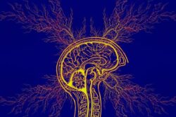
Image created using Canva AI.

This post was published originally in January 2015.

Mommy Brain: Pregnancy and Postpartum Memory Deficits

15931 views

Pregnancy and postpartum memory problems are common complaints amongst new moms. Are they real? Some research says yes, other research says no.

In graduate school I studied this issue and completed several studies on pregnancy and postpartum mood and memory changes. In one such study, I ran a full battery neuropsychological tests on a group (n = 28) of highly educated, healthy, medication free, first-time moms. We tested in late pregnancy and within 10 days following the delivery of the child. We also measured a range of hormones (progesterone, DHEAS, testosterone, estrone, estradiol and estriol) to determine what relationship the radical hormone changes of pregnancy and childbirth had on a woman’s cognitive ability. We knew from animal research that steroid hormones could affect learning in very significant ways. It wasn’t that difficult to suspect the same would be true of human women.

The study was never published, rejected from at least three, maybe four journals and has been sitting in a file ever since (along with a number of other studies). With the open access and open science movements growing, I decided it was time for this research to see the light of day. I will be self-publishing much of my research over the coming weeks and months. Here is the first study. Understanding Maternal Cognitive Changes: Associations between Hormones and Memory.

Is the Mommy Brain Real?

More importantly, are hormones to blame?  The answer is yes on both counts. We found that pregnant and postpartum women exhibited detectable cognitive deficits across multiple domains. The deficits were worse in late pregnancy and mostly improved postpartum. These memory problems were linked to both the excessively high hormones of late pregnancy, the low hormones following delivery, and the large changes in hormone concentration from pregnancy to postpartum.

What Types of Cognitive Deficits?

Pregnant and to a lesser degree, postpartum women had difficulty sustaining focus – this may be the mommy brain fog that many women complain of. We also found that during pregnancy especially, women were unable to manipulate and organize incoming information effectively. This presented as poor performance across a number of tests that assessed both short and long term memory.

In the case of verbal memory, these highly intelligent (estimated average IQ was 114 – 119) and educated (average years of education was 16 years) women tested in the low single digit to the 20th percentiles across multiple IQ-adjusted verbal recall measures. Even when estimates of IQ were not used to adjust scores, the participants performed poorly compared to normative standards. This was surprising given that many of these women had advanced degrees and were working in professional capacities.

The verbal tests involved remembering lists of words; words that could be grouped into meaningful categories that would improve memory significantly. Most of the study participants had difficulty grouping the words into categories. Instead, they would attempt to remember by rote sequence, which is always much more difficult. They also exhibited high numbers of intrusions – recalling words that were not in the original lists and repetitions – repeating words.

Similarly, and more strikingly visible, visual- spatial memory was marred by the inability to group bits of information and perhaps even to see the groupings in the first place. In this test, the study participants were given a complex figure to copy (shown below). They were not told that they would be asked to recall and redraw the picture later. When asked to redraw the figure, the inability to see the totality of the picture, to group bits of information was apparent.

Visual – spatial memory deficits as assessed by the Rey Complex Figure Test. Marrs et al. 2013, © 2013 Lucine Health Sciences, Inc. All rights reserved.

Does Memory Improve Postpartum?

Interestingly, while spatial memory improved significantly from pregnancy to postpartum, verbal memory did not. And this is probably what troubles women the most, the perceived deficits in verbal memory. Most of us think in words, when our ability to find words, retain words, organize information effectively is compromised, we notice.

Hormones and Memory

Both high levels of late pregnancy estrogens, (estrone, estradiol and estriol – we measured all three) and the low levels these estrogens postpartum were correlated with multiple measures of diminished memory, attention and processing. Additionally, the larger the change in the circulating levels of estrogens from late pregnancy to early postpartum was associated with poor memory postpartum. Indeed, women who had higher postpartum estradiol and estriol specifically, performed better on measures of verbal memory than those who did not.

Progesterone, long thought to be associated with cognitive function, primarily because of its sedative properties, was not associated with any measure of cognitive function at either test time, although large changes in progesterone were associated with some performance measures. DHEAS and testosterone, not often measured in pregnancy, postpartum or even in women’s health in general, were also associated with a few measures of cognitive functioning.

What This Means

Ladies, you are not imagining the pregnancy memory problems. They exist and they are related to the hormones. Most women knew this already, but it took a while for science to catch up. Not to worry though, the memory problems do resolve as the hormones stabilize (my next study to be self-published – a long term follow-up). Read the full study for all the details: Understanding Maternal Cognitive Changes: Associations between Hormones and Memory.

We Need Your Help

More people than ever are reading Hormones Matter, a testament to the need for independent voices in health and medicine. We are not funded and accept limited advertising. Unlike many health sites, we don’t force you to purchase a subscription. We believe health information should be open to all. If you read Hormones Matter, like it, please help support it. Contribute now.

Yes, I would like to support Hormones Matter. 

This article was originally published on Hormones Matter on March 26, 2013.

Tackling the Contraceptive Conundrum: Questions and Answers

3953 views

Last weekend I had the privilege of speaking at a much overdue conference on hormonal contraceptive safety – the Contraceptive Conundrum. I was charged the unenviable task of giving the ‘overview of everything’ talk and providing a framework through which to view these medications; not easy in a 45 minute presentation. Needless to say, there was a tremendous amount of information omitted from my talk. I will be sharing some of this information in series of blog posts over the coming weeks. The presentation was videotaped and I will post it when it becomes available. For the time being, however, I would like to offer up the power point (below) and answer some of the questions posed by audience members that I was unable to address or address fully given the time constraints.

Best Medical Journals

One of presumably less controversial questions I was asked was which medical journals I prefer. As it turns out, even this question inspires indignation on social media. I am strong proponent of open access journals and the entire open data movement. I believe that health research should not be hidden behind a paywall and the raw data behind drug safety trials ought to be readily available for independent analysis and scrutiny. Indeed, all science should be in the public sphere and a part of public discourse. As a matter of course, science should not be available only to the privileged few. The mere suggestion that I prefer open access journals, however, ignited a heated debate on Twitter; the instigators of which suggesting this preference supersedes attempts to access paywalled articles. Let me assure you it does not. I always track down primary sources. Nevertheless, for the reasons stated above and many more, my preference is for open access journals.

Hormonal Contraceptives and IVF

Another audience member asked about the research and risks associated with the use of hormonal contraceptives and IVF. I should preface my response with a disclaimer: I am no expert in IVF, however, I have written about fertility medicine on a number of occasions (here, here, here), mostly with regard to this specialty’s hubris and egregious lack of insight or concern regarding the longer term consequences of many of their practices. As a point of consideration, I write about the hubris and lack of research that pervades all of women’s healthcare. Those are my biases, do with them what you will.

As far as the use of hormonal contraceptives and IVF are concerned, the research is mixed at best and unacceptably limited in scope. The reasoning for using oral contraceptives in advance or in conjunction with IVF treatments ranges from the ease of cycle scheduling to a purported increase in oocyte yields. From an IVF expert:

In my view, it is not only acceptable, but even ideal to take the BCP [birth control pills] for at least one cycle prior to starting COH [controlled ovarian hyperstimulation] in preparation for IVF. Doing so allows one (without prejudice) to better plan and time cycles of IVF. Furthermore, since the BCP also suppressed LH, it is often especially advantageous in older women, in women with diminished ovarian reserve and in those with PCOS (in whom high LH levels can compromise egg/embryo quality). 

Despite the perceived utility of these medications, some research suggests that perception diverges from reality. In fact, the use of oral contraceptives in IVF may not be beneficial in increasing oocyte yields or pregnancy outcomes, especially in older women with limited oocyte reserve. A recent study, Does hormonal contraception prior to in vitro fertilization (IVF) negatively affect oocyte yields? – A pilot study found that even in young women with sufficient oocyte reserve, combined oral contraceptives diminished the number of oocytes retrieved compared to women who were not given oral contraceptives. The androgenic contraceptives were most deleterious. This comes on the heals of a Cochrane Review that found that not only was there limited research on the topic, but oral contraceptives resulted in poorer pregnancy outcomes. Missing from these data are the very real risks to maternal health mediated by the cocktail of hormones used in IVF (Lupron being top among them, followed by dexamethasone) and the potential long-term consequences to the health of the children born from IVF. Despite the lack of data and the often contradictory research findings, the practice of using oral contraceptives in IVF is well entrenched.

Hormone and Other Differences Between Oral Contraceptives, Depo Provera, NuvaRing and the IUDs

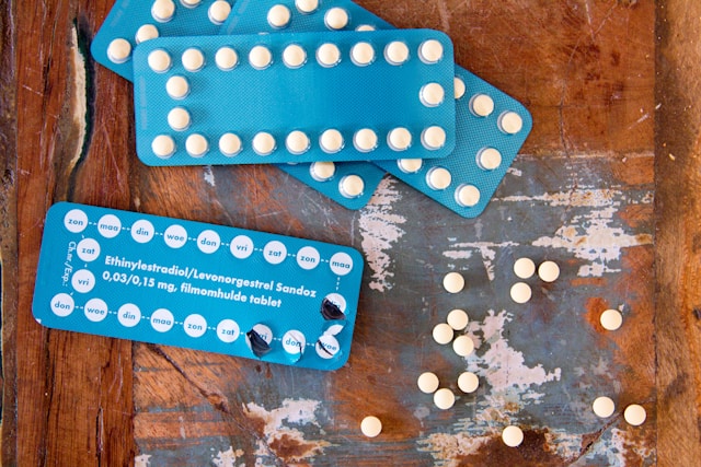
From the hormonal perspective, the various forms of contraceptives differ mostly by the type of synthetic progestin used. Oral contraceptives use a variety of progestins (see here), while Depo Provera contains medroxyprogesterone, hormonal IUDs utilize levonorgestrel and NuvaRing uses etonogestral. Most of the oral contraceptives contain the synthetic estrogen, 17a-ethinylestradiol, as does NuvaRing. Depo Provera is a progestin only, injectable form of birth control while the hormonal IUDs are a slow-release progestin only contraceptives. In addition to the differences in formulation and dose, each of these methods utilizes a different different delivery mechanism. The delivery mechanism will affect how much of the drug is absorbed and bioavailable, how quickly, the duration of availability, and those variables (along with several others), then affect the risk for side effects. Videos on pharmacokinetics and pharmacodynamics can be viewed here (dynamics video follows).

How Do Oral Contraceptives Affect Mitochondrial Morphology and Replication?

While there is a noticeable lack of data in this area, there are clear indicators that ethinylestradiol induces both structural and functional damage to mitochondria in the liver and the kidney, at least in rodents. Liver biopsies of women using oral contraceptives have also demonstrated structural changes in mitochondria. I would suspect similar changes in mitochondria throughout the body.

Indirectly, we know that reduced endogenous estradiol concentrations (herehere, here) damage mitochondria and that women who use oral contraceptives have lower endogenous estradiol concentrations. We also know that oral contraceptives deplete vital nutrients that are critical for mitochondrial functioning. And we know that the metabolism of 17a ethinylestradiol, the estrogen used in hormonal contraceptives, oral and otherwise, does not follow the same path as endogenous estradiol, and thus, likely damages mitochondria. (Ethinyl estradiol metabolism produces what are called catechol estrogens. Catechol estrogens are both directly (DNA adducts) and indirectly (mitochondrial reactive oxygen species – ROS- evoked as a byproduct of the metabolism) implicated in animal models of cancer.) Complicating matters, however, endogenous estradiol depending upon the concentrations, can have both pro – and anti-oxidant properties and impact mitochondrial functioning both positively and negatively. Nevertheless, I would argue that the synthetics derail the balance of endogenous hormones and because of their very real structural and functional differences, evoke a number of processes that are not only distinct from those of the endogenous estrane hormones but are likely damaging in ways we have not yet begun to understand.

Presentation

We Need Your Help

More people than ever are reading Hormones Matter, a testament to the need for independent voices in health and medicine. We are not funded and accept limited advertising. Unlike many health sites, we don’t force you to purchase a subscription. We believe health information should be open to all. If you read Hormones Matter, like it, please help support it. Contribute now.

Yes, I would like to support Hormones Matter. 

Photo by Simone van der Koelen on Unsplash.

Migraines and Hormones: Behind the Curtain

7194 views

Before puberty, migraines are three times more frequent in males than in females but after puberty the tides turn and females are more likely to suffer from migraines than males. An Oxford study found that females are twice as likely to have migraines and that

“brains are deferentially affected by migraine in females compared with males. Furthermore, the results also support the notion that sex differences involve both brain structure as well as functional circuits, in that emotional circuitry compared with sensory processing appears involved to a greater degree in female than male migraineurs.”

The overwhelming belief is that the connection is clear: the hormones kick in for women at puberty and that must be the reason. This begs the questions: 1) Do males have the same hormonal problems before puberty as females do after puberty? If hormones are at root of the problems, then there must be some similarities, right? 2) If female hormones are responsible for migraines, do all females have migraines when they reach puberty? 3) Do migraines cease when hormones stop changing after menopause? 4) What about pregnancy or postpartum, how do hormones impact women then? And finally, 5) Do men stop having migraines after puberty?

Some of the answers to these questions will surprise you and may make you wonder if hormones have anything to do with migraines at all. In this post, I show you that while there are some connections between hormones and migraine they might not be the primary drivers of migraine. The relationship between hormones and migraine is not in the presence of hormonal changes but what those changes require in terms of brain energy, the lack of which causes migraines.

First, I would like to respond in quick the five questions I asked earlier: 1) Do males have the same hormonal problems before puberty as females do after puberty that causes them migraines? The answer to this is no. 2) If female hormones are responsible for migraines, do all females have migraines when they reach puberty? The answer to this also is no. 3) Do migraines stop after menopause? Many women have more migraines and some even start migraines in their menopause, so the answer is no. 4) Do migraine increase or decrease during pregnancy or postpartum? The answer is no during pregnancy, but yes postpartum. 5) Do men stop having migraines after puberty? No they do not.

It is not obvious that the cause of migraines must have anything to do with female monthly cycles and their associated hormones. Given also that many women have migraines after puberty, we are safe to assume that some other factors may play a role. It would be hard to envision a world full of children in which our evolutionary road took women to necessarily experience migraines with their menstrual cycles. So what is the connection to hormones; how do women end up with migraines; and why?

Rather than listing all the hormones that activate throughout the monthly cycle of a woman, let’s take a look at what is happening in the body of that woman backstage, during the hormonal changes. First, in a small review I cover in a few sentences what a migraine is.

Migraine is a collection of symptoms that have an underlying physiological mechanism based on chemical (ionic) imbalance in the brain. Migraine is a neurovascular event that Dr. Charles at UCLA called “spectacular neuro-physiological event” that changes the neurophysiology or chemistry of the brain itself. This can be seen using fMRI technology where oxygenation of brain regions shows where activity occurs during migraine—albeit this does not show why it occurs. The same article also suggests that though medications are available to treat the pain associated with migraines, half the sufferers do not receive any pain relief benefit from the drugs. I find this statement alone interesting because if migraine was truly understood, the pain medication would work for all. This clearly is not the case. To understand what is happening, we must think out of the box and leave behind the hormonal theory of migraines.

Moving Beyond the Hormone Migraine Theory

We now visit the female body all through a month. Let’s start two days after her menstrual cycle has ended. As female, we feel great, no pain, no bleeding, life is awesome. But what we don’t see works hard in the background using up important energy: the brain. Our hormonal changes are happening every moment of the day only we don’t feel it—hormonal changes are directed by the brain. Because we don’t feel the changes, we are ill-prepared for the inevitable day when it reaches a threshold point of not enough brain energy and the migraine starts. This typically happens 2-4 days prior to menses. I do not think migraines are caused by hormones, but rather they are triggered by the lack of energy available to the brain as the hormones cycle. When the brain runs out of energy, a wave of cortical depression begins in some part of the brain. This is what we feel as a migraine.

What actually happens that uses all that energy? After the menstrual cycle is over, the female body immediately prepares for the next menstrual cycle. There is no downtime for rest. The brain turns off one group of hormones and turns on others thereby manipulating how women see the world prior to and during estrus (fertile time). After a menstrual cycle is over, the brain turns on the estrogen to do a few things:

  1.  Prepare the uterus with a new fertile lining to accept the fertilized egg should one arrive and start a new life.
  2. In order to make such fertilized egg happen, the egg must be prepared in the ovaries so hormones initiate the ripening of a new egg.
  3. The woman’s body goes through amazing visible changes at this time of the month. If she had pimples, they magically disappear. If she was bloated, her bloating goes away. Her face becomes the most symmetrical it possibly can; the more symmetrical the more sexually appealing she becomes to the opposite sex.
  4. She becomes extremely attracted to high testosterone males requiring her pheromones to change and to be able to sense a high testosterone pheromone male’s presence. This high testosterone attraction changes after estrus to attraction to low testosterone males for the safety of the child, should mating end in a baby.

With all this activity going on in the female body that she cannot feel, she is in danger of exceeding the threshold of brain energy-shortage without prior notice or preparation. The cost of all of these activities behind the curtains in the female body is very high in terms of brain energy and hydration.  These are sex-hormonal functions that only exist for a certain period of time during the female life. Females are known to be born with all of their eggs they will ever ripen for possible babies. Only these eggs are not “ripe” at birth. Every month one egg ripens in one of two ovaries (sometimes in both and sometimes in none). This egg breaks out of the ovary and starts its journey down the ovarian tube where it either gets fertilized by a sperm or not. If the egg is fertilized, it attaches to the wall of the uterus lining—later to become the placenta of the baby—and a new life cycle begins in the mother-to-be. If however there is no sperm able to penetrate the egg, while it descends in the ovarian tube, the egg will have to be cleared from the uterus together with the nutritious blood vessel rich lining created. This happens with the menstrual bleeding. This we can see and feel.

My Theory: Why Hormone Changes are not the Cause of Migraines

As shown earlier, migraines are not equally present in everyone’s life. Other factors, such as genetic predisposition to sensory organ hyper sensitivities (SOHS) that require more energy, may be the cause. Recent research hints at ionic balance (meaning energy available for use) is crucial in maintaining optimal function and the slightest imbalance can cause major problems (Wei et al.).

When the body is tasked with demanding activities the cells responsible for completing those extra tasks are doing extra chores and need extra energy. The brain regulates the creation of extra hormones for the menstrual cycle. The brain manages the clearing of the uterus after the fertile layer was not used.

By the third day after the cycle, the brain is ordering an egg to ripen—this takes extra energy. This is a once a month event. The brain must have extra energy to complete this task. Ever tried to run a marathon on empty or run your car the extra mile without fuel in your tank? Not possible. Something must break. The brain is the logical one for those who are predisposed to SOHS. If their brain runs out of energy, the neurons cannot generate voltage and stop creating neurotransmitters that instruct the production of hormones in the body. This leads to cortical depression and migraine.

Migraine during Pregnancy

Hands up: how many of you had migraines during pregnancy? Up to 75% of migraineurs do not have migraines during pregnancy. Why you may ask? There is more than one reason for this. The first and most important reason is that while the mom-to-be is pregnant, she has no menstrual cycles so the brain has no monthly cyclical job and it need not use extra energy. Even if the pregnancy comes with a menstrual flow here and there—as it sometimes happens—there is no egg that ripens and there is no uterus layer to remove. It is only a bit of bleeding but no extra energy was needed by the brain for this menstrual flow.

The second important factor is that during pregnancy the mom-to-be seems is more cognizant of what her and her baby-to-be needs. She eat more, tends to eat what she craves and is less likely to be good-looking-body conscious during this time. Pickles with ice cream are famous cravings of women. All the nutrients the brain craves for re-creating energy and feed the brain to prevent migraines: salt, calcium, magnesium, and fat that converts to sugar in the brain.

Migraine during Postpartum

After giving birth nearly, nearly all women immediately revert to eating for a good looking body, lose all the baby fat, and get back into the size zero genes. They stop eating brain-healthy after pregnancy (they never realized they ate brain healthy the first place). Nearly all women return to their migraines postpartum as they return to their old dietary habits.

Post-Menopausal and Menopausal Migraines

We are often told that after we enter menopause or are post-menopausal, our migraines will disappear. Yet, I talk to many women, who have more migraines after their fertile period of life has passed. I am one of those women who experienced more migraines in menopause than in early life. Thus, being no longer fertile, no longer ‘hormonal’ does not mean that we become migraine free; further pointing to the lack of connection of migraines to hormonal fluctuations. In menopause, many women are still very body conscious and watch their dress size more than their health. Others, however, recognize the value of a body supporting diet that may not create a body to fit into such small jeans but may be healthier for an older woman. This second group probably stops experiencing migraines (like I did) whereas the first group remains dehydrated and lacks brain nutrition to work those SOHS brains. They end up continuing their migraines as they had them before.

Of course, we already know from my previous posts that migraines are genetic so not everyone abusing her body will end up as migraineur. To be migraine free, everyone, male or female, must follow the rules of brain fuel.

Fuel for Migraines (Hormonal or Not)

What exactly is the fuel for migraines of any kind? I am leading you back to the first post on migraine that tells you what nutrition the brain needs to return to energy and fuel-filled comfortable homeostasis. The brain works on electricity, which requires specific charge differences inside and outside the cell’s membrane. This voltage is created by salt (sodium and chloride) in ample supply. Sodium also retains water inside the cells for hydrations and opens the sodium-potassium gate to allow nutritional exchange. I am also linking you back to the second post on migraines that explains the anatomy of migraines and what actually happens when the brain in not in homeostasis. How a migraine starts is now visible in fMRI. If you follow the posts I linked to and read the book on how to prevent and fight migraines, chances are, you may never have to face another migraine in your life.

Sources:

  1. Fighting the Migraine Epidemic; A complete Guide. An Insider’s View by Angela A. Stanton, Ph.D. Authorhouse, February 2014. https://www.amazon.com/Fighting-Migraine-Epidemic-Complete-Migraines/dp/154697637X/ref=tmm_pap_swatch_0?_encoding=UTF8&qid=1518636023&sr=8-1 
  2. Why Women Suffer More Migraines Than Men by Patty Neighmond, NOR April 16, 2012 3:17 AM ET http://www.npr.org/blogs/health/2012/04/16/150525391/why-women-suffer-more-migraines-than-men
  3. Her versus his migraine: multiple sex differences in brain function and structure by Maleki et al. BRAIN. 2012: 135; 2546–2559, http://brain.oxfordjournals.org/content/brain/135/8/2546.full.pdf
  4. Hormones & desire Hormones associated with the menstrual cycle appear to drive sexual attraction more than we know. American Psychological Association By Bridget Murray Law. March 2011, Vol 42, No. 3 Print version: page 44 http://www.apa.org/monitor/2011/03/hormones.aspx
  5. Human Oestrus by Steven W Gangestad, Randy Thornhill. The Royal Society, Proceedings B May 2008  http://rspb.royalsocietypublishing.org/content/275/1638/991
  6. Ovulating Women are STRIPPING Men of their Money. Cal Poly Bio 502 class lecture notes article. A blog about human evolution, economics, and sexual physiology. Why do strippers make more money at different times of the month? By Hayley Chilton http://physiologizing.blogspot.com/2013/01/ovulating-women-are-stripping-men-of.html
  7. Migraine and Children. Migraine Research Foundation http://www.migraineresearchfoundation.org/Migraine%20in%20Children.html
  8. Prevalence and Burden of Migraine in the United States: Data From the American Migraine Study II; Richard B. Lipton, MD; Walter F. Stewart, MPH, PhD; Seymour Diamond, MD; Merle L. Diamond, MD; Michael Reed, PhD. Journal Headache; 646:657
  9. Population-based survey in 2,600 women. Karli et al., The Journal of Headache and Pain October 2012, Volume 13, Issue 7, pp 557-565 http://link.springer.com/article/10.1007%2Fs10194-012-0475-0
  10. Multisensory Integration in Migraine Todd J. Schwedt, MD, MSCI. Curr Opin Neurol. Jun 2013; 26(3): 248–253
  11. Unification of Neuronal Spikes, Seizures, and Spreading Depression. Wei et al., The Journal of Neuroscience, August 27, 2014 • 34(35):11733–11743 • 11733

Estrogens Doth Make the Heart Grow Sweeter

4354 views

For years researchers have postulated the cardio–protective effects of the endogenous estrogens, namely estradiol.  Research published in 2012 in the journal Hypertension identifies a mechanism by which the estrogens regulate heart function. It appears that systemic estrogen receptors (ER alpha to be specific) and by association estradiol, the hormone that binds to the estrogen receptors, are necessary to regulate cardiac glucose metabolism. Glucose is critical for maintaining heart contractility and mass.

Using both ovariectomized (ovaries removed) and knockout mice (genes that regulate the receptors are knocked out or removed, producing animals without estrogen receptors), researchers found that cardiac glucose metabolism was significantly impaired. When a drug that increases estradiol actions was given to the ovariectomized animals, cardiac glucose function was restored.

Estrogen Receptors and the Heart Muscle

In another study researchers found that the number of estrogen receptors located on the heart nearly doubles during end-stage cardiac disease. Moreover, the patterns and locations of these estrogen receptors differ significantly between males and females. Since males die from heart failure more frequently and more rapidly than females, researchers speculate that the increase in cardiac estrogen receptors is a protective, compensatory reaction that slows down and maybe even prevents heart failure.

Estrogen Receptors, Glucose and the Healthy Heart

Under normal circumstances, healthy hearts derive most of their energy from free fatty acids with only a smaller percentage from glucose metabolism. During ischemic events or heart attacks, the metabolic balance switches and glucose metabolism increases significantly. Since estrogen receptors appear to mediate cardiac glucose metabolism, it is likely that circulating concentrations of estradiol, the hormone that binds to the estrogen receptor, also plays a role in heart health or, more specifically, in its ability to survive and recover post heart attack. The estrogen receptor-cardiac glucose connection may be the mechanism leading to the higher survival rates for women, especially women with higher circulating estradiol (pre-menopause).

Diabetes and Estradiol

Interestingly, in diabetic patients the metabolic pattern is reversed. Rather than an increase in glucose metabolism post heart attack, free fatty acid metabolism increases making it difficult for the heart to generate sufficient energy to recover. Researchers speculate that this may account for the increased rate of heart failure in individuals with diabetes. Indeed, in experimental (rodent) models of diabetes, research show that diabetics have lower estradiol levels. The research in humans is mixed.

Hormones Matter

These studies point to the importance of steroid hormones beyond their role in reproduction. The interplay between steroid hormones and cardiac function is but one example of many where the traditional view and nomenclature of reproductive, sex, or female hormones has become outdated and likely limits our understanding of health and disease.

We Need Your Help

More people than ever are reading Hormones Matter, a testament to the need for independent voices in health and medicine. We are not funded and accept limited advertising. Unlike many health sites, we don’t force you to purchase a subscription. We believe health information should be open to all. If you read Hormones Matter, like it, please help support it. Contribute now.

Yes, I would like to support Hormones Matter. 

Photo by Tim Mossholder on Unsplash.