immune system

PPARs and Oxalate Metabolism: Do They Intertwine?

2676 views

In a world where disease states characterized by high levels of inflammation are increasing, more and more scientists are taking a closer look at the connection between oxalate and these conditions.

Oxalate is an inflammatory molecule, that when bound up to minerals in the body, can crystallize and accumulate in organs and tissues. By binding minerals, oxalate can induce nutrient deficiencies impairing many important systems and processes in the body.

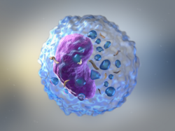
High levels of oxalate will also trigger an inflammatory cascade where the NLRP3 inflammasome, which is like our innate immune system’s alarm bell, becomes over-activated increasing NF-kB activity and increasing the release and expression of inflammatory mediators and cytokines like TNF-a, IL-1B and IL-6.

This over-activation can compromise our innate immune system’s ability to jump into action when the next “immune attack” comes along. Whether it is a pathogen-associated molecular pattern (PAMP) triggered by something like influenza A or Covid-19 or a damage-associated molecular pattern (DAMP) triggered by something non-infectious, like uric acid crystals or hyaluronan fragments or oxalate, chronic immune activation precipitated by oxalate will threaten the immune system’s ability to fight the next illness.

The other problem with constant over-activation of the NLRP3 inflammasome is that we now have elevated inflammatory cytokines, which will increase with the next attack, potentially driving us into a cytokine storm syndrome. So, keeping this type of inflammation low is the goal for having a strong immune response when it is needed.

Genes Influencing  Oxalate Metabolism

Inborn errors in three particular genes can contribute to faulty glyoxylate metabolism leading to hyperoxaluria – excessive oxalate storage. The glyoxylate pathway is where oxalate metabolism takes place.

  • A problem with alanine glyoxylate aminotransferase (AGT/AGXT) will cause someone to convert less glyoxylate to glycine, creating a buildup of glyoxylate which can convert to oxalate. This is characterized as primary hyperoxaluria type 1 (PH1).
  • An issue with glyoxylate reductase/hydroxypyruvate reductase (GRHPR) will lead to a lesser conversion of glyoxylate to glycolate and less push by hydroxypyruvate reductase of glyoxylate towards gluconeogenesis, again leaving an accumulation of glyoxylate available to convert. This is referred to as primary hyperoxaluria type 2 (PH2).
  • Primary hyperoxaluria type 3 (PH3) involves SNPs in 4-hydroxy-2-oxoglutarate aldolase 1 (HOGA1) and over activity can form excessive glyoxylate from hydroxyproline.

A loss of function mutation in any of AGT, GRHPR or HOGA1 will allow glyoxylate to pool and become available for lactate dehydrogenase (LDH) to convert to oxalate.

Non-genetic Pathways to Poor Oxalate Management

In the Trying Low Oxalates group we see a lot of people without defects in these genes still having issues with oxalate. Here, we have to consider other factors that contribute to elevated oxalate. Some main factors include, but are not limited to:

  • Endogenous production (can be caused by both genetic mutations and/or nutrient deficiencies in specific B vitamins)
  • Exogenous food sources, particularly in dietary extremes
  • Certain supplements that convert or can be degraded to oxalate
  • An unhealthy microbiome and estrobolome in general with fewer oxalate degrading microbes and potential hormonal imbalances, often spurred on by past antibiotic use but this is not the only cause

I believe the loss of peroxisome proliferator-activated receptor activity might also play a key role. This family of nuclear receptors are important in many different areas but particularly inflammation and immunity.

What Are Peroxisome Proliferator-Activated Receptors and Why Are They Important?

Peroxisome proliferator-activated receptors (PPARs) are ligand dependent transcription factors that regulate the expression of many genes involved in inflammation, fatty acid metabolism, energy homeostasis and metabolic function. There are a number of ways that PPARs might impact oxalate issues. The glyoxylate cycle itself occurs within the peroxisome. So from a purely mechanical perspective, any derangement of peroxisome function would potentially impact oxalate metabolism.

Additionally, a quick look at studies show that PPARs may alter activity levels of some key genes in oxalate metabolism (AGT, GRHPR). Third, PPARs play a key regulatory role on (intestinal) tight junctions (here, here). Tight junctions help prevent hyper-absorption of oxalate through the gut, while leaky junctions (leaky gut), allow oxalate to disperse into the bloodstream and travel to different tissues and organs where they cause damage.

Lastly, PPARs regulate macrophage polarization and therefore crystal phagocytosis (clearance of oxalate). They also increase antioxidant activity (here, here).

PPAR activity seems directly important to oxalate metabolism due to its control over the genes, tight junctions, macrophages and antioxidant status. A lack or loss of PPAR activity might also affect our glyoxylate cycle and levels, leaving plenty available for conversion to oxalate. Perhaps the lack of PPAR activity plays a role for those who do not have obvious SNPs in the important oxalate genes, yet still wind up in oxalate overload.

PPAR Activation and Inflammation

PPARs play a critical role in inflammation. They have a regulatory effect on both crystal-related enzymes and pro-inflammatory enzymes such as iNOS, metalloproteinase MMP-9 and COX-2, but their main function in inflammation is to promote the inactivation of NF-kB thereby decreasing the production of inflammatory cytokines (here, here). The exact mechanism behind how these are reduced is not clear though. I believe that the reduction of highly inflammatory oxalate in plasma contributes to this. The research seems to support this hypothesis. It is a bit technical, so bear with me.

  • Alanine glyoxylate aminotransferase (AGT) is positively regulated by PPAR-alpha (PPAR-a). This means that in absence of sufficient PPAR expression, AGT activity will be low. This allows glyoxylate to accumulate and potentially be converted to oxalate.
  • PPAR-a activation is crucial in inducing transcriptional activation of glyoxylate reductase hydroxypyruvate reductase (GRHPR) in mice but in humans GRHPR expression was shown to be PPAR-a independent due to promoter reorganization during primate evolution. (here, here). This means that without proper PPAR activation GRHPR activity will be lower, again allowing glyoxylate to accumulate. However, there is uncertainty if this works the same way in humans as it does mice due to the promoter reorganization that has occurred in man.
  • Mice deficient in PPAR-a present higher plasma levels of oxalate and as expected, administration of a PPAR-a ligand reduces plasma oxalate levels.
  • PPAR-gamma (PPAR-y) activation suppresses calcium oxalate crystal binding and oxalate-induced oxidative stress.
  • Yet oxalate itself impairs PPAR-y expression and phosphorylation creating a very important “negative feedback loop”.
  • PPAR-y agonists enhance barrier function through the upregulation of tight junction molecules claudin-1 and -4, occludin, and tricellulin at the transcriptional level, and will thereby be protective against hyper-absorption of oxalate through the “leaky gut”. PPAR expression also leads to a significant increase in tight junction strands, which will create a stronger barrier between the apical and basolateral membrane domains limiting the excess passage of oxalate and other proteins and lipids (here, here).
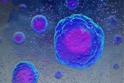
  • Classically activated M1 macrophages (which are inflammatory and cause tissue damage) facilitate renal crystal formation while alternatively activated M2 macrophages (that are anti-inflammatory and focused on tissue repair) suppress it (here, here). Macrophages have an important role in crystal phagocytosis, which is the process where phagocytes engulf and destroy foreign substances, like oxalate. PPARs play a key role in regulating this macrophage polarization to ensure that the anti-inflammatory macrophages are activated and assisting with tissue repair and crystal phagocytosis.
  • PPAR-y agonists can regulate TGF-β1 and HGF/c-Met to exert antioxidant effects against hyperoxaluria and alleviate crystal deposition and can exert an antioxidant effect through the PPAR-γ-AKT/STAT3/p38 MAPK-Snail signaling pathway.

Nutrients That Influence PPAR Activity

We know nutrient deficiencies play into oxalate issues so it is not surprising that B vitamins (B1, B2B3, B5, B6,  biotin, folic acid, and B12) activate PPAR-a and PPAR-y.

There are many natural ligands that can activate PPARs, like essential fatty acids and eicosanoids, but as indicated in this study they are required in extremely high concentrations of up to 100 uM for PPAR activation. A quick glance at ctdbase.org reveals that a wide variety of nutrients are capable of activating PPARs, but again, I would guess they would be required in huge pharmacological doses.

A couple notable ligands (but certainly not limited to) are free fatty acids with a preference for long-chain polyunsaturated fatty acids (PUFA’s) like arachidonic acid, alpha linolenic acid, EPA and DHA, linoleic acid, carnitine, alpha tocopherol, various carotenoids, endocannabinoids, PGJ2, leukotriene B4 and more. Ligands worth noting that have inhibitory effects are caffeine. There are other nutrients that have seem to have both activating and inhibitory effects, like zinc and copper.

Beta carotene appears to have both inhibitory and activating effects on PPAR’s depending on concentration, but BCMO1 is also transcriptionally regulated by PPAR-y, so might be worth digging into for anyone with SNP’s there (here, here).

Folic acid appears to have differential effects on PPAR-a and PPAR-y activity, so could be valuable to consider for those with MTHFR SNPs (here, here).

PEA and PPARs

I have wondered how much palmitoylethanolamide (PEA) comes into play. PEA is a fatty acid amide produced endogenously by neurons and glial cells in the central nervous system and also contained in egg yolks, peanuts and soybean. PEA is involved in the neuroprotective mechanisms that are activated following tissue damage and inflammation. In the cell, PEA is hydrolyzed (broken down) by fatty acid amide hydrolase (FAAH). Upregulation of this FAAH gene can cause us to rapidly break down PEA in the cell making it unavailable for its anti-inflammatory and analgesic effects.

In the body, it is produced on demand and acts locally, and its production is increased in areas of inflammation. PEA has been shown to activate both PPAR-a and PPAR-y. In doing so, it regulates inflammation and crystal-related genes and in this way helps to quench inflammation. So, elevated activity of FAAH may work against us with regards to inflammation by too rapidly breaking down this helpful endogenous fatty acid amide.

Genes involved in FAAH activity:

  • rs324420 A allele will cause lower and C will cause higher FAAH activity
  • rs2295633 G allele will cause likely higher activity
  • rs3766246 G allele will cause possibly higher activity

PEA is a ligand for the PPAR nuclear receptors, but it is not clear if it is PEA’s direct effect that is most beneficial or the “entourage effect” that improves PPAR activity. In the entourage effect, PEA competitively inhibits FAAH’s hydrolysis of and provides a sparing effect of anandamide (AEA). AEA is known as the “bliss molecule” as it can bind to cannabinoid receptors, providing an analgesic effect and creating feelings of happiness and wellbeing.  AEA is the more potent ligand for the PPAR’s so it is believed PEA’s prevention of AEA breakdown is also important for better PPAR activation.

It is also possible to have mutations in the 3 PPAR’s themselves which can lead to a loss of function, so they may be worth looking at too.

Conditions that Overlap with Oxalate and PPAR Activity

There are some key conditions that have traditionally been recognized as overlapping with oxalate overload. Notably, these include: COPD and respiratory disorders, cystic fibrosis, chronic kidney disease, thyroid disorders, vulvodynia, and autism.

We are also beginning to see that oxalate intersects with many other conditions. Studies show and we have learned through discussions on TLO that there is a clear tie between oxalate and conditions like metabolic syndrome, diabetes, obesity, mast cell activation syndrome, chronic fibromyalgia type pain, mood disorders, fatty acid oxidation issues and more. It seems that dysfunctional PPAR activity is central to most of these, as well.

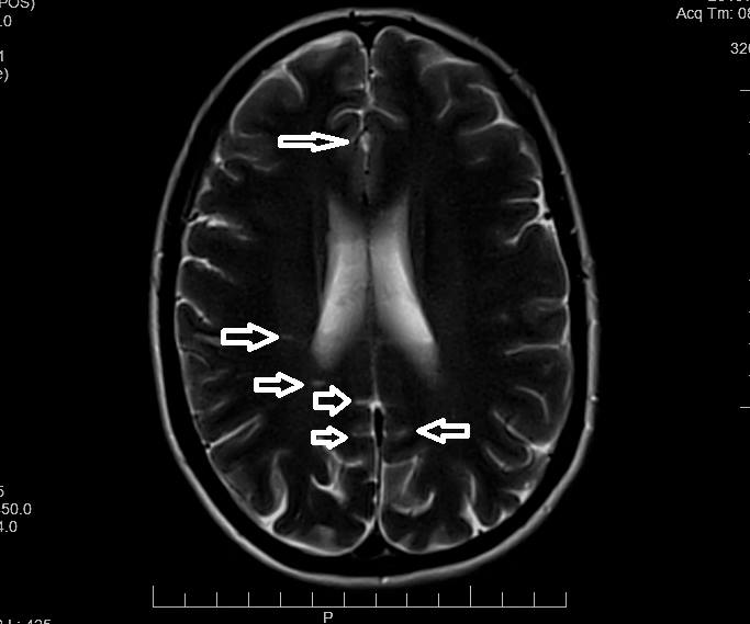
Inadequate PPAR expression may affect overall oxalate concentrations and allow many of these conditions to develop alongside oxalate overload. This is one of the reasons that inflammation is finally being recognized as a clear contributor to neurological and mood disorders. We know oxalate can also be causative for some of these conditions, so there is a viscous cycle of inflammation that needs to be broken. I hope that factoring in PPAR activity and its effect on oxalate metabolism might be helpful to your own research and health journeys.

In the next article, I will take a closer look at some of the conditions discussed here and how oxalate and the PPARs might both play a role.

We Need Your Help

More people than ever are reading Hormones Matter, a testament to the need for independent voices in health and medicine. We are not funded and accept limited advertising. Unlike many health sites, we don’t force you to purchase a subscription. We believe health information should be open to all. If you read Hormones Matter, like it, please help support it. Contribute now.

Yes, I would like to support Hormones Matter. 

Natural Killer Cells – Nutritional and Physical Biomodulation

2271 views

Natural Killer (NK) cells are an essential component of the human immune system and serve as part of innate immunity. These cells are built to seek and destroy virally infected cells, or cells displaying peculiar cancerous activity. NK cells’ development and function depend on complex networks of nutritional, biochemical, and environmental signals. Optimization of these signals through careful biomodulation will result in optimal NK cell development and functional activity against pathogens or tumors. The term biomodulation here will be defined as a change of cells or tissue in response to a therapeutic or pathologic stimulus. These modulations include nutrition, and systemic bodily perturbations.

A Short Primer on the Immune System

The human immune system is composed of a convoluted array of dynamic and static defenses. These include the skin, biochemical agents (e.g., antimicrobial defensins), and specialized cells that form innate and adaptive immunity. This intricate web of defense produces an environment built to protect the host from continuous onslaught by external, or opportunistic pathogens. Cells of the immune system are also involved in surveying bodily tissues for damaging insults acquired from the environment. Harmful insults could be chemical or radiation-induced bio-alterations that could lead to cancerous cell formation.

A dynamic faction of the immune system is the White Blood Cells (WBC) component – categorized under innate or adaptive status. WBC circulate through the blood network where they enter/exit tissues for surveillance. WBC form within the bone marrow from hematopoietic stem cells. For example, lymphoid precursors form adaptive lymphocytes, T and B cells. On the other hand, neutrophils, mast cells, and monocytes are formed from myeloid progenitor cells. Some WBC undergo further development stages, such as in the Thymus for T cells, before they are released. The latter allows for detection of cells that are either self-reactive (autoimmune), or non-reactive (do not function as well). T cells that do not pass these tests (receptor signals) are commanded to undergo apoptosis, or programmed cell death. All immune cells depend on a host of nutritional factors for development, activation, and effector functions. Malfunction or dysregulation of the immune network will result in autoimmune disorders, external and opportunistic infections, and formation of multiple types of cancers.

Innate and Adaptive Immunity – The Two Arms of Immunity

Innate immune cells are the first line of defense against infections. Innate cells harbor specific receptors which allow recognition of conserved pathogen patterns, or pathogen-derived chemicals. These receptors are termed germline-encoded pattern recognition receptors (PRRs). An example of PRRs would be Toll-like receptors (TLR) that recognize bacterial and viral components such as flagellin, lipoproteins, lipopolysaccharides (LPS), and ribonucleic acid (RNA). The responses of innate immune cells are relatively rapid and will occur within four hours of activation. However, although innate immune cells are essential, some pathogens can overcome their frontline defenses. Thankfully, adaptive immune cells eventually become involved so that they can “adapt” to these infections wherein long-term solutions are formed, in other words, formation of immune memory. However, before adaptive cells can form memory, they first need to be primed through an initial infection. In this case, adaptive cells (T and B cells) work in concert with innate cells to be primed and to form long-term memory against re-invading pathogens. This includes expansion of pathogen-specific T cells, termed clonal expansion. Importantly, B cells become antibody producing cells. The result is a reservoir of long-term circulating memory cells and antibodies poised for a fast response to infections – generally without the need of the initial discomfort when encountering new pathogens (fever, disease, etc.).

Natural Killer and Natural Killer T Cells

Natural Killer (NK) and Natural Killer T (NKT) cells are categorized under innate immune cells. NKT are special because they are viewed as a mixture of NK and conventional adaptive T cells (hence NK-T cells). NKT cells, however, are different from adaptive T cells because their T cell receptor can be activated by glycolipids. This is in contrast to conventional adaptive T cells that are activated by peptide (protein) complexes. NK/NKT cells play an important role in the defense against virus-infected and tumor cells. They patrol the body while integrating a diverse array of signals from cells and their environment. Once stressed cells are found (tumors or infections), NK/NKT cells become activated. Here, these innate cells destroy tumor or infected cells through direct receptor engagement, utilizing cytolytic granules and death receptors – the latter causing apoptosis.

Another important role for NK and NKT cells is to strengthen adaptive responses in-part by secreting immune-modulating compounds. The latter include cytokines [tumor necrosis factor-α (TNF-α), interferon-γ (IFN-γ), granulocyte macrophage colony-stimulating factor (GM-CSF)], and chemokines (CCL1, CCL2, CCL3, CCL4, CCL5, and CXCL8). Nutrition is an essential component for the optimal function of NK and NKT cells. As we will see below, exposure of NK/NKT to nutritional compounds allows for better movement, optimized secretion of immunomodulatory chemicals, and enhancement of killing activity. For example, the importance of magnesium for NK cell function has been discussed previously on Hormones Matter. Herein, we focus mostly on NK cells with some insights for NKT cells.

Modulating natural killer cells
Figure 1. Biomodulation of natural killer cells.

Vitamin D Antiviral and Anti-Tumor Actions

The secosteroid, vitamin D3, is synthesized in the skin from 7-dehydrocholesterol upon exposure to electromagnetic waves in the ultraviolet-B region. Vitamin D2 is produced by irradiation of fungi and yeast, and both D3/D2 forms can be obtained from diet. Several modifications by cytochrome p450 enzymes in other tissues including, the liver, kidney, and lung epithelial cells produce the active forms, 1,25-dihydroxyvitamin D3/D2 ( 1,25D), which circulates as a hormone in the blood. The effects of 1,25D are mediated through binding of nuclear Vitamin D Receptor (VDR). Numerous cell types and tissues express VDR including the lung, kidney, bone, and lymphocytes. Upon activation by 1,25D, VDR influences calcium/phosphate homeostasis and metabolism.

The 1,25D/VDR axis results in cell signaling conductive to anti-viral and anti-tumor responses. Binding of Vitamin D to its receptors generates anti-microbial peptides by neutrophils and macrophages, and to enhance T cell activation via the T cell receptor. VDR signaling blocks the proliferation of human cancer cells, reduces lung metastasis in animal models, and is a preventive factor in the metastasis of lung cancer in humans. Interestingly, observational studies demonstrated an inverse correlation between circulating 1,25D or ultraviolet-B exposure and the incidence of lung cancer. In addition, increased dietary 1,25D improves results for lung cancer patients after surgery. Therefore, Vitamin D is a crucial component mediating anti-tumor responses through direct suppression of tumor cells, but also through the modulation of immune cells.

How does Vitamin D signaling affect NK cells? Mariani et al. showed a functional relationship between the numbers of cytolytic/cytotoxic NK cells and levels of Vitamin D. Although this is a correlation, we know that NK cells express abundant levels of VDR and can respond to 1,25D stimulation by upregulating and downregulating multiple genes. Therefore, we can see that vitamin D-induced stimulation directly affects NK cells. Other studies demonstrated that VDR signaling augments tumor susceptibility to NK cell mediated lysis. Interestingly, the cancerous growth studied were human melanoma cells. Here, 1,25D modulation increased specific proteins (FAS) on the cell surface of melanoma cells which then allowed for a better NK cell detection followed by destruction of the cancerous cells. Next, a clinical trial in ICU patients showed significant increase in NK and NKT cell counts after weekly treatment of 60,000 IU 1,25D. Another study showed that treatment with the active form of Vitamin D caused an increase in NK cell cytotoxicity after 1 month of oral treatment. The authors suggest the use of this secosteroid for immunomodulation in patients with low NK cell activity. We can extrapolate from these and multitude of other studies on Vitamin D, that increasing 1,25D levels during an infection produces a positive immunomodulatory effect for NK cells.

What About NKT Cells?

NKT cells are a major player in the anti-tumor response. For example, numerical defects in NKT cells are considered a poor prognostic marker in patients with colorectal cancer and neuroblastoma. In other words, lower numbers of NKT cells lead to worse cancer outcomes. Because they are so effective against cancer, NKT cells were used in immunotherapy, and these treatments were shown to enhance innate and adaptive immune response to tumors, and to increase serum levels of the immune-boosting cytokine IFN-γ in patients with various metastatic malignancies. IFN-γ is one of the cytokines released by both NK and NKT cells.

Do NKT cells receive the same beneficial effects from Vitamin D? Indeed, the effects of Vitamin D seem to even be stronger for NKT cells. The effects are seen from the developmental period whereby VDR knockout mice have fewer peripheral NKT cells due in part to a requirement for VDR signaling during development. In addition, VDR signaling regulates the expression of chemokine receptors which control NKT cell recruitment to different tissues. All of this highlights the importance of “indirect” VDR signaling for NKT cell function. Direct 1,25D stimulation can augment NKT cells’ ability to secrete cytokines through in-vivo VDR stimulation. Specifically, mice were fed 1,25D for 1 week and then injected with NKT cell stimulating compounds. The authors found that NKT cells from Vitamin D fed mice had significantly more IFN-γ and IL-4 production. Similarity, mice who had their VDR knocked out, had significantly less amounts of these cytokines. We can conclude here that vitamin D is important for the development and function of NKT cells, and that lower vitamin D levels will affect individuals in terms of reduced ability for development of strong and functional NKT cells.

Zinc Enhances NK Activity

Immune cells can regulate the influx and outflux of minerals depending on signals received from the environment. For example, in adaptive T cells, once their T cell receptor is activated, adaptive T lymphocytes begin by mobilizing sizable cytoplasmic and nuclear influx of multiple ions such as calcium, magnesium, and zinc. This activity leads to expression of numerous genomic pathways, including the secretion of IL-12 cytokine by WBC. Interestingly, one of the roles of IL-12 is to enhance NK cell activity. Therefore, zinc indirectly affects NK cells.

A 2018 study demonstrated that zinc supplementation enhances NK cell activity through increasing the expression of their cell-destroying proteins. Zinc also increased the effects of IL-2 on these cells, thereby acting as a synergistic molecule to this important survival cytokine. On the other hand, zinc deficiency reduced NK cell activity and dampened the effects of IL-2. Another study showed that mice deficient in zinc had poor control of liver tumors. In contrast, too much zinc showed an inhibitory effect on NK cell cytotoxicity – demonstrating a potential zinc-induced imbalance (possibly through reduction of copper).

How does zinc regulate immune cells? By scoping the genomic level, we can find that zinc is an integral component of how the cell regulates transcription from DNA. Transcription is the process of making messenger RNA (mRNA), and other types of RNA, so that the cells can respond optimally to danger signals. Here, zinc is part of specialized protein structures termed Zinc-Finger protein domains. These domains require zinc ions in order to stabilize the amino acid structure so that the proteins maintain an appropriate 3-dimensional form. These Zinc-Fingers are part of larger proteins that bind with DNA, some of which can be named transcription factors. Together, these zinc-based structures play an essential role in DNA regulation. For example PLZF, a zinc-finger transcription factor, was shown to be involved in NK and NKT cell development. This is so important that immune cells make sure to express specific zinc transporters to capture this ion inside the cell, so that enough ions are available for protein function when danger signals arrive.

Copper, Ceruloplasmin and Hematological Dysfunction

We have previously discussed the importance of copper with regards to thyroid and mitochondrial function – all of which are important for proper immune function. Copper deficiency is an established cause of hematological abnormalities and can present in an almost identical pattern to myelodysplastic syndromes. Myelodysplastic syndromes are a group of cancers involving immature blood cell formation. When copper levels become chronically depleted, these blood abnormalities can show up as mono- bi- or pancytopenias (lower than normal red or white blood cells), with the neutrophil (innate cells) lineage sometimes being the most susceptible to low copper reserves. But is there evidence for copper-mediated mechanisms directly on NK cells?

Let’s first take a look at a case study for copper deficiency. Bhat et al. recently presented a case of a 2-year old patient exhibiting symptoms of Menkes disease (low serum copper and ceruloplasmin, hypotonia, kinky hair, and developmental regression). Menkes disease was then confirmed by genetic assessment. The treating team commenced on a regimen of copper chloride, which produced positive results (more alertness, and better movements). This patient had suffered from recurrent respiratory infections, in-part due to a substantial dysfunction of NK cells. Given this observation, they postulated that copper is needed for NK cell activity (and probably development). The latter has been directly shown in a 1987 study whereby rats fed a copper-deficient diet had lower antibodies and NK cell cytotoxicity. Certainly, it would make sense to assume here that these defects are due to a reduction of energy and mitochondrial function, needed for proper NK cell function.

Clues about mechanisms behind copper-mediated regulation of NK cells come from another research group, Banha et al. They demonstrated that NK cells express the multi-copper protein, ceruloplasmin. Ceruloplasmin (also named ferroxidase I) carries around 95% of the copper found in plasma. It is required for the oxidation of ferrous iron to the ferric forms. This process allows the incorporation of ferric iron into the protein, transferrin, and eventually hemoglobin. However, relative to other immune-related proteins, not a lot is not about ceruloplasmin in general, other than its role in binding and transporting copper.

Banha et al. showed another potential role for this copper binding protein by performing mRNA and protein studies on human derived WBC. Their experiments revealed a higher density expression of ceruloplasmin specifically with NK cells, compared to other WBC. Even the closely similar NKT cells did not display as much ceruloplasmin expression. Interestingly, the experiments revealed that human WBC express the secreted and membrane bound versions of ceruloplasmin. These experiments highlight a secondary role for ceruloplasmin outside of the liver and plasma. Observation with NK-associated ceruloplasmin could elucidate why the Bhat et al study with the 2-year-old child showed a dysfunction of NK cells, as ceruloplasmin is a major copper binding/transport protein. Taking the two studies together we can postulate here that immune cells utilize ceruloplasmin to harbor and release copper around infection sites in order to boost immune activity in a local infection or tumor sites.

Movement and Exercise Improve Immune Function

There is little doubt that movement and exercise are one of the best health-improving tools. For example, in sprints, just the jarring of the skeleton produces a stimulus in bone forming cells. The shock-waves and force implemented when bone and ground meet alter the back-and-forth dynamics between the osteoblast (bone forming cells) and osteoclasts (bone absorbing cells) – inducing bone density increase. In addition, the body is frantically signaling to itself to increase mitochondrial power and bio-genesis.

The benefits of movement are not solely limited to bodily power and output. The immune system, and in particular NK cells, receive some of that energy from exercise. In fact, NK cells are one of the most responsive cells to exercise with regards to mobilization from body tissues into circulation. NK cell mobilization is associated with exercise-dependent protection against cancer. Given their strong anti-tumor activity, mobilization of these cells likely allows for better trafficking with optimized seek & destroy activity. Of note, exercise seems to increase only the mobilization and not necessarily the production of new NK cells.

Just how much exercise is needed to receive these benefits? Although different types of movements can produce this mobilization effect, movement can be as little as a small flight upstairs. Millard et al utilized 29 volunteers and drew their blood before and immediately after exercise to study the frequency of their WBC. They showed that as little as 150 stair steps will increase the numbers of NK cells in the blood by 6 fold. Interestingly, females had a larger increase in the numbers of NK cells than males. Another work by Timmons et al showed that maximal NK cell mobilization occurred at 30 minutes of endurance training, after which no increase was observed. This effect on NK cells by exercise has been attributed in-part to the release of catecholamines (epinephrine and norepinephrine). Direct evidence of catecholamines-induced NK mobilization was displayed, when systemic administration of catecholamines produced similar effects as exercise-induced increased of NK cell. What is also important is that mobilized NK cells had higher cytotoxicity – so they performed better against tumors. Why do NK cells react so well to catecholamines? It is because they express the highest amounts of beta-adrenergic receptors (receptors for catecholamines) of all WBC.

It does not all stop with catecholamines, however. Myokines, which are cytokines derived from muscles during exercise, become involved in regulation of mobilized NK cells. Myokines are known to stimulate immune cells and they include IL15, IL7, and IL6. Thus, NK cells are involved in an intriguing immune-to-muscle crosstalk during exercise. The mobilization and optimization of functional activity of NK cells by myokines during exercise is believed to explain why exercise can be so beneficial in a cancer setting.

Magnetic Forces to Harness NK Potential

Environmental impacts of NK cells are not limited to just different forms of exercise. Magnetic forces can be utilized to harness the power and potential of NK cells. Modulatory effects of static and dynamic magnetic forces on many cell types, have been well known for decades. With respect to NK cells, their activity is potentiated by static magnetic fields. A relatively recent work by Lin et al. showed that perturbation by a 0.4 Tesla static magnetic field increased the viability and activity of human NK cells. If you are wondering, 0.4 Tesla can be classified as moderate level static magnetic field which is below two other levels (strong & ultra-strong fields). Magnetically treated NK cells showed significantly more killing activity compared to non-magnetized cells, after incubation with K562 tumor cells. The effects of magnetism on NK cells have been attributed to increased receptor activation pathways (MAP Kinase). These results indicate to us that proper magnetism can be beneficial to a variety of diseases (including cancer).

Manipulations by static and dynamic magnetic fields on all cell types, though no doubt produces measurable effects, have nevertheless been conflicted with varying and sometimes opposing results. The issues stem mainly from researchers using different experimental procedures and protocols. The latter includes, field strength, frequency and duration of the magnetic fields, target cells, and distance from magnets. In addition, cells tend to respond differently depending on their stages of growth and types/strength of magnetic fields. For example, testing on guinea pigs showed suppression of killing activity of NK cells when treated with a 50 hertz electromagnetic field. Interestingly, the same strength electromagnetic field causes DNA perturbation resulting in activation of latent Epstein Barr Viruses, a prime target of NK cells! Therefore, some frequencies not only activate viruses, but also result in suppressing the same cells required for removal of these pathogens. On the contrary, another study revealed positive effects on rat NK cells when treated with electromagnetic waves. Specifically, NK cell cytotoxic activity increased by 130-150%. Other types of immune cells have also shown a similar behavior when activated under magnetic fields. For example, moderate level static magnetic field perturbation caused human CD8+ T cells (adaptive immune cells) to increase mitochondrial respiration, ATP production, and increased immunomodulatory secretions.

There is no doubt that electrical and magnetic forces affect how our cells behave. It seems that moderate level static magnetic fields tend to promote beneficial activity for innate and adaptive immune cells. However, much more research is needed for specifying the mechanisms of these forces and to decipher beneficial and harmful parameters. Important parameters to elucidate would be A) how do perturbations of static and dynamic fields differ in their effects, and B) what doses are appropriate. Overall, our cells clearly respond to electromagnetic signals, but more work is needed to show how we can use these fields to promote immunity.

Conclusion

The role of NK cells in protecting our cells from viruses and tumors is clearly established. To support these cells in these important functions, we must provide important nutritional and physical ingredients. A deficiency in zinc not only affects several bodily and structural functions, but also suppresses the immune system, including NK cells. Similarly, both copper and Vitamin D are needed for development and promotion of NK cell-mediated anti-tumor activity. Production and anchoring of ceruloplasmin on NK cells’ membrane, is an indication of the importance of copper for NK cell function. Other, not so clearly elucidated mechanisms, such as magnetic fields seem to be an important component in the convoluted web of signals that NK cells receive. The above discussion also highlights how too much or too little of anything can compromise this web of signals. Too much zinc will alter copper metabolism, and the wrong electromagnetic signals can promote viral expression and suppression of immune cells. The reader is therefore advised to embark on a journey to optimize the immune system through implementation of calculated nutritional and physical mechanisms.

We Need Your Help

More people than ever are reading Hormones Matter, a testament to the need for independent voices in health and medicine. We are not funded and accept limited advertising. Unlike many health sites, we don’t force you to purchase a subscription. We believe health information should be open to all. If you read Hormones Matter, and like it, please help support it. Contribute now.

Yes, I would like to support Hormones Matter. 

Feature image purchased from iStock, #18165875. 


The Cytokine Storm as an Inflammatory Reflex

8219 views

Relatively recent research has revealed a truly extraordinary connection between the brain and the body; between the vagus nerve, the immune system, and the autonomic system. All of which depend upon energy produced by the mitochondria and thus are dependent on thiamine. Before I embark on a description of this research, a few definitions are required.

  • Vagus nerve: This is the longest nerve in the body. There is one on each side and the right side nerve has a different action from the left side nerve. Both start in the brain and both go to the spleen, the lung, and the intestine. It is able to send signals to and from the brain to the body. The name comes from the Latin word meaning “wanderer”.
  • Cytokine: There are many of these in the body. They are small proteins, some of which cause inflammation and some inhibit it.
  • Cytokine storm: If and when the inflammatory cytokines get out of control their profusion is called a cytokine storm.
  • Inflammation: A localized physical condition induced by cytokines in which the affected part of the body becomes reddened, swollen, hot, and often painful, especially as a reaction to injury or infection. We now know that the inflamed joints in rheumatoid arthritis are caused by inflammatory cytokines and might justify that disease as a form of dysautonomia.
  • The spleen: An organ that is located in the upper left part of the abdomen. It produces lymphocytes which are important elements in the immune system. It is the largest lymphatic organ in the body.
  • Macrophage: This is a type of white blood cell of the immune system that engulf and digest cellular debris, foreign substances, microbes, and cancer cells, in a process called phagocytosis.
  • The autonomic nervous system (ANS): It has been emphasized in many posts that we have two different nervous systems. The voluntary system responds to free will whereas the ANS acts automatically. It is controlled by the lower part of the brain and this will be important as we develop this post.
  • Acetylcholine: A chemical substance known as a neurotransmitter. Without this substance, the vagus nerve will not function.
  • Citric acid cycle: This is a complex series of chemical substances that occur in nearly every cell in the body. It is the “engine” of the cell, manufacturing the energy required for the cell to function. This energy is stored in another chemical substance known as adenosine triphosphate (ATP). Although the analogy is imperfect, the nearest thing to explain its function is “a battery”. So you might think of the relationship as being like a continual recharge of a battery. Consumption of energy for function must be met by a continual production. It is the deficit between energy production and its consumption that causes the symptom that we call fatigue.

The Inflammatory Reflex, an Autonomic Response

The reason that I have called this a reflex is because we now know that inflammation, a defensive mechanism, is controlled by the brain through the vagus nerve. It is an automatic and reflexive response controlled by the autonomic system. The inflammation of selected parts of the body results from the release of inflammatory cytokines by the action of this nerve. That is why I think of rheumatoid arthritis as a form of dysautonomia (abnormal ANS function).

All of us live in an essentially hostile environment. We have to cope with an attack by microorganisms such as bacteria and viruses, different types of trauma, the weather, and virtually any outside or inside change that demands a response. In other words, we live our lives from one end to another in a state of attack and defense. I have indicated many times that any kind of response to some form of attack requires energy to run the machinery that acts in our defense. When energy is insufficient, the ANS begins to fail and defense mechanisms collapse or become distorted in their action. If severe enough, death may follow. Another cause of disease is faulty genetics but that is also partially energy dependent because of the science of epigenetics.

The ANS works all the time and is monitoring our well-being. It is able to detect an attack by a microorganism or other forms of “stress”. This results in a message, sent via the vagus nerve to the brain. It is essentially a notification that some form of attack has been detected. What happens then is so well described in an abstract from a published paper that I quote from it liberally below.

The ‘cytokine theory of disease’ states that an overproduction of cytokines can cause the clinical manifestations of disease. Much effort has been expended to determine how cytokines are regulated in normal health. Transcriptional, translational and other molecular control mechanisms protect the host from excessive cytokine production. A recent discovery revealed an unexpected pathway that inhibits macrophage cytokine production. The inflammatory reflex is a physiological pathway in which the autonomic nervous system detects the presence of inflammatory stimuli and modulates cytokine production. Afferent signals to the brain are transmitted via the vagus nerve, which activates a reflex response that culminates in efferent vagus nerve signaling. Termed the ‘cholinergic anti‐inflammatory pathway, efferent activity in the vagus nerve releases acetylcholine (ACh) in the vicinity of macrophages within the reticuloendothelial system.

Some of the cytokines cause inflammation while others inhibit inflammation. A growing body of clinical data suggests that a cytokine storm is associated with Covid-19, suggesting that it might be an important component for rescuing patients with severe Covid 19.

The Energy Immune Response Hypothesis

I have done my best to describe the vitally important relationship between the body and the brain in defending ourselves from the attacks of environment. By far the most important phenomenon seems to be how we produce energy and I learned many years ago that beriberi was the prototype for failure of energy production. Thiamine was blamed as the cause of this disease and in spite of the importance of other members of the B complex, it seems to be the dominant vitamin in the consumption of glucose to provide energy to the brain. We have good reason to believe that an insufficiency of this vitamin is all too common in the United States and is responsible for a huge amount of unrecognized disease. Because the lower part of the brain that controls the ANS is particularly sensitive to this deficiency the commonest result is different manifestations of dysautonomia. However, patients are not visibly affected, as seen by others, often visiting their family physician where a common false diagnosis is psychosomatic disease. The general effect is a lack of fitness that makes them more susceptible to the more severe results of any infection.

The Cytokine Storm, the Vagus Nerve, Acetylcholine and Thiamine

If cytokine pathology is responsible for the symptoms of Covid 19, stimulation of the inflammatory reflex is an obvious method of treatment. This can be done by electrical stimulation of the vagus nerve. However, if thiamine deficiency is a common finding in the American public, it would result in a deficiency of acetylcholine, the neurotransmitter essential to the vagus nerve. It strongly suggests that thiamine, used as a drug, might be successful in suppressing the cytokine storm. The most effective thiamine derivative would be thiamine tetrahydrofurfuryl disulfide (TTFD). Because my years of study have demonstrated to me that there is no toxicity arising from it, it could be used empirically in the emergency room, although if given to someone with long-standing chronic thiamine deficiency, there is a possibility of refeeding syndrome. If this is the correct solution to the symptoms of Covid 19, it may well be that thiamine deficiency is responsible for those that are generated in the so-called Longhaulers. The chronicity of symptoms is the same as those that occur in beriberi.

We Need Your Help

More people than ever are reading Hormones Matter, a testament to the need for independent voices in health and medicine. We are not funded and accept limited advertising. Unlike many health sites, we don’t force you to purchase a subscription. We believe health information should be open to all. If you read Hormones Matter, like it, please help support it. Contribute now.

Yes, I would like to support Hormones Matter.

A New Model for Medicine

3754 views

What is a Medical Model?

 In the Oxford English dictionary the word model is defined as “design to be followed, style of structure”. Then it follows that there must be a model to distinguish health from disease, that differentiates the two states of being. No disease can be treated without knowing exactly what caused it. Let us go back to Hippocrates, 400 BCE, who said “let food be your medicine and your medicine be your food”. What Hippocrates was saying was essentially that nutrition was the core issue in the maintenance of health. At this time and throughout the Middle Ages there was no model for the cause of disease. Consequently, treatment was extremely primitive and almost purely empirical. In the time of ancient Egypt it was believed that mental illness was caused by the presence of evil spirits in a person’s head. They bored holes in the skull to let the evil spirits out. If you think about that, perhaps it relieved the occasional headache because of increased pressure in the skull caused by a brain tumor. Hence, a few successes might have caused it to be retained as beneficial. During the Middle Ages, the only treatment that seems to have been used is bloodletting. It might have been temporarily useful in people with high blood pressure. A few successes yielded the conclusion that it was beneficial for all disease.

The First Controlled Experiment

Semmelweis was a 19 th century Hungarian physician. In those days, the incidence of puerperal disease (childbed fever) was absurdly high. Semmelweis made the observation that doctors, delivering their patients, entered the delivery room and went directly to their patients without changing their garments or washing. He came to the simple conclusion that the doctors were bringing something in with their hands that caused the problem. The obstetric ward consisted of a number of beds on each side of the room and Semmelweis directed that doctors delivering their patients on one side should wash their hands in chlorinated lime, while doctors on the other side of the room would continue in the old way. Of course, the incidence of childbed fever was so different that it did not need a statistician to document the difference. Semmelweis’s observations conflicted with the established scientific and medical opinions of the time, particularly as he was unable to explain what was on the hands of the doctors. Some doctors were even offended at the suggestion that they should wash their hands. It is truly an amazing vision of human behavior. Innovation carries with it loss of reputation for the innovator, no matter how successful the innovation. Well, of course everyone today knows that it was microorganisms on the hands of the doctors that caused the disease, but they had not yet been discovered. Poor Semmelweis wound up in a lunatic asylum and died in his 40s after a beating by attendants. Today, he is regarded as the first person to introduce antiseptic medicine.

The First Paradigm in Medicine: Microscopic Organisms

Most people are aware that the invention of the microscope, and the work of historical figures like Louis Pasteur, led to the discovery of organisms, that could only be seen with the microscope, caused what we now call infection. We are all familiar with the fact that a tremendous number of diseases are due to infection by bacteria, viruses or fungi. It was a perfectly logical conclusion that the development of treatment should be aimed at killing these organisms. This was the first paradigm in medicine, meaning that it was accepted by all. A glance at history will tell us that the search for medication that would kill these organisms was hard won. It was difficult to find something that would kill the germs without killing the patient and many patients lost their lives as a result of this search. The discovery of penicillin represented a dramatic change in perspective as it gave birth to the antibiotic age. Millions of lives have been saved. However, we are now entering an era where the development of antibiotic resistance is becoming an increasing problem. More and more potentially damaging antibiotics have been synthesized that present their own problems in therapy.

The Second Paradigm in Medicine: Immunity

It has been said that Louis Pasteur made the statement on his deathbed, “I was wrong: it is the defenses of the body that matter”. I believe that this may well become the second paradigm in medicine. So what are we talking about? Everyone recognizes that we have immunity but the average person has only the vaguest idea of what this really means. In fact, body defenses against infection are exquisitely complex and incredibly efficient when the immune system is healthy. The primary mechanism for health maintenance is exactly what Hippocrates said, not only the quantity but the quality of nutrition. By recognizing this, the concept is offered that preventive medicine, the use of nutrients based on a knowledge of the biochemical machinery that give our cells function, is the second paradigm.

Presently, we stimulate our immunity by the use of vaccines. However, each vaccine gives a protection to a specific microorganism, perhaps the best example being the flu. Most of us are aware that there are many strains of the flu virus and it may not be possible to predict the particular strain responsible for the “next epidemic”. Natural immune defense mechanisms recognize most invaders as “enemies”. Those whose adaptive/immune mechanisms cannot respond will succumb to the infection. Assisting the immunity mechanisms by making energy synthesis as efficient as possible and killing the “enemy” with maximum safety to the patient might just be the way of the future.

How the Body Responds to Environmental Stressors  

Each one of us comes with a “blueprint” derived from our parents in the form of genes that carry a code called DNA. This code is unique for each person and provides the structure that makes up a living person. The body is composed of 70 to 100 trillion cells, all of which have to cooperate to produce what we call function. I think of it being like an orchestra where all the organs are made up of cells, each one of which has a specific specialty to provide its contribution. Like instrumentalists in an orchestra, the cells within each body organ have to work together. This requires a conductor, a function that is performed by the subconscious brain. Coordination is administered through an automatic (autonomic) nervous system and a bunch of glands known as the endocrine system that produce messengers called hormones.

Consider what happens when a person is attacked by a pathogenic Streptococcus, for example. The throat becomes sore, the marker of inflammation. Controlled and executed through the brain, it increases local blood supply, bringing white blood cells into the area and is part of a defensive process. Glands in the neck become enlarged and this is also a defensive process, designed to catch and destroy the germs beginning to spread. Body temperature becomes elevated because disease producing bacteria are most virulent at normal body temperature and their efficiency is reduced at a higher body temperature. A standard procedure in medicine for many years has been to reduce the fever and it has always seemed to me to be a disadvantage, based on this explanation. We sweat when the environmental temperature is high and evaporation from the skin results in cooling. When the environmental temperature is low, we shiver and the muscular activity produces heat to maintain body temperature. These are examples of how we are able to adapt to changes in our environment that threaten our well-being. All of this is purely automatic and the only thing to complete the picture is how our food (fuel) is used to create energy. Maximum efficiency of brain metabolism is mandatory. Assist and protect the “conductor”.

How We Create Energy: Enter the Mitochondria

Because any form of burning is the union of oxygen with the fuel, in the body it is termed oxidation. The process is complex and many vitamins and minerals are involved, besides calories. It has long been known that thiamine (vitamin B1) deficiency is the cause of beriberi, the disease that had plagued humanity for thousands of years. Because this deficiency affects every cell in the body, it can degrade the efficiency of virtually any organ. But because different tissues have their own rate of metabolism and the brain and heart are the two tissues that require fast and efficient oxidation, it is the cells in those tissues that are most affected. Therefore, thiamine deficiency has its major effect in the brain and heart, but they are not exclusive.

Glucose is the main fuel, but like any other fuel used to produce energy, it has to be ignited. Thiamine, much like a spark plug in a car, processes this ignition. All simple sugars taken in the diet are broken down to glucose.  But before this happens in the body, dietary sugars have two effects. The first is a signal from the tongue to the pleasure zones of the brain. It is this sweet taste that makes sugar addictive. The second is that this excess of sugar overwhelms the capacity of thiamine to oxidize glucose to create energy. A person may have a perfectly normal thiamine level in the blood that is inadequate to meet the demand. It is the ratio of “empty carbohydrate calories” to the concentration of available thiamine that counts. I have called this “high calorie malnutrition” that seems to be an oxymoron since malnutrition is generally considered to be on the way to starvation. The patients with this form of malnutrition may be obese, remain relatively active, do not look ill and multiple symptoms are regarded by their physicians as “psychologic, or psychosomatic”. There appears to be no reason to seek laboratory evidence of malnutrition and the patient is written off as a “problem patient”. It is hardly surprising that the patient leaves the doctor’s office angry and tells friends that “the doctor told me that it was all in my head”.

The irony is that it IS in the patient’s head, but because of electro-chemical changes in brain metabolism. It has always seemed odd to me that physicians often consider that “psychological issues” are somehow “invented” by patients without thinking that every thought, every action, has a mechanism produced in a chemical “machine” called a brain. Distortions are the result of a combination of cellular energy deficiency (malnutrition), coupled with a potential genetic risk and perhaps a stress factor such as an otherwise mild infection/injury, or an inoculation. Any one of the three factors may dominate the clinical presentation, but in most cases the other two are involved.

A New Model: Genetics, Nutrition, and Stress

Throughout life each of us depends on our ability to survive in an essentially hostile environment. The first thing that it depends upon is our genetic inheritance that I have called “the blueprint”. But we also know that the “engines” of our cells, known as mitochondria, have their own genes in which the DNA is more susceptible to damage than our cellular genes. A new model must consider the fact that any stress requires energy in an adaptive response to any form of environmental attack resulting from a mental or physical problem or infection. The only way that we can protect the structural components of our bodies is by the use of the natural ingredients of nutrition, the ancient teaching of Hippocrates. The new science of epigenetics finds evidence that nutrition and lifestyle can make changes to our genes that might be beneficial or not, according to the circumstances. If a person has become sick from an excess of empty calories and refuses to change, the only way to treat that person would be by increasing the concentration of the missing nutritional ingredient in the form of a supplement. It is of paramount interest that in 1962 a paper was written in a prestigious medical journal. The author had found 696 medical journal manuscripts that reported 250 different diseases that had been treated with supplementary thiamine, with varying degrees of success. This suggests the possibility that health is produced by a combination of genetic influence, how we meet the daily impacts of stress and the quality of our nutrition. Disease results from, either genetic failure (cellular or mitochondrial), failure to meet stress because of energy deficiency, malnutrition, or combinations of the three elements.

We Need Your Help

More people than ever are reading Hormones Matter, a testament to the need for independent voices in health and medicine. We are not funded and accept limited advertising. Unlike many health sites, we don’t force you to purchase a subscription. We believe health information should be open to all. If you read Hormones Matter, like it, please help support it. Contribute now.

Yes, I would like to support Hormones Matter.

Photo by Sebastian Unrau on Unsplash.

What Constitutes Disease?

2124 views

Survival for any animal or plant throughout the world is a lifelong battle between the environment and the constitutional design of the organism. Health is defined in Webster as “physical and mental well-being; soundness; freedom from defect, pain, or disease; normality of mental and physical functions”. But it might well be seen as the ability to adapt to whatever the environment throws at us. Disease is defined as “any departure from health”. If then, life can be broken down into attack and defense, the first thing to consider is the nature of an attack.

Stress or Attack on the Body

The word stress has many meanings and can be used in different ways. I have chosen the definition in the Oxford Dictionary, “pressure or tension or compulsion; times of stress when much energy is needed”. Using the word this way means that our defenses are being tested by the nature of the stress. This can be mental or physical. The stresses that acted on our ancestors were much more physical. Civilization protects us from most of the physical forces that they experienced. The majority of the stress we face today taxes our mental ability and is often long-term. Physical stress is imposed by trauma or the attack of organisms, most of which we cannot see without a microscope.

The Body’s Natural Defenses

For the purposes of this discussion, think of the human body as being made up from between 70 and 100 trillion micro-organisms we refer to as cells. Each group of cells has evolved with a specialized duty, congregated together to form organs. As an analogy, the body must imitate a well-functioning orchestra. Each instrumentalist within a group of instruments is a competent musician who has to conform to the musical script dictated by the conductor. Think of body organs as being like banks of orchestral instrumentalists all playing their parts in the “symphony of health”. The “conductor” is in the brain. An infection is recognized as an attack by a microorganism and the brain initiates a defensive program. The patient feels ill, driving him or her to take rest. Body temperature is increased because the microorganism is less efficient in the presence of a higher  temperature. Lymph glands may be increased in size to capture the organisms and white cells are released into the circulation to go to war with them. We refer to this as an illness, whereas we should appreciate the fact that this is organized in our defense, under the command of the brain. Many people will remember that trying to bring the temperature down with aspirin in children with a virus infection brought on a lethal condition known as Reye’s syndrome.

What Impedes the Body’s Defenses?

Genetic Variation

Each of us comes into the world with a “blueprint” or genome derived from our parents. If the DNA that forms this blueprint is perfect, we are gifted with  machinery that operates the first line of defense. We know, however, that perfection is impossible and we can expect imperfections that come under the heading of genetic risk. Or the genetic mistake might be so severe that it alone will cause disease. The vast majority of genetic mistakes (changes in DNA) constitute risk rather than being absolute as the sole cause of a given disease. Some form of stress activates the brain that puts out a call for mental and physical action. As the action is consummated, the whole organism goes into defensive mode and the genetically determined fault produces its own effects. As an analogy, imagine the performance of an eight cylinder car if one cylinder becomes dysfunctional. The effect would become more obvious when more energy is required from the engine. For example, type I diabetes, generally considered to be genetically determined, actually occurs as a result of genetic risk. The very first symptoms of the disease may not appear until middle age, often precipitated by a stress factor such as a viral infection, trauma or even bad news. Huntington’s disease is caused by a genetic defect, but the symptoms are usually delayed until later life. If the gene were the sole cause of the disease, we would expect it to operate at birth. It could be the effects of aging itself or some form of life stress that “jump-starts” it. We are looking at a state of balance between attack and defense. The probable reason is that aging is an example of a gradual decline in the efficiency of energy production, thus all the components of the defensive reaction are weakened. The weakest link is more likely to be exposed

Mitochondrial Genetics

Each cell produces energy, required for its internal function. Energy is defined by Webster as “internal or inherent power; capacity for vigorous action”. This is so fundamental to life that its lack of consideration in modern medicine until recently has been truly amazing. Mitochondria are literally the “engines” of each cell, where food is converted into energy. They have their own genes in addition to the cellular genes inherited from both parents. The mitochondrial genes from the mother are the only ones to pass to both her male and female children, constituting a form of maternal inheritance. Paternal mitochondrial genes are lost at the time of conception. The energy requirements vary from cell to cell and those that are most demanding are in the brain and heart. Mitochondrial genes govern the machinery that carries out this energy synthesis, so a mistake in their DNA will cause disease in the affected individual, mainly because of lack of cellular energy. We now know that many different diseases, particularly those affecting the brain and nervous system, have a mitochondrial defect as the underlying, genetically determined risk factor. Some form of environmental stress such as a viral infection, mild trauma or even an inoculation can trigger the first symptoms of the disease. The question remains as to whether such a disease always has inherited risk, activated by stress, or whether environmental factors alone can damage the mitochondrial DNA. Energy demand must keep up with energy consumption. The sensation known as fatigue is felt in the brain and constitutes a warning that energy synthesis requires a rest. The non-caloric nutrient components (vitamins and minerals) go to work in the process of reconstitution.

Nutritional and Oxygen Deficits

Many people take for granted that we breathe but are often ignorant of the reason. The fact is that mechanisms in the lung extract oxygen from the air and place the oxygen in the blood stream. Red blood cells, coated with hemoglobin, transport the oxygen to the tissues, where it is unloaded into the mitochondria, the oxygen combines with glucose in the presence of nutrients to produce energy. This process is known as oxidation and is exactly the same in principle as burning gasoline in the cylinder of a car. The importance of thiamine is that it is the major nutrient, without which oxidation cannot take place and may be seen as the equivalent of a spark plug in a car engine. Since the greatest consumption of energy occurs in the brain, nervous system and heart, it is these organs that are the first to suffer from lack of any one of the three components necessary for oxidation. Obviously, a complete lack of glucose, oxygen, or thiamine would be lethal.

Glucose

All simple sugars are broken down in the body to glucose that is then used as the fuel. It has given rise to a common fallacy that taking sugar in any of its unnatural forms will provide “quick energy”. The manufacture of glucose in the body from food is very complex and does not rely on sugar in the many artificial ways it is consumed today. The only source should be fresh fruit, vegetables, nuts and seeds.

Oxygen

Everyone recognizes the necessity of oxygen. However, physicians often use oxygen administration in sick people without thinking about the nutrients required for its consumption. Is the suffering patient short of essential vitamins?

Non-Caloric Nutrients

There are well over 40 nutrients that are required from our food other than calories. Because there is such a huge consumption of empty carbohydrate calories, particularly in Western civilization, oxidation is compromised in much the same way as an excess of gasoline in the cylinder of a car would compromise oxygen consumption. Imperfect combustion would give rise to excessive smoke from the exhaust pipe, representing unburned hydrocarbons. The symptoms would be poor performance in the car. The symptoms experienced by someone in a state of high calorie malnutrition would be caused by poor performance in the brain. The lower part of the human brain is peculiarly sensitive to thiamine deficiency and “poor performance” could be represented as an exaggerated emotional reaction, since our emotions are the result of action in this part of the brain. An emotional reaction may give rise to depression, violent or nonviolent anger or any other emotion of which we are capable, and may be elicited with a stimulus that might otherwise be suppressed if the state of the brain was biochemically normal.

Ineffective oxidation, first perceived in the brain, might give rise to a sense of anxiety (or panic) fast beating of the heart and sweating. All of these symptoms are those of a reflex known as fight-or-flight. This is a guardian reflex, activated to prepare us for physical and mental action when confronted by danger. If such a phenomenon is experienced without an obvious external stimulus, it may result in a visit to a physician. The most likely diagnosis would normally be “a panic attack”, since the physician is not encouraged in the modern practice of medicine to consider ineffective oxidation as the basic cause. In most cases, a prescription for some form of tranquilizer would be provided, because the sense of anxiety would be regarded as the “psychosomatic” cause.  This perception by the cells in the brain would constitute a dangerous state and activate the fight-or-flight reflex. The brain in this case is activating a survival reflex but does not tell the sufferer which of the missing components is responsible for ineffective oxidation. That has to be interpreted. Many forms of so-called psychosomatic disease are really examples of this. It is only the interpretation of the symptoms that enables us to perceive the reality of the situation and should not be pure guesswork.

Reconsidering Disease

An attacking agent might be traumatic injury, a microorganism (a virus or bacterium), a poison, or an environmental weather change. Each one demands a defensive response. That means that the adaptive machinery is activated: (a change from ease to dis-ease). For example, a Streptococcus attacks the throat. The defensive response is inflammation, fever, swollen glands, feeling ill (demands rest), and an overall increase in circulating white cells to attack and kill the microorganisms. All are designed to defeat the enemy, so to use a drug like aspirin to bring the fever down assists the attack.  Another example is the response to environmental temperature change. If we are exposed to cold, we shiver and if we are exposed to heat we sweat. Shivering is an automatic compensatory process in the muscles that generate heat while sweating is a method of losing heat as the moisture evaporates from the skin to produce cooling. If the defensive machinery is in perfect order, the Streptococcus doesn’t even bother to attack or it is quietly subdued by the defensive machinery without a fuss. We call that good immunity. As in any form of attack, the defense has to be coordinated and that is done by the lower part of the brain. Because that is the most oxygen requiring tissue in the body, its requirement for nutritional elements, particularly thiamine, is extremely demanding. Health therefore can be defined as sufficient cellular energy to meet the demands of a hostile environment.

This puts a very different face on an “illness” (loss of ease) because the outcome depends on the virulence of the attack versus the strength of the defense. They can only be one of three outcomes: defensive victory meaning a return to wellness (ease), death from defeat by the enemy, or stalemate that might be seen as chronic disease. Ensuring that nutritional elements are present throughout life, providing the body with the ingredients it cannot make is by far and away the best prevention. If, however, an “attack” results in an “illness”, killing the “enemy” should be exercised only if it is not dangerous to the machinery of defense. The most important treatment is to provide an excess of the noncaloric nutrients to make sure that the energy supply is adequate to meet demand. This is particularly important in the treatment of chronic disease that I have represented as “stalemate”.

We Need Your Help

More people than ever are reading Hormones Matter, a testament to the need for independent voices in health and medicine. We are not funded and accept limited advertising. Unlike many health sites, we don’t force you to purchase a subscription. We believe health information should be open to all. If you read Hormones Matter, like it, please help support it. Contribute now.

Yes, I would like to support Hormones Matter.

Photo by julien Tromeur on Unsplash.

Vitamin D3 and Influenza

5807 views

It’s that time of year again: Signs advertising flu shots dot the commercial landscape. Retail pharmacy stores conveniently sell flu inoculations while shopping. Flu-shot kiosks at airports are common. Pharmaceutical companies produce flu vaccines in nasal-spray form for younger people and high-dose flu shots for older folks. When I think about this ambitious marketing campaign, my reaction is the same: adequate vitamin D3 levels may protect us from influenza as effectively as flu vaccines.

The “flu” is a highly contagious, respiratory disease caused by a type (or strain) of influenza virus. Influenza A, the most common flu virus, usually prevails during the autumn and winter seasons when the least exposure to ultraviolet B sunlight occurs. Seasonal flu vaccines comprise a mixture of the most predictive influenza viruses. However, the effectiveness of flu immunization can be called into question due to the uncertainty about which flu strain will emerge during the season.

Activated vitamin D3 has a profound impact on the immune system. Vitamin D3’s anti-viral and anti-inflammatory functions may lower of the risk of contracting or dying from influenza. To strengthen the immune system, activated vitamin D3 produces two peptides called cathelicidin and defensin that combat viruses. John J. Cannell, M.D., founder and Executive Director of the Vitamin D Council, and colleagues published a paper in the British journal Epidemiology and Infections that proposed low vitamin D3 levels are why the flu occurs more often during the winter. They also suggested that adequate daily vitamin D3 supplementation may reduce influenza symptoms. Subsequently, Dr. Cannell led a team of researchers who further examined vitamin D3’s mechanisms of action on epidemic influenza. Published in the February 2008 issue of the Virology Journal, the researchers confirmed the association between vitamin D3 deficiency and the seasonality of influenza.

From December 2008 through March 2009, researchers conducted a randomized, double-blind, placebo-controlled trial involving over 300 Japanese schoolchildren. Children who took a daily 1,200 IU supplement of vitamin D3 benefited from up to a 60 percent reduction in the influenza A infection rate during the darkest months of the year. Four times as many children in the placebo group developed the flu compared to the vitamin D3 group. (Note: A daily dose of 1,200 IU is quite low compared to current recommendations of vitamin D experts.)

More than 186,000 persons died from the H1N1 “swine flu” (a strain of Influenza A) pandemic in 2009-10. Months after the initial outbreak of the virus, University of Virginia researchers published an article in the Journal of Environmental Pathology, Toxicology and Oncology strongly recommending that “all healthcare workers and patients be tested and treated for vitamin D deficiency to prevent” the spread of the H1N1 virus.

A 2012 article published in the journal Critical Reviews in Microbiology reviewed data from randomized, controlled clinical trials to examine the impact of vitamin D3 supplementation in infectious diseases including influenza. The Dutch scientists indicated that vitamin D3 supplementation may prevent or possibly treat influenza viruses but noted that the optimal daily dosage regimen of vitamin D3 has yet to be determined.

A study published in the September 27, 2012 issue of the European Journal of Nutrition examined laboratory results of the treatment of bronchial cells infected with influenza A virus, specifically the H1N1 strain, with vitamin D3. The Indian researchers found that vitamin D3 reduced the severity of H1N1influenza.

Sales of vitamin D3 supplements have dramatically increased over the past several years.However, for the first time in a decade, worldwide sales of influenza vaccines decreased over $4 billion in 2011, according to Kalorama Information, a healthcare market research publisher. Could vitamin D3 awareness and consumption have contributed to the decline in the flu vaccine markets? Given the research, some in the medical community believe that vitamin D3’s antiviral and anti-inflammatory effects on the immune system may prevent influenza as well as potentially alleviate flu symptoms.

Copyright ©2012 by Susan Rex Ryan
All rights reserved.

What is Immunity?

3620 views

Arguably, my primary intellectual concerns around modern day infectious disease management and “prevention”, is an acute awareness of how little we know about our relationship to microbes in and around us, and about our immune system. We are just beginning to appreciate the role of the several trillion bacteria that inhabit our guts, dictate our immune responses, and synthesize nutrients. Add to this very steep learning curve, multiple layers of epigenetic expression and biochemical individuality and we have a recipe for disaster with a one-size-fits all vaccination schedule and rampant application of antibiotics.

Antibody-Response: Is that Immunity?

We have been led to believe that antibody-response to vaccine administration is in any way equivalent to protection from illness. This sadly rudimentary model of “immunization” is antiquated beyond acceptability, and in no way encompasses what we have learned about the relevance of the innate immune system, cytokines, and the role of nutrient sufficiency in vulnerability to infection.  Beyond the well-documented incidence of outbreaks of illness such as pertussis, mumps, measles, tetanus, polio, rotavirus, and chicken pox, in highly vaccinated populations, we have also learned that antibodies often play no role in the course of infectious diseases such as lethal vesicular stomatitis virus, discussed here. We also know that agammaglobulinemics (those born without limited capacity for immunoglobulin antibody production) contract and recover from measles in the usual fashion. So, it seems like we may have fundamentally misunderstood the role of antibodies in immunity.

This would be an excusable and understandable step in the evolution of biological sciences if we weren’t wielding the application of this misunderstanding in a lethal and morbid way. Room for primary vaccine failure based on fundamental misattribution of disease-protection to antibody production (which is always temporary) is one thing, but inducing chronic disease, atopy, neurodevelopmental delay, inflammation, autoimmunity, and death as a part of this effort, is quite another.

Auto-immunity and Evolving Theories of Immune Function

We are witnessing epidemic rates of autoimmunity in the American population and we are learning that vulnerability is more than genes + environment.  In fact, theories of immunity have evolved considerably since the 1950s when it consisted only of self vs non-self mechanisms.  The most all-encompassing theory is called the Danger theory, which posits that the immune system targets self-tissues when there is a “danger signal” or inflammation from the tissue itself.  Here is where the role of oxidative stress and inflammation play into immunity and autoimmunity in a significant way, and why the “terrain” is, in fact, everything and the germ is, in fact, nothing.

Evaluating the Safety and Efficacy of Vaccines and Medications

The fact that there is such an evolving conceptualization of immunity and one that only begins to account for the role of diet, environmental toxins, and gene expression variation should serve as a serious wake up call to those who believe that modern-day physicians and pharmaceutical companies are in any position to make recommendations, let alone mandates, about how we, as individuals, should manage our risks of infection. The truth is, once interventions such as vaccines and antibiotics have perturbed our natural mechanisms, there is very little that Western medicine can to do help. Chronic disease and autoimmunity are not the forte of the average doc, so gambling with that potential risk should certainly be done with thought and care.

To that end, there are so many tremendous resources out there, but the latest and greatest is Dissolving Illusions, which takes you on a meticulously documented tour of the role of hygiene and diet in the epidemiology of infectious disease and the misconceptions surrounding vaccinology and health.

For more practical tips, Saying No To Vaccines is an important guide for new parents to educate yourselves about each and every vaccine, because each and every one is a major medical intervention that should be scrutinized independently.

We need to remain humble about what we don’t know, measured in our assumptions about the safety and efficacy of our pharmaceutical interventions, and reliant on time-tested ways to support natural immunity through nutrient dense diet, minimized environmental chemical exposures, and stress reduction. We need to lose the fear we have been conditioned to bring to conversations about infectious disease.

After all, germs are all around and within us, we need them, and they need us.  We’ve spent quite a long time developing a sophisticated language with which to communicate, and we are only beginning to decode it.

About the author. Dr. Brogan is an M.I.T/Cornell/Bellevue-trained psychiatrist specialized in holistic women’s health. She is a mother of two and has a busy practice in Manhattan. A passion for understanding the intersection between health, nutrition, and the environment are the bedrock of her wellness approach with patients and at home. Visit her site at: Kelly Brogan, MD, Holistic Women’s Health Psychiatry.


Poor Nutrition Stress: The Enemy of Health

4374 views

In previous posts, I have indicated that stress can initiate or exacerbate disease and medication or vaccine adverse reactions. Read that statement, you might think I am attributing the onset of serious disease and adverse reactions to a psychosocial cause. That is not the case. Stress comes in a myriad of forms, some external, some internal, and although much of what we call stress relates to psychosocial responses to perceived threats, I think stress encapsulates so much more. At its most fundamental level, stress represents a physical state where the body is performing less than optimally. Let me explain.

What is Stress?

I define the word “stress” as a physical or mental force that is acting upon you. An example of mental or psychosocial stress might be an insult from a person, meaning that the stress comes from a source outside the body. On the other hand, it might be the realization that a deadline has to be met, a mental source from within. Any form of injury is an obvious source of physical stress. Physical action such as shoveling snow is another form of stress, demanding energy consumption imposed by the individual who wishes to get rid of the snow. Being infected with a virus or by bacteria is a form of stress that demands a defensive reaction. In each of these instances, the body reacts to the inflicting stressor. Sometimes, when the resources are available, it reacts efficiently. Other times, when the resources are not available or when additional factors intercede, the body’s response to the stress is ill-adapted.

Your Body is Your Fortress, Your Immune System the Soldiers

Perhaps an analogy might help to provide an explanation for the remarks that follow. I imagine the body as being like an old fashioned fortress. The people living within it go into action when the fortress is attacked by an enemy from outside. It would be of little use if the defense soldiers went to the eastern battlements if the attack came from the west and so there had to be a central figure that would coordinate the defensive reaction. The nature of the attack would be spotted by a guard on duty and the central figure informed by messenger.

The body represents the fortress and the lower part of the brain represents the central figure that coordinates the defense. The cells in the blood known as white cells can be thought of as soldiers, armed with the necessary weapons to meet the nature of the enemy. Suppose, for example, a person’s finger is stuck by a splinter carrying a disease bearing germ. The pain, felt in the brain, recognizes its source and interprets it as a signal that an attack has occurred. White cells in the area can be regarded as the “militia under local command” and a “beachhead” is formed to wall off the attack. The white cells sacrifice themselves and as they die, they form what we call pus. If the beachhead is broken and the germs manage to get into the bloodstream, it is then called septicemia and the brain/body goes into a full defensive reaction where high fever is the most obvious result. Such an illness is an attack/defense battle.

The symptoms that develop from such an infection represent the evidence for this defense, feeling ill, pain and developing a fever are excellent examples. Micro-organisms are most efficient at 37° C, the normal body temperature. The rise in body temperature, initiated by the brain, makes the microorganisms less efficient and may kill some of them. One therefore has to question the time honored method of reducing the fever, during illness, as being an example of good treatment. While reducing fever improves the symptoms caused by the infection, it also reduces the efficiency of the immune battle raging within.

The outcome against the stressor is death or recovery; although it is possible sometimes to end up in a kind of stalemate, represented by prolonged symptoms of ill health. Chronic illness may be viewed as the immune system’s inability to eradicate fully the stressor.

Poor Nutrition and Stress

As I have emphasized in previous posts, the autonomic (automatic) nervous and endocrine systems are used to carry the messages between the body and the brain that enable the defense to be coordinated. This demands a colossal amount of cellular energy, no matter the nature of the stress. That energy to fight stress comes from oxidation of the fuel that is provided from nutrition. Of course, the greater the stress the greater the energy demand, but in the end the equation is quite simple. If the energy required to meet the stress is greater than the energy that is supplied, there must be a variable degree of collapse within the defensive system. That collapse presents as intractable symptoms, where the body is unable provide the energy needed to sustain health. This is the secret of the autonomic dysfunction in the vitamin B1 deficiency disease, beriberi. It is also the secret behind the initiation of POTS because both conditions are examples of defective oxidation. You can read more details regarding thiamine deficiency, beriberi, POTS and other health issues from previous posts on this website

High Energy Demands Equal High Nutritional Demands

Nutrient density of diet might appear to be perfectly adequate for a given individual, but inadequate to meet the self-initiated energy demands of a superior brain/body combination in a highly active individual such as an actively engaged student or athlete. Our genetic characteristics, the quality of nutrition and the nature of life stresses each represent a factor that all combine together to give us a profile for understanding health and its potential breakdown.

Epigenetics and Mitochondria: The Stress of Our Parents

Epigenetics, the science of how our genes are influenced by diet and lifestyle, is relatively new. Epigenetics considers the possibility that genes can be activated and deactivated by nutrition and lifestyle. Stress can come in many forms, from psychosocial trauma, poor nutrition, environmental and medical toxin exposures, to infections. Stress impacts how our genes behave. Even though one may inherit a hard-coded genetic mutation from a parent, that mutation may not be activated unless exposed to a particular type of stress. Similarly, an individual who may have no obvious illness-causing genetic abnormalities but stress, in the form of nutritional depletion, exposures or trauma, can turn on or turn off a set of genes that induce illness. What is remarkable about epigenetics is the transgenerational nature of the stressors. The memories of stressors affecting our parents and even our grandparents can affect our health by activating or deactivating gene programs.

We also have to consider the state of our mitochondria, the “engines” in each of our cells that produce the energy for cellular function (to learn more about mitochondria and health, see previous posts on this website). Mitochondria have their own genes that are inherited only from the mother. Damage to the DNA that makes up these genes sometimes explains the similarity of symptoms that affect a given mother and any or all of her children. For example, although this damage may be inherited, we also have scientific evidence that thiamine deficiency, known to be the result of poor diet, can damage mitochondria. A bad gene might be the solitary cause of a given disease, but even where this is known as the cause, the symptoms of the disease are sometimes delayed for many years, suggesting that other variables must play a part. A minor change in cellular genetic DNA might be alright to meet the demands of normal living, but impose a risk factor that could be impacted by prolonged stress or poor nutrition, and disease emerges.

Nutrition is the Only Factor that We can Control

The imposition of stress on any given individual is variable, most of which is accidental and out of our control. Therefore, if we represent these three factors, genetics, stress and nutrition as three interlocking circles, all of which overlap at the center of such a figure, there is actually only one circle over which we have control and that is nutrition. We now know from the science of epigenetics that nutritional inadequacy can affect our genes. By examining the mechanism by which we defend ourselves against stress, we can also see the effect of poor nutrition.

Poor Nutrition Equals a Poor Stress Response

Using these three variables, perhaps we can begin to understand several unanswered questions. Why does a vaccination negatively affect a relatively small percentage of the total population vaccinated? Or why do some medications negatively impact only some individuals? It might be because of a genetic risk factor or because of a collapse of the coordinated stress response related to quality of nutrition or a combination of both. Why does a vaccination tend to “pick off” the higher quality students and athletes? Again, the same kind of answer; high quality machinery demands high quality fuel. Since the limbic system of the brain has a high energy demand and represents the computer that coordinates a stress response we can understand the appearance of beriberi or POTS and cerebellar ataxia, all examples of a deviant response to stress. Nutrition, therefore, should not be looked at as supplement to good health, but as the foundation of health. When disease or medication and vaccine reactions emerge, efforts to identify and then restore nutritional deficiencies must be the first line of immune system health. Without critical nutrients, the body simply cannot mount a successful stress response and the battlefield will expand and eventually fall.

We Need Your Help

More people than ever are reading Hormones Matter, a testament to the need for independent voices in health and medicine. We are not funded and accept limited advertising. Unlike many health sites, we don’t force you to purchase a subscription. We believe health information should be open to all. If you read Hormones Matter, like it, please help support it. Contribute now.

Yes, I would like to support Hormones Matter. 

Image by Pedro Figueras from Pixabay.