menopause

The Myth of Menopausal Muscle Decline

14548 views

One of the more pervasive, and frankly, annoying myths about menopausal women is that they cannot build muscle after a certain age. The argument goes that as certain hormones decline, so too does the ability to build muscle. On the surface, that seems like a reasonable argument. Indeed, there have been an endless number of studies that suggest hormone decline, in both males and females, negatively influences muscle mass and strength. Ditto for the opposite – increasing certain hormones increases muscle development for both males and females. Why else would we have entire industries devoted to the development of hormones for use in competitive sports and sub-industries whose sole purpose is to find ways to circumvent the detection of those products? Yes, all else being equal, certain hormones impact muscle development more positively in higher concentrations and more negatively in lower concentrations. Does that mean however that hormones are the sole contributors to muscle development, or as the menopausal research always seems to conclude, that the state of diminishing hormone concentrations as we age and move through menopause is enough to hamper or prevent muscle development in women? No. Absolutely not.

Like so many aspects of women’s health research, the connection between declining hormones and declining muscle is spurious at best. It relies on equal parts latent (and not so latent) biases towards women and surrogate markers that may or may not equate directly with muscle and strength. The bias holds that women, in general, have difficulty building muscle compared to men based upon their unique hormonal makeup e.g. lower androgens and higher estrogens, a difference that is magnified with aging. This bias leads to research questions that essentially presume the answer in the framing of the question. That is, the research asks ‘why women have difficulty building muscle’ across menopause or compared to men and not ‘do women have difficulty building muscle.’ The research assumes that women have this issue, and thus, proceeds show us why. It then uses surrogate markers of this supposed muscle-building difficulty, neither defining clearly what constitutes muscle development nor measuring actual muscle development in women who train to build muscle. When the associations between these surrogate markers and the hormone in question is found, usually estradiol, are found, as they so often are, causation is inferred and PR campaigns begin.

This bugs me to no end for a number of reasons, not the least of which, is because it is an asinine way to conduct research, but mostly, because it is logically fallacious. We have made the assumption that women have difficulty building muscle and proceed to demonstrate why. Of course, we are going to find some reasons. We can do that with any research design that assumes the answer in the question. It would be no different than assuming all men are idiots and designing research to find out why. It is offensive and it is wrong, and yet, this is the standard course of most research. What is that saying – assumptions make asses out of you and me? Well, research like this makes asses out of us all.

menopausal muscle
What academics think older women lift.

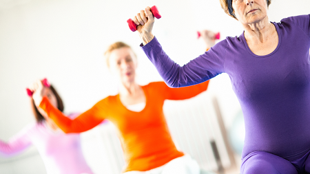
So where is all of this going? Well, yet another study illustrating why menopausal women have difficulty building muscle crossed my desk, replete with the expected PR piece showing grey-haired women lifting the cute and oh-so-cheerfully colored 1lb dumbbells.

To say that I had a negative reaction to it would be putting it mildly. I am not sure why this particular study set me off more than any other though. I mean these types of studies and their associated PR are dime a dozen in this field and this one was not any worse than any of the others that I have read. In fact, it had some cool technology that allowed them to assess scientifically sexy surrogate markers, the muscle stem cells called satellite cells. They ‘discovered’ that muscle stem cells in women do indeed respond to hormones, as they do in men, and as all cells do. And they do indeed diminish and die off absent those hormones, both in cell culture and in ovariectomized rodents, making recovery from injury difficult. They also found that in four of the five older women tested, none of whom was reported to engage in muscle-building activities, muscle stem cells numbers naturally declined across time, as did estradiol concentrations. There was no information on the fifth woman. Groundbreaking right? This explains why women have difficulty building muscle as they age, right? Sure, if you don’t know anything about how hormones work or how muscle gets built.

Hormone Basics

To understand why this and studies like it that attribute the actions of a single hormone like estradiol to a more global function like muscle development fall short, we have to understand a thing or two about how hormones work in real life versus how we measure them in the lab. I would argue that much, if not all, single-hormone-to-single-variable research fails, no matter the hormone or the variable because the actions of a single hormone are fundamentally incongruent with how hormones behave in real life. Hormones never, ever, ever act in isolation. Each hormone’s existence is entirely dependent upon its sister hormones up and down the metabolic chain, and thus, they are all in a constant and interdependent state of communication and activity. Not only that, hormones interact with the totality of one’s internal chemistry, responding to and signaling information about things like nutrient availability/unavailability, immune and inflammatory changes, and most importantly, mitochondrial energetics. So no, a single hormone study will never come close to approximating the complexity of interactions in real life. And it doesn’t take an advanced degree to understand this. A quick Google image search hormone synthesis shows us quite clearly how much more complicated hormone to variable interactions are. Here is one of many illustrations.

Figure 1. Steroid hormone synthesis in the adrenal cortex.

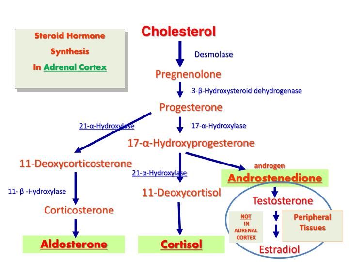
This is an oversimplified version of what is really happening. Take a look at this image to appreciate the complexity of steroid hormone synthesis. Even so, from the image to the left, we can see that all steroid hormones are synthesized from cholesterol (yes, that cholesterol, the one we are all so happy to block with drugs) to pregnenolone (in the mitochondria), and then to progesterone and on through a complicated network of enzymes to the more commonly recognized steroid hormones like testosterone and estradiol.

These figures should tell us a few things about hormones. First, hormone metabolism is damned complicated. While understanding the mechanisms of action of a single hormone is an important endeavor, we must always remember that hormones never act in isolation and so any results we may attribute to that single hormone, are necessarily limited and often flawed. Second and following from the first point, all steroid hormones are connected to each other in some way or another. That means, the concentration of one hormone invariably affects and is affected by, everything else around it, both its downstream metabolites and through feedback control mechanisms, its upstream precursors.

Third, neither progesterone nor estradiol are exclusively female hormones. Progesterone is a precursor hormone for everything else and so, yes, men have it too, and estradiol derives directly from testosterone, and so both men and women have both testosterone and estradiol. What determines how much of each and what directions these metabolic pathways take is determined by enzyme activity, which is determined first by genetics and then by where, what tissue or gland, the enzyme is located within the body. In the ovaries, we get more pregnane (progesterone) and estrane hormones (the estrogens collectively), and in male gonads, the paths shift towards the androgens, but in neither are those the ‘only’ hormones produced by those glands. Moreover, in both males and females, the adrenal glands produce a full complement of androgens, some estrogens, and what are called glucocorticoids (cortisol) and mineralocorticoids (aldosterone). Fat cells synthesize, store, and react to estrogens, in males and females. While muscle cells synthesize and respond to a full complement of hormones, not just androgens, and do so, in what is called a sexually dimorphic manner. Interestingly, after a single bout of exercise testosterone is upregulated in both male and female muscle, while estradiol production is increased in males only. So how is it that estradiol is solely responsible for muscle in women?

I should also note that the brain synthesizes and responds to steroid hormones. Not only does the brain have the entire collection of enzymes to synthesize its own hormones from scratch, but because steroid hormones are lipid-soluble, those produced in the body easily cross the blood-brain barrier (and for the more nerdy among us, even the less fat-soluble sulfated hormones cross via transporters). Then, of course, are all of the environmental estrogens that we are exposed to on a daily basis, but that is another topic for another day. Needless to say, hormones can be synthesized, and thus act, anywhere.

Finally, steroid hormones do things, this perhaps, is the most important point. They can turn things on or off directly or they can fuss with the volume of a signal, turning it up or down. How they do this involves binding to receptors. Some of these receptors are on the cell membranes (fast-acting, volume control) and others are inside the cell nucleus (slower acting, gene regulation). Steroid hormones are promiscuous in their choice of receptors. While they have preferences for and tend to bind more tightly to their own receptors (estradiol to estrogen receptors, for example), they may also bind to other receptors (progesterone to mineralocorticoid receptors or deoxycorticosterone [substrate for cortisol] to androgen receptors, for example). See here for a brief description. Bottom line, we all have the same hormones, just different concentrations relative to genetic sex, enzyme activity, and gland presence/absence.

I point all of this out because laboratory studies tend to focus their work on the actions of a single hormone using a variety of common methodologies. They expose cells to hormone and hormone-free media measuring the differences between the two or they remove the hormone-producing gland, knock out a gene or two involved in its synthesis or the synthesis of its receptors, and again measure effects without the hormone and then added back. While experimentally valid approaches, in vivo, that is in real life, hormones do not act in isolation, and barring some very specific genetic issues or medical treatments, single hormones are never reduced to zero. So when we see studies that claim to show that X hormone is responsible for Y behavior using these methods, we have to remember that while it may be implicated in said behavior, the real relationship is far more complicated than experimental design allows.

It’s Never Just One Hormone

With all of this in mind, let us consider the role of estradiol in health. Estradiol, like many hormones, is a necessary component of human physiology. We have estrogen receptors all over the body and the brain. Both men and women produce estradiol and have estrogen receptors, though men to a much lesser degree. Cells exposed to estradiol are going to do something, usually turn something on, but also, especially when concentrations get too high, turn something off. An interesting side note, the estrogen receptor is considered to be the first hormone receptor to evolve, dating back some 450 million years ago. The progesterone receptor evolved next. Both receptors appear to have evolved well after their respective hormones. How exactly that worked, I am not sure. Importantly though, estradiol does stuff. So studies that tell us cells do something in response to estradiol and die off in its complete absence are interesting, but not necessarily indicative of anything else but what is happening in that particular experimental environment.

Similarly, the common ovariectomy and estradiol add-back protocols used in research with female rodents, though unfortunately closer to reality, as hundreds of thousands of women are castrated annually, still fall short but for different reasons. Castrating an animal or a human woman causes a violent, immediate decline in hormones (not just estradiol, but all of the hormones produced by the ovaries), and as we would expect it to, that violent decline in hormone availability causes problems -everywhere. Resources have to be reallocated for survival. In the immediate aftermath of castration, cell death is to be expected. Again, and of course, the add-back therapy will diminish some of the symptoms and prolong the life of the cells in question. Remember though, estradiol receptors are everywhere and are required for cell function (and mitochondrial function). Period. If we remove it, cells die. If we add back, they don’t die as quickly. We could do the same thing with other steroid hormones and get the same result to varying degrees because those receptors exist too and therefore impact cellular function as well. It is never just one hormone. Keep repeating that. It is never just one hormone.

Does any of this suggest that estradiol is THE HORMONE involved in female muscle development? No. It suggests what we expect it to suggest, that estradiol is involved in muscle development, just like a whole bunch of other variables are also involved but rarely measured in combination. Other hormones, diet, and the single most overlooked variable it seems, actual strength training, play role in muscle development. As such, we would expect any one of these variables, alone or in combination to positively affect any number of the surrogate markers tested within these protocols.

Will You Get to Muscle Part, Already?!

Now that we know estradiol does stuff but not in isolation, can we really attribute the menopausal decline in estradiol to this seemingly entrenched idea that women of a certain age have difficulty building muscle? No, we cannot for the reasons stated above, but also, because we have yet to define either what building muscle constitutes or what ‘difficulty’ means. Does it mean that with the appropriate weight training, menopausal cannot increase muscle size? Does it mean that with training they cannot build strength? Or maybe it means that older women will never be as muscularly defined or as strong as their younger counterparts? Nope, not according to much of the research. That would be too logical. What these studies seem to mean by difficulty building muscles is that their particular surrogate marker(s) for muscle diminishes naturally across time, in the absence of specific muscle-building activities, and as estradiol, declines(here, here, here, here, here, and here). Well, duh, on so many counts. Of course muscle mass and strength decline across time with disuse or absent activities designed to build muscle and of course estradiol declines with age. We don’t magically build muscle when we are younger just because we are younger, save perhaps when we are children and still growing. That is not how muscle grows in adults. Neither do we magically grow muscle just because we have more estradiol or even testosterone absent training. One has to train the muscle to grow it and train it in specific ways.

As I dug into the literature, it became clear that many of these studies, even when humans were included, which often they are not, never measured anything close to actual muscle development. Currently, the preferred surrogate marker for muscle development is accelerometer readings. Yes, accelerometers. Study participants wear accelerometers that sense change in activity level, and using some entirely-too-complicated metrics, calculate energy expenditure based upon the change from sedentary to active. From there, researchers would estimate what is deemed ‘moderate to vigorous physical activity’ (MVPA). From what I can tell, one can achieve this desired MVPA with about ~40 minutes daily of general exercise including brisk walking, heavy cleaning or gardening, biking, dancing, or other aerobic activity. This is not strength training. It’s just not.

In the past and in less well-funded studies, surveys of physical activity were used. I should note that these two metrics do not correlate. Grip strength is another favorite (here, here), along with various measures of isokinetic strength, using mostly cross-sectional designs. Then there are the slew of studies whose sole purpose appears to convince us that menopausal muscle decline requires hormone replacement (HRT). These too, fall significantly short both in design and their intended purpose of promoting HRT. None of these studies involved any actual strength training, just random assessments of, what I would argue, are random surrogate markers of ‘strength’ (here, here, here, here). In one large study assessing ‘muscle strength’ (N=654 men and women ages 20-93) the researchers noted:

“Only a very small percentage of subjects (<1%) participated in any type of regular resistive exercise, and there was no significant difference in participation by age or gender.”

Less than one percent of study subjects participated in any sort of resistance training and yet the study concludes that both men and women lose strength as they age. Well yes, of course, they do. Disuse across time, no matter the age equals a loss of strength. That does not mean though that age is the variable of concern. Rather, it indicates that if we do not use our muscles, they atrophy.

Of the few studies that I could find with protocols involving strength training in actual older women, we do see muscle growth but it is not as significant as one would expect and perhaps hence, the ‘difficulty building muscle’ conclusion is reinforced. Here again, though, the strength training protocols employed, are not typical of what we would see used to build maximal strength, even when the titles of the studies suggest otherwise. The programs involved using lower weights, machines, and rep schemes that do not accord with strength training programs used in younger or even older lifters. They do not even accord with hypertrophy or bodybuilding programs. It is as if in the very design of the program, the bias that older women are weaker and thus require fundamentally different types of exercises to build strength than younger women or men, determines program design. Admittedly, age plays a role in how one trains, but not in the way that seems to be perceived by the research community.

If we define building muscle as a measure of strength, then we have to design programs to build maximal strength, not just everyday activity strength. If we define building muscle as a bodybuilder might in terms of definition and size, then we have to design the programs to develop muscle in that capacity. If, however, we define muscle building more amorphously, as something to do with overall fitness or functional life capacity, then we are not necessarily ‘building muscle’. We are building overall health with some muscle capacity along the way. This is not necessarily a bad thing, especially compared to a sedentary lifestyle, but it does not answer the question of whether older women can build muscle or strength. It simply does not. And it should go without saying, we must include nutritional protocols as well, particularly protein intake. Training and nutrition cannot be separated if we are building muscle, and unfortunately, nutrition is rarely addressed in these studies.

Can Menopausal Women Build Muscle?

As an older female who lifts heavy and continues to get stronger and more muscular year after year despite my waning hormones, the answer is yes. Menopausal women, when trained appropriately can build muscle – a lot of it.

I came to strength sports late in life, just a few months shy of my 48th birthday, in that perimenopausal period where the rate of muscle loss is purported to increase. As a lifelong swimmer and water polo player, who worked out in these and other sports 6-8 hours a week for most of my life, I was never sedentary for very long. Aerobically, I was in great shape, a little overweight, but I could hold my own in the pool in the fast-paced, high-intensity sport of water polo. By the standard measures used to quantify muscle-building activities in the research cited, I was well above the norm for my age, but was I strong? I thought I was, but when I first stumbled into a CrossFit gym, I found out just how wrong I was. The first time I swung a kettlebell, it was 15lbs and I thought it was heavy. I was genuinely afraid my grip would loosen and I would throw the kettlebell across the room. The first time I deadlifted, 65lbs about killed me. Remember, by the measures cited above, I should have been strong. I wasn’t. My muscles had not been trained for strength.

Fast forward 4 1/2 years or so, I have a little over three years of powerlifting training under my belt and at 52 years of age, I regularly swing 100lb kettlebells and my deadlift currently stands at 342lbs. I also have every intention of pulling 400lbs in a few years. Contrary to the expectations of the academic community who suggest a rapid decline in muscle capacity as we age and as we lose estradiol, I have no difficulty building muscle or strength, despite my declining estradiol concentrations, and though I continue to get older, I am getting stronger. Why? Because I train for maximal strength. It is not as radical as it seems. If one trains to get stronger, strength happens. Of course, I eat right, but mostly, it is because I train and I train hard.

Arguably, an N of 1 is little more than anecdotal evidence. It tells us nothing. I could be unique, an outlier. I am not. I am not even that strong compared to other women in my age group and older. I know 60-year-old women who can rep my max weight like it is nothing. How do I know this? Well, from multiple sources, none of them academic and none of them constitutes evidence as of yet but they are all worth considering as each chips away at the paradigm of inevitable menopausal muscle decline.

The first bit of evidence, master’s powerlifting is a rapidly growing sport for women. A quick glance at an organization that tracks powerlifting records across federations, we see that some phenomenally strong masters’ age women, lifting ridiculously heavy weights. In my age and weight, drug-tested class, the top female lifter deadlifts over 500lbs, and many pull over 400lbs. Remember, these are women in their 50s. Menopausal muscle decline? I don’t think so. Across all weight classes, there are over 3100 female lifters over the age of 50, the oldest listed, is 92 years of age. If this many women are competing, how many others are not? Well, if we look at the CrossFit demographic data, we see that women aged 45 and older make up about 7% of their market. With approximately 4 million CrossFit enthusiasts worldwide, this equates to about 280,000 peri-menopausal women lifting heavy barbells. I suspect many of those women have built significant muscle and strength, certainly more than the women used in academic research or the regular gym-going women whose trainers abide by the menopausal weakness bias and limit their clients to those silly 1-5lb dumbbells.

National Record deadlift 220.5 lbs.
What real women lift. Rosemary Nichols, age 80, 220lb deadlift.

The second bit of evidence, though again non-traditional, about a year ago, I started a Facebook group and website for older female lifters. We currently have over 5100 women from all over the world. We post our workouts, and our meets, and generally help each other navigate lifting while older and female. The largest segment of the group is women in their fifties, followed closely by women in their sixties.

Among the members of this group, we have women who began their lifting journeys late into their 60s and sometimes even in their 70s. We have many 70-year-olds who can deadlift over 200lbs, some are approaching 300lbs. We even have a 120lb, 80-year-old woman, who began lifting in her 70s and now can deadlift over 200lbs with a fair amount more ease than most 20-year-olds. And oh, most of us squat, bench too. Many of the women in our group compete in the Olympic lifts too and we have others who compete in Strongwoman events. Given the opportunity, older women can and do lift heavy.

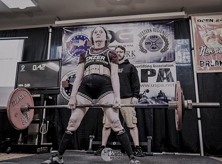
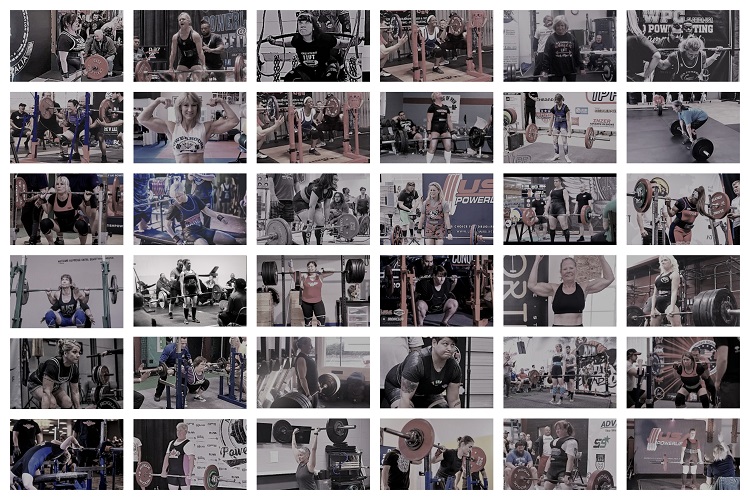
Figure 2. Older women who lift heavy.

Menopausal muscle

If any one of these women were tested for muscle density and capacity change across time, we would see legitimate and likely statistically significant changes in both. We would also see that while hormones, nutrition, and other factors co-vary and interact, the most important variable would be the training itself, and the most important aspect of the training is that it is done to increase maximal strength. It is not done for general fitness or weight loss, though those are often happy side-effects, the training we do is to lift as much as possible. Period. Measure that and the myth of menopausal muscle decline goes by the wayside. I would argue that while muscle decline coincides with aging and hormonal decline, it is more attributable to disuse than either of those two factors. The capacity to build muscle exists, even as we age and even after longstanding disuse. It just has to be trained.

Bottom Line: Use It or Lose It

It’s really that simple. Menopausal women can build muscle and strength. We are only limited by whether and how hard we are willing to train. So, if you are an older woman considering lifting weights, do it. Find a coach, put in the time and you’ll be pleasantly surprised just how strong you are. All of us at old ladies lift are here for you. Find us on Facebook @oldladieslift or online at OldLadiesLift.com. If you are a coach or trainer, don’t dismiss the older woman who wanders into your gym and says she wants to lift heavy. She can and she will. Need more proof, take a look at some of these videos. Finally, if you are a researcher who wants to study strength changes in older women, contact me. It’s time to put the myth of menopausal muscle decline to rest.

We Need Your Help

More people than ever are reading Hormones Matter, a testament to the need for independent voices in health and medicine. We are not funded and accept limited advertising. Unlike many health sites, we don’t force you to purchase a subscription. We believe health information should be open to all. If you read Hormones Matter, and like it, please help support it. Contribute now.

Yes, I would like to support Hormones Matter.

Uterus and Ovaries: Fountain of Youth

7765 views

Numerous studies have shown a strong correlation between removal of both ovaries / bilateral oophorectomy (castration) and accelerated aging as measured by an increased risk of chronic health conditions. Hysterectomy / uterus removal with preservation of both ovaries is also associated with some of these chronic conditions. These include heart disease, stroke, metabolic syndrome, osteoporosis, hip fracture, lung cancer, colorectal cancer, dementia, Parkinsonism, impaired cognition and memory, mood disorders, sleep disorders, adverse skin and body composition changes, adverse ocular changes including glaucoma, impaired sexual function, more severe hot flushes and urogenital atrophy. Wow, that’s quite a list!

Ovaries: Health Powerhouses

This 2016 article titled “Study: Remove ovaries, age faster” sums up the findings of Mayo Clinic researchers proving yet again the harmful and unethical practice of ovary removal. The study found that ovary removal (oophorectomy) is associated with a higher incidence of 18 chronic conditions and should be discontinued in women who are not at high risk for ovarian cancer. Although this study cites the increase in chronic conditions in women who undergo oophorectomy before age 46, other studies have shown that oophorectomy even after menopause does more harm than good. Here is one that showed that to be true up to age 75.

The ovaries have both reproductive and endocrine functions as detailed in this International Menopause Society article. After menopause, the ovaries produce mostly androgens, some of which are converted into estrogen. Testosterone levels are more than 40% lower in women without ovaries compared to intact women. Women without their uterus likewise have lower levels but not as low as women without ovaries per this article. Estrogen therapy mitigates some but not all of the increased health risks of oophorectomy. But estrogen further reduces androgen levels increasing risk of osteoporosis and fracture. Nothing can replace the lifelong functions of the ovaries (and uterus).

The Uterus / Ovaries / Tubes Connection

The harms of ovary removal would also apply to ovarian failure that commonly occurs after hysterectomy and some other medical treatments. As previously cited, women who have had a hysterectomy have lower levels of testosterone. According to this 1986 publication, 39% of these women showed signs of ovarian failure. This study showed a nearly 2-fold increased risk of ovarian failure when both ovaries were preserved and nearly 3-fold when one was preserved. This likely explains the increased risk of heart disease and metabolic conditions as shown by multiple studies including this recent Mayo Clinic one. However, per this 1982 study, the uterus itself protects women from heart disease via the uterine substance prostacyclin. Loss of bone density is another harm of hysterectomy as shown by multiple studies such as this one.

Removal of even one ovary (unilateral oophorectomy) without hysterectomy is also harmful. Studies out of the Mayo Clinic showed increased risks of cognitive impairment or dementia and parkinsonism. Colorectal cancer is another increased risk according to this Chinese study and this Swedish one.

The Fallopian tubes appear to impair ovarian function to some degree as evidenced by Post Tubal Ligation / Sterilization Syndrome. This study shows an increase in Follicle Stimulating Hormone (FSH) after tube removal (salpingectomy).

Ovarian impairment after hysterectomy or salpingectomy is thought to be the mechanism of the reduced risk of ovarian cancer which is already rare.

The Uterus: Anatomy, Sex, Cancer Prevention

Hysterectomy is associated with other harms besides impaired ovarian / endocrine function. The uterus and its ligaments / pelvic support structures are essential for pelvic organ integrity as well as skeletal integrity. The effects on these structures and functions are detailed here and here. This article shows the many hysterectomized women lamenting their “broken bodies” – changes to their figures, back, hip and midsection pain, pelvic pain, bladder and bowel issues, and effects of severed nerves and blood vessels.

The uterus and associated nerves and blood vessels play a key role in sexuality and vibrancy. You can hear the desperation in women’s comments about the devastating sexual losses and feelings of emotional emptiness.

There is an increased risk of renal cell, thyroid, and colorectal cancers after hysterectomy. How ironic when cancer fear tactics are commonly used to market hysterectomy and/or oophorectomy.

Adhesions that commonly form after these surgeries can cause serious problems especially in the long term. Surgical complications – nerve injuries, bladder, bowel and ureter injuries, vaginal cuff dehiscence, a too short vagina, infections, hemorrhage – are more common than indicated by gynecologists.

Although “The Miraculous Uterus” article fails to mention the anatomical harms, it is otherwise “spot on.” It talks about the “ovarian conservation scam” and that “passion, love, ecstasy, the emotional essence that drives human achievement, forever after elude them.” This explains why “there’s no effective outrage against the barbarism of hysterectomy.”

Compelling Evidence of Harm

Clearly, there is compelling medical evidence that both hysterectomy and oophorectomy are destructive surgeries. Unfortunately, some hysterectomy forums censor negative posts giving a slanted view of the life shattering effects. Here is a sampling of women’s experiences on the Gyn Reform site.

The medical literature on the harms of these surgeries dates back over a century. Listed below are a small number of the numerous publications (minus the ovarian failure studies cited above). The Gyn Reform website has a fairly comprehensive list of resources on oophorectomy. Its Ovaries for Life sister site provides a good overview of the lifelong importance of our ovaries.

1912 – The Physiological Influence of Ovarian Secretion

1914 – Nervous and Mental Disturbances following Castration in Women

1958 – The controversial ovary

1973 – Osteoporosis after Oophorectomy for Non-malignant Disease in Premenopausal Women

“Oophorectomy before the age of 45 years was found to be associated with a significantly increased prevalence of osteoporosis within three to six years of operation.

1974 – Endocrine Function of the Postmenopausal Ovary: Concentration of Androgens and Estrogens in Ovarian and Peripheral Vein Blood

1978 – The emotional and psychosexual aspects of hysterectomy

1981 – Premenopausal hysterectomy and cardiovascular disease

1981 – Sexual response after hysterectomy-oophorectomy: Recent studies and reconsideration of psychogenesis

1981 – The role of estrogen and oophorectomy in immune synovitis

1982 – Prostacyclin from the uterus and woman’s cardiovascular advantage

1989 – The effects of simple hysterectomy on vesicourethral function

“The results show that simple hysterectomy is associated with a significant incidence of post-operative vesicourethral dysfunction and that there is an identifiable neurological abnormality incurred at operation which is pertinent to the subsequent disordered voiding.

1990 – Effects of bilateral oophorectomy on lipoprotein metabolism

1994 – The climacteric ovary as a functional gonadotropin-driven androgen-producing gland

1996 – Urinary incontinence in older women: who is at risk? Study of Osteoporotic Fractures Research Group

“Urinary incontinence is a common problem in older women, more common than most chronic medical conditions. Of the associated factors that are preventable or modifiable, obesity and hysterectomy may have the greatest impact on the prevalence of daily incontinence.

1997 – Bladder, bowel and sexual function after hysterectomy for benign conditions

1998 – Ovaries, androgens and the menopause: practical applications

1998 – Impairment of basal forebrain cholinergic neurons associated with aging and long-term loss of ovarian function

1998 – Influence of bilateral oophorectomy upon lipid metabolism

1999 – Estrogen and movement disorders

2000 – The hypothalamic-pituitary-adrenal and gonadal axes in rheumatoid arthritis

2000 – Risk of myocardial infarction after oophorectomy and hysterectomy

2000 – Hysterectomy, Oophorectomy, and Endogenous Sex Hormone Levels in Older Women: The Rancho Bernardo Study

2005 – Ovarian conservation at the time of hysterectomy for benign disease

Ovarian conservation until age 65 benefits long-term survival…. There is sustained, but decreasing, benefit until the age of 75, when excess mortality for oophorectomy is less than 1%.

2007 – Ovarian conservation at the time of hysterectomy for benign disease

Approximately 78% of women between the ages of 45 and 64 years have prophylactic oophorectomy when hysterectomy is performed for benign disease. Therefore, the decision to perform prophylactic oophorectomy should be approached with great caution for the majority of women who are at low risk of developing ovarian cancer.”

2009 – Ovarian conservation at the time of hysterectomy and long-term health outcomes in the nurses’ health study

In no analysis or age group was oophorectomy associated with increased survival.

2010 – Current indications and role of surgery in the management of sigmoid diverticulitis

A previous history of hysterectomy is a valuable clinical clue to the correct diagnosis as colovaginal and colovesical fistulas are rare in females with their uterus in place, as the uterus becomes a screen interposed between the inflamed colon and the bladder and vagina.”

2012 – Oophorectomy for whom and at what age? Primum non nocere

2016 – Study: Remove ovaries, age faster

2017 – Cardiovascular and metabolic morbidity after hysterectomy with ovarian conservation: a cohort study

A Harmful Practice That Won’t Die

Ovary removal / castration was introduced by Robert Battey in 1872 and “was practised widely for several decades….. Better insight into female physiology and ovarian function finally pushed the sinister operation of Robert Battey from the scene.” This publication refers to Battey’s operation as “barbaric.”

Despite the long-standing and compelling evidence of harm, these surgeries continue at alarming rates. Publications are misleading in that they report inpatient surgeries despite the large majority being outpatient (70% in 2014). This 2008 article reported that oophorectomies “more than doubled in frequency since the 1960’s.” According to results of a FOIA request by Ovaries for Life, there are over 700,000 oophorectomies every year despite there being only ~22,000 cases of ovarian cancer. Hysterectomy figures obtained by Ovaries for Life are also shocking at 830,000 in light of less than 70,000 cases of endometrial and cervical cancers.

Many media reports have questioned the high rate of these surgeries since gynecologic cancers are rare. The oldest one I could find was dated 1969. I found about three articles per decade in the mainstream media since then. According to the Athena Institute, half of U.S. medical schools in 1986 “had changed their suggestions and were now recommending a reconsideration of the common practice of ovariectomy.” Evidently, that never took hold.

Congress held two hearings on hysterectomy, one in 1976 and one in 1993. The 1993 transcripts state that the hysterectomy rate increased 250% in women ages 15 to 24 and 186% in ages 25 to 34 from 1965 to 1984! Despite these shocking statistics, it appears that no action was taken after either hearing.

According to this “Reassessing Hysterectomy” article, the Agency for Healthcare Research and Quality sponsored research and conferences on the overuse of hysterectomy in the 1990’s. This article is packed with information on the prevalence and harms of hysterectomy and oophorectomy as well as alternative treatment options. Yet, the high rate of hysterectomy has continued such that 45% of women will end up having one. Citing 2006 data, the oophorectomy rate was 73% of the hysterectomy rate.

How to End the Harm?

I’ve been researching this subject for over 10 years and sharing my experience and knowledge on various websites. It’s shocking how many women are misled and deceived into these surgeries. Age doesn’t seem to matter; younger and younger women are undergoing these surgeries. This appears to be the biggest surgical racket and women’s healthcare con as discussed here.

There are a number of issues that perpetuate the gross overuse of these harmful surgeries. These include:

  1. These surgeries and “forever after” care are very lucrative.
  2. The public has been led to believe that the female organs are disposable after childbearing is complete.
  3. Medical education and decades of practice have made these surgeries “a standard of care.”
  4. Informed consent is seriously lacking.
  5. Gynecology consent forms are open ended giving surgeons “carte blanche” to remove organs unnecessarily.
  6. We still live in a climate of gender disparity / male dominance.

As you can see from the list of publications above, some study authors have called out the practice of ovary removal as unethical. Numerous professional societies have issued guidelines discouraging its use in most women. But most have been silent on the overuse of hysterectomy despite its many harms.

Why has our government not stepped in to address this egregious harm? Women who have contacted their legislators have been met with indifference. Gyn Reform reported on their experiences with legislators and other authorities who can effect change. The non-profit HERS Foundation has been educating women and advocating for informed consent legislation since the 1980’s.

Why do insurance companies approve so many of these surgeries that are rarely necessary? Not only are the surgeries themselves expensive, treatments for the chronic after effects are costly. Reining in unnecessary treatments especially those that cause lifelong harm would go a long way towards making healthcare more affordable.

Why has Graduate Medical Education (GME) not changed their surgical requirements to favor organ preservation? Each resident must do at least 70 hysterectomies but there is no requirement for myomectomy (fibroid removal). Residents don’t need to do any cystectomies (cyst removals) either which is partly why so many women lose ovaries for benign ovarian cysts. Here are the GME ob/gyn requirements.

A popular mantra at Tufts in the 1970’s – “There’s no room in the tomb for the womb” – reflects this culture of the disposable uterus and gynecologists’ obsession with its removal. Insurance reimbursement rates are also to blame as they incentivize hysterectomy and oophorectomy over myomectomy and cystectomy. In many cases, medical management versus surgery is the appropriate course. The “Reassessing Hysterectomy” article cited above lists a number of treatment options for gynecologic problems. Revamping reimbursement rates to strongly favor organ preservation should eventually force GME to change their requirements. But how do we make that happen?

We Need Your Help

More people than ever are reading Hormones Matter, a testament to the need for independent voices in health and medicine. We are not funded and accept limited advertising. Unlike many health sites, we don’t force you to purchase a subscription. We believe health information should be open to all. If you read Hormones Matter, like it, please help support it. Contribute now.

Yes, I would like to support Hormones Matter. 

Lucas Cranach the Elder, Public domain, via Wikimedia Commons

Menopause: New Insight Into Hot Flashes and Night Sweats

6408 views

Menopause is defined as the time after a woman’s final menstrual period, and is confirmed after one year with no periods. For many women, this is a natural, gradual process, that begins with perimenopause: the four to eight years before the final period. The time through perimenopause, the final menstrual period, and a few years after the final period is known as the menopausal transition. Many women go through this transition naturally as they age, but other women go through induced menopause, caused by surgical removal of the ovaries or cancer treatments.

Symptoms Related to Menopause

The average age of menopause in North America is 51 years, with most women going through menopause between the age of 40 and 58. During the menopausal transition, hormonal changes may result in symptoms that range from mildly bothersome to downright unbearable. Vasomotor symptoms, or hot flashes and night sweats, are one of the main menopause-related symptoms that women seek medical treatment for, and are experienced by about 80% of women during the menopausal transition. Some women also experience vaginal dryness and/or pain with sexual intercourse during menopause.

During perimenopause, many women will start to experience hot flashes and night sweats. Other symptoms of perimenopause include vaginal dryness, irregular periods, sleep problems, mood changes, weight gain, thinning hair, and dry skin (sounds awesome, right?). However, the North American Menopause Society correctly points out that the menopausal transition often coincides with a number of midlife stressors such as relationship issues/divorce, caring for aging parents, career issues, and struggles with adolescents (is anyone else’s hair going grey from teenage daughter stress?). These stressors may also contribute to symptoms a woman experiences at this time.

Hot flashes and night sweats (vasomotor symptoms or VMS) can be troublesome to many women. As with other issues in women’s health, lack of research led to the long held false assumption that these symptoms are generally experienced for only a few years right around the final menstrual period. However, new research has shown that in some women, these symptoms last for a decade or longer. In addition, the pattern of these symptoms, or when they occur relative to the final menstrual period, is not the same in all women.

New Insights into Frequency and Timing of Hot Flashes and Night Sweats

A recent, large study of ethnically diverse women going through natural menopause across the U.S. has provided some new insights into how women experience vasomotor symptoms during menopause and who is at risk for longer lasting or more severe symptoms. This study followed women from 12 years before the final menstrual period to 15 years after. The median duration of vasomotor symptoms was 7.4 years and the median persistence of symptoms after the final period was 4.5 years. In this study, frequent vasomotor symptoms lasting more than seven years occurred in more than 50% of women. Starting to have vasomotor symptoms at a younger age was predictive of having a longer duration of symptoms, and surprisingly, of having a longer persistence of symptoms after the final period.

This study also found that vasomotor symptoms during the menopausal transition fall into four distinct patterns. In about 27% of women, called the early onset, low frequency group, there is a low probability of vasomotor symptoms across the whole menopausal transition, with a slight increase around the time of the final menstrual period. In 26% of women, there was a  high frequency of vasomotor symptoms throughout the entire menopausal transition. In the 18% of women who had an early onset, there was a higher frequency of symptoms, which decreased right after the last period. And 29% of women had a late onset of higher frequency symptoms, with a sharp increase just after the final period.

The study also examined whether certain health or lifestyle measures were associated with particular patterns of vasomotor symptoms and some associations were found. Relative to the women in the low frequency group, women with early onset, higher frequency symptoms were older at the final menstrual period, were in poorer health, and were more likely to suffer from depression and anxiety. Women with late onset, higher frequency symptoms were more likely to be African American/black, more likely to be smokers, and less likely to be obese. And women in the high frequency group were more likely to have less education, be in poorer health, have an increased consumption of alcohol, have depression and anxiety, and be African American/black.

These “co-variables” were all independently associated, meaning, for example, that someone with poor physical health, or depression and anxiety, was more likely in this study to be in the early onset, high frequency group, or in the group with high frequency across the whole transition. The other variables did not need to be present in addition, to create the association.

Hormones Levels During the Menopausal Transition

It has generally been thought that declining estrogen levels during the menopausal transition are responsible for vasomotor and other menopausal symptoms. However, not surprisingly, this view is too simplistic. This same study measured the concentrations of one of the major estrogens in the human body, estradiol, throughout the study, and found that estradiol concentrations have four different patterns during this time as well. However, these patterns did not correlate perfectly with the different groupings of vasomotor symptoms.

In general, women with a consistently lower concentrations of estradiol were more likely to have frequent symptoms across the menopausal transition. However, the study also found that fluctuations in estradiol around the time of the final menstrual period may be associated with vasomotor symptoms at this time. A greater rise in follicle-stimulating hormone (FSH) was also associated with a greater likelihood of vasomotor symptoms. The conclusion of the study was that estradiol alone was not solely responsible for vasomotor symptoms; that other hormones varying during this time may also play a role. For example, some adrenal steroid hormones increase transiently during menopause.  DHEAS, a steroid hormone that generally declines with age,  increases transiently in some women during menopause.

Are Cardiac Risks Related to Vasomotor Symptoms?

After menopause, a women’s risk of certain serious conditions increases, including cardiovascular disease and osteoporosis. Some preliminary results suggest that the pattern of vasomotor symptoms may be correlated to an increased risk for cardiovascular disease. In one study, carotid intima media thickness (IMT) was measured, which can detect the presence of atherosclerotic disease before cardiovascular symptoms appear. Women with early onset vasomotor symptoms had higher IMT compared to women with low frequency of vasomotor symptoms. This could indicate a higher risk of cardiac events for women who fall into this group. In another study, late vasomotor symptoms were found to be associated with an increased risk of major cardiovascular events, stroke, and death from all causes, and this association was not accounted for by other risk factors. However, because of the size of the study, these results are considered preliminary. More research may allow doctors to predict, based on a woman’s pattern of vasomotor symptoms, her risk of cardiac and other serious events, and may allow doctors to direct lifestyle and other early interventions to those most at risk.

Altogether, this research on vasomotor symptoms during the menopausal transition is helpful because it can help women understand what to expect. It may also help guide treatment decisions for women who are seeking treatment for symptoms; for example, if they start having frequent symptoms early, this may be a sign that these symptoms will persist. In addition, if risks for serious health problems are associated with distinct patterns of vasomotor symptoms, again, this information could help guide early interventions, and help women be alert for early warning signs of a problem.


What Causes Ovarian Cancer?

6601 views

Ovarian cancer is the most difficult to diagnose of all the gynecologic cancers, which are uterine, fallopian, vulva, and cervical. The symptoms of bloating, back or pelvic pain, digestive issues, changes in bowels, urinary frequency, and fatigue are the most commonly reported. Other symptoms include vaginal bleeding or unusual discharge, change in weight, painful intercourse, and menstrual changes. Any one of these can be symptomatic of other diseases or disorders such as Irritable Bowel Syndrome, Gastritis, Back Strain, or Menopause to name a few.

Risk Factors for Ovarian Cancer

Scientists do not know what exactly causes ovarian cancer, but according to the American Cancer Society, there are “some factors that make a woman more likely to develop epithelial ovarian cancer,” which is the most common form of ovarian cancer. The three most prominently researched factors include:

  • Estrogen exposure – synthetic and endogenous
  • Genetics
  • Family history

Ovarian Cancer and the Estrogens

Estradiol is a female hormone that plays an important role in normal sexual and reproductive development. It is one of many estrogenic hormones produced by the ovaries. The ovaries produce most of the estrogenic hormones, though a far lesser amount comes from the adrenal glands. After menopause, the adrenal glands produce the majority of estrogens.

Estradiol and the other endogenous estrogens affect skin, hair, mucus membranes, pelvic muscles, breasts, bones, urinary tract, heart and blood vessels, and the brain. Perhaps more importantly, the estrogens affect mitochondrial energy metabolism in the heart, but also in the brain, and everywhere else as well. In light of that, it is understandable that synthetic estrogens might influence these same systems.

Oral contraceptives. According to the Center for Disease Control and Prevention, “there is no known way to prevent ovarian cancer.” However those who used birth control pills “has consistently been found to be associated with a reduced risk of ovarian cancer….Oral contraceptive formulations with high levels of progestin were associated with a lower risk ….”  Various studies have shown a 10-12% decrease after just one year of use and as much as a 50% decrease after five years.

Some research suggests that total duration of exposure to estrogenic hormones, whether endogenously produced or synthetic (and there is some debate about this), decreases a woman’s risk for ovarian cancer. With this line of reasoning, some researchers have argued that the use of oral contraceptives may reduce the risk of ovarian cancer because contraceptives prevent ovulation. The theory is that each time she ovulates the woman is exposed to more hormones. According to researchers at Johns Hopkins “It is hypothesized that the longer a woman is exposed to estrogen, the higher her risk of ovarian cancer…. The longer the woman menstruates, the higher her risk.”  This may not be the case for all women, however.

For those who carry the BRCA1 and BRCA2 mutations, oral contraceptives may or may not reduce the risk for ovarian cancer, but pregnancy does. A study published in 2001 stated that “We believe that it is premature to prescribe oral contraceptives for the chemoprevention of ovarian cancer in carriers of a BRCA1 or BRCA2 mutation, particularly in the light of the report of a possible increased risk of breast cancer in such women.”   In contrast, a study in May of 2014 the Journal of the National Cancer Institute wrote, “Although we were not able to undertake meta-analyses of the existing data, it is likely that oral contraceptives are associated with ovarian cancer risk reduction. For those who carry the BRCA1 or BRCA2 mutations, oral contraceptives did reduce the risk for ovarian cancer.” More research is required to fully determine if or how oral contraceptives affect the occurrence of ovarian cancer, for those women with or without BRCA1 or BRCA2 mutations.

Other researchers speculate that it is not the estrogens but exposure to androgens that increases risk. Regardless of the mechanisms by which oral contraceptives may or may not confer protection against ovarian cancer, they are not without other risks. When a woman takes birth control pills she is also increasing her risk for breast, cervical, and liver cancer. Therefore, it is imperative that she understand and discuss with her physician these risk factors, as well as her personal and family history.

Since the introduction of oral contraception in the 1960’s there have been many studies on its relationship to various cancers. The studies have been on birth control pills of estrogen only, estrogen-progesterone, and those with androgenic properties (testosterone effects). How much a factor estrogen alone is in causing ovarian cancer requires more research.

I chose to not take birth control pills because I did not believe it was healthy to control when a woman was to have or not have a period. In other words, control her hormones and menstrual cycle. I am the mother of three healthy sons. If I had taken the birth control pills between pregnancies, would I now have ovarian cancer? No one knows for sure. I made my decision many years ago believing it was best for my health so I do not have any regrets.

Hormone Replacement Therapy. For many years women have used Hormone Replacement Therapy (HRT) to help relieve the hot flashes, mood swings, and other symptoms due to menopause. According to a study published in the Lancet in February, 2015, a study of 21,488 postmenopausal women with ovarian cancer, concluded an increase risk from the use of both estrogen-only and progesterone and estrogen combinations of Hormone Replacement Therapy. “Women who had taken HRT for at least 5 years were still at increased risk of ovarian cancer 10 years later.”

An interesting article about HRT and its affects on ovarian and breast cancers, a historical review of HRT, and post oophorectomies written by Chandler Marrs raises the question if HRT is “largely or wholly causal in ovarian cancer.”  More research is needed to answer the questions raised by Dr. Marrs.

There are additional factors for the woman and her physician to take into consideration if HRT is right for her. Has she had a hysterectomy? What are her menopausal side effects, and how severely are they affecting her quality of life? Has she had breast cancer, or have a family history? “Based on the WHI (Women’s Health Initiative) study, taking EPT is linked to a higher risk of breast cancer.”  It is recommended that a woman be on as low a dose and for as short a period of time as possible to reduce her risk of ovarian cancer. There are also over-the-counter herbs and supplements that might help reduce or eliminate the menopausal side effects.

Once again I chose not to take any HRT for three reasons: one, I did not want to manipulate my hormones; two, I had a family history of breast cancer from one aunt; and three, menopausal symptoms of hot flashes and insomnia were not severe enough for me to take any HRT. I used over the counter meds for the insomnia.

Another theory suggests that tubal ligation or hysterectomies might lower the risk of ovarian cancer. This is based on “some cancer-causing substances may enter the body through the vagina and pass through the uterus and fallopian tubes to the ovaries.” This theory also requires more research.

Finally, there appears to be a link between polycystic ovarian syndrome (PCOS), an imbalance of the female hormones, and ovarian and endometrial cancers. Any personal or familial breast cancer history also puts women at higher risk. More research is required for this complex and familial disease.

Ovarian Cancer and Genetic Risk Factors

Is ovarian cancer caused by some defect in our DNA or genes? Our DNA carries instructions for each cell in our body. Any defect in our genes can cause or lead to any type of cancer. What causes the genes to be defective is not completely known. Most often the body is able to correct any damage to a gene, but due to not fully understood factors sometimes the mutated gene is not reversed. “Usually, it takes multiple mutations over a lifetime to cause cancer” which is why aging is a high risk factor for ovarian cancer.

  • Lynch Syndrome, an inherited cancer of the digestive tract causes a 12% risk increase for ovarian cancer because of the mutation in DNA repair genes.
  • BRCA1 and BRCA2 are inherited genes that produce tumor suppressor proteins. If these genes are mutated they increase the risk of developing breast and ovarian cancers. Unfortunately women of Eastern Jewish descent (Ashkenazi) are at greatest risk. Norwegian, Dutch and Icelandic peoples also have a higher incidence of these mutations. BRCA1 and BRCA2 mutations account for about 15% of ovarian cancers overall. BRCA1 and 2 carriers also have a higher concentration of female hormones. Research is examining how estrogen affects the Fallopian tubes where most ovarian cancers begin. Many women with BRCA1 and 2 gene mutations choose to have their breasts and ovaries removed, which is a drastic and difficult decision.

Most DNA mutations related to ovarian cancer are not inherited but instead occur during a woman’s life….So far, studies haven’t been able to specifically link any single chemical in the environment or in our diets to mutations that cause ovarian cancer. The cause of most acquired mutations remains unknown.”

Genetic testing is recommended. The process will include your family health history and environmental factors as well as the gene test. The information can be valuable for the patient, family members, and future generations. If tested positive, family members have the opportunity to make prophylactic decisions as to their health care options.

I chose to be genetically tested though I did not have any of the ethnic risk factors associated with BRCA1 and BRCA2. I wanted to be sure I had not passed on a mutated gene. I am glad to say that I tested negative.

Family History and Ovarian Cancer

How much of a factor is family history? Any family history of ovarian, breast, or colon cancers might raise a red flag to the physician. How close the relative is to the patient is a question to be considered.

My aunt on my mother’s side had breast cancer and we do not know if she had a mutated gene (other than BRCA) that got passed on to me. My great grandfather had colon cancer but it was decided that he was too far removed from me to be a factor. My mother died from stomach cancer and my uncle who was a habitual smoker died of throat cancer. To sciences present knowledge there is no connection with my ovarian cancer.

Do you know your family history? This knowledge may be important along with the presenting symptoms for your physician in determining a diagnosis. From a small, non-scientific, and anonymous questionnaire of gynecologic survivors, here are some unfortunate and interesting statistics:

  • Did your physician(s) ask about your family history?  Yes from 35 out of 131MD’s
  • Did your physician(s) ask about your symptoms? Yes from 34 out of 131 MD’s

Only about 25% of the doctors discussed the family history or symptoms. The women mainly saw their family physician or gynecologist for an initial appointment with presenting symptoms. Here are the most common symptoms:

  • 55%–Bloating
  • 42%–Abdominal pain
  • 28%–Digestive problems
  • 28%–Fatigue
  • 20%–Frequent urination
  • 16% –Constipation
  • 13%–Vaginal bleeding
  • 11%–Weight change
  • 11%–Back pain
  • 8%–Painful intercourse
  • 4%–Shortness of breathe
  • 4%–Menstrual issues
  • 2%–Temperature
  • 2%–Overweight

Here are some comments from the women who completed the questionnaire:

“My family doctor only did testing because I demanded it after she sent me home the first time and I still didn’t feel better after 2 months later.” (Diagnosed Stage IIIC)

“Had I not demanded the ultrasound my diagnosis would have been missed….” (Diagnosed Stage IIIC)

I look back and I do believe I did have subtle symptoms, but my male doctor ignored my complaints….with a family history I feel I should be been looked at more closely.” (Diagnosed IIC)

“I found that the best doctor is the one that listens.”  (Diagnosed Stage IC)

The following patient had a hysterectomy 14 years prior to diagnosis, removing only her uterus. “…I was on Estrogen only for all those years.” (Diagnosed IIIC)

It is important to recognize that there are many possible risk factors that have not been substantiated in various case studies. Such factors are infertility drugs, talcum powder, smoking, aspirin and NSAIDS, and dietary considerations. According to the National Cancer Institute (May 15, 2015) more research is needed for these possible contributors.

Early Detection Is Key

In closing, early detection & diagnosis are a woman’s best opportunity to treat gynecologic cancers. From Johns Hopkins, “…the combination of bloating, increased abdominal size, and urinary problems was found in 43% of women with ovarian cancer…. Women presenting with non-specific symptoms, particularly if severe intensity or rapid onset, should be thoroughly evaluated for the possibility that the symptoms are due to an ovarian mass.”

When we experience any of the symptoms of ovarian cancer for two weeks without any relief despite medications, women need to seek out medical attention quickly. We must advocate for ourselves that the physician listen to our symptoms, discuss our family history, and insist that an abdominal ultrasound and CA125 be parts of the diagnostic tools.

Outshine Ovarian CancerAbout the author: Karen Ingalls is the author of the award-winning book, Outshine: An Ovarian Cancer Memoir, which discusses the symptoms, risk factors, and statistics of this lesser known disease; shares her journey; and how she used traditional medicine and complementary therapies together. She writes how “the beauty of the soul, the real me and the real you, outshines the effects of cancer, chemotherapy, and radiation.”

Corrections. On June 26, the word ‘increases’ was changed to ‘decreases’ in the following sentence:  ‘…whether endogenously produced or synthetic (and there is some debate about this), decreases a woman’s risk for ovarian cancer.’  The 2014 study was added to the discussion of BRCA mutations and oral contraceptives. 

HRT ‘Largely or Wholly Causal’ in Ovarian Cancer

2938 views

Think about that quote for a minute. Hormone Replacement Therapy or HRT may be largely or wholly causal for a significantly increased risk of ovarian cancer in women, according to authors of a recently published study, Menopausal hormone use and ovarian cancer risk: individual participant meta-analysis of 52 epidemiological studies. ‘Largely or wholly causal’ is very strong statement; one that makers of HRT are sure to dispute, but one that nevertheless was supported by data.

Ovarian Cancer Risk with HRT Use

Based upon prospective (17 studies; 12,110 cases) and retrospective (35 studies; 9,378 cases) epidemiological data reviewed by the Collaborative Group of Epidemiological Studies of Ovarian Cancer in Oxford, England and published in the Lancet, out of the 21,488 post menopausal women with ovarian cancer, 9303 had used HRT, most more than 5 years. From a statistical standpoint, the risk for ovarian cancer was significantly greater in women who had ever used HRT versus those who had never used HRT and even greater in those who were currently using HRT or had recently used HRT than those who had ceased HRT years prior. In other words, HRT use accounted for almost half of the cases of ovarian cancer. Moreover, the risk for HRT induced ovarian cancer was greatest while on the medication and soon after ceasing, but declined as time passed. This is an big finding.

Historical Associations between HRT, Cancer and Disease

Since the Women’s Health Initiative (WHI), debate about the safety of HRT has been forefront among millions of women. WHI demonstrated an increased risk of breast cancer and all sorts of other adverse reactions to menopausal hormone replacement therapy. Despite the findings of the WHI, drug companies and supporters were quick to point out that participants in the WHI were largely older, thus skewing the data. In fact there were and continue to be studies and critiques suggesting only minimal risk if younger women were to utilize HRT immediately upon menopause and for a shorter duration. Ovarian cancer was not considered among those risks.

The current study dispels the notion that younger women (those around the age 50) can use HRT safely and adds to the growing constellation of HRT mediated cancers and ill-effects. Indeed, age did not contribute to the overall risk for ovarian cancer, neither did other commonly considered factors like weight/BMI, smoking or oral contraceptive use. Only use versus non-use and recency of HRT usage were found to increase the risk of ovarian cancer.

Another common argument in support of HRT suggests that estrogen only HRT medications are more strongly associated with negative outcomes and that by adding a progestin, the risk for these side effects is minimized. While that may be plausible for some negative side effects, it was not true for ovarian cancer. Both types of HRT, estrogen only and estrogen plus progestin had equally high rates of ovarian cancer.

Utilizing the data reviewed in this study, the authors calculated the relative risk for ovarian cancer and death from HRT in England. The numbers are striking. For every 1000 women who utilize beginning at around 50 years of age and for approximately five years, we can expect one additional case of ovarian cancer and 1/1700 death rate, per year. When HRT is used more chronically (10 years), that risk increases significantly – one in every 600 women will develop ovarian cancer with death in one in 800 of those cases, per year.

An additional finding was the type of ovarian tumors most influenced by HRT. Ninety-eight percent were epithelial, the majority were serous tumors, followed by endometrioid tumors.

HRT, Breast Cancer and Other Risks

When combined with the increased risk of breast cancer (19 in 1000 per the Million Women Study), stroke, embolism, heart attack, gallbladder disease (Cochrane Review, 2012), one wonders why these medications are yet on the market. They are, however, and 6 million women in the US and UK use them regularly for years. From a statistical standpoint, the risk for any one of these side effects is relatively low. With breast cancer for example, HRT use accounts for 8-12% of the total cases each year.

By any standards, a 0.08% increased risk of breast cancer for each year of HRT use is extremely low. After 10 years of use, the cumulative 0.8% increase in risk is still low, but it has become a reasonable fraction of the 8% to 12% total risk of breast cancer diagnosis. Nationally, with millions of women taking HRT, many thousands will presumably have HRT-associated disease…Ken Muse, MD 

Many thousands, indeed, will develop HRT induced breast cancer or HRT induced ovarian cancer each year and some will die. The question one has to ask, is the need to decrease hot flashes, night sweats and vaginal dryness and other symptoms of menopause greater than the increased risk for breast or ovarian, heart attack and stroke and the host of other side effects associated with HRT? Can we not come up with safer therapeutic options to temper menopausal symptoms without increasing the risk for cancers and other diseases. Menopause is a temporary state of hormonal fluctuation, cancer, heart attack and stroke are more permanent.

HRT Post Oophorectomy

Perhaps, even more importantly, what are the risks for cancers and other disease processes in women who have had their ovaries removed and who must replace the hormones lost using HRT? Unlike natural menopause, where hormones decline gradually over years, with oophorectomy, hormones decline rapidly and almost completely (other sources of hormones synthesis are yet available, though generally insufficient to account for the loss of the ovaries). Certainly, their risk for ovarian cancer sans ovaries is eliminated but what about their risks for HRT-induced breast cancer, cardiovascular disease, gallbladder disease, dementia and cognitive decline? Have we removed one risk only to add five others?

The answers to these questions may be especially troubling in those women whose ovaries were removed without consent and/or because of some ill-conceived notion of protection against ovarian cancer. Prophylactic oophorectomy as archaic as it sounds is still quite common. Does this practice predispose an otherwise healthy woman to an exponentially increased risk for cancer and other diseases? It might. Without even the limited production of endogenous ovarian hormones to temper the onslaught of synthetic HRT, I fear we have increased the risk for many disease processes, of which we know little.

Quick News: HRT, Gallstones and Gallbladder Disease

5998 views

Hormone replacement therapy or HRT, used by millions of women worldwide to minimize the severity of menopausal symptoms, is associated with increased risk of gallbladder disease necessitating surgery to remove the gallbladder. A recently published, very large (+70,000 women), longitudinal study assessed the risk for gallstones, gallbladder disease and gallbladder removal in women who used synthetic HRT medications either in patch or oral form.

The researchers found a significant increase in cholecystectomy – the surgical removal of the gallbladder as a result of complications related to gallstones in the women who used synthetic HRTs. The risk was was so high that researchers estimated that over five years, 1 in every 150 women who use HRT would require a surgery.

Over five years, about one cholecystectomy in excess would be expected in every 150 women using oral estrogen therapy without a progestagen, compared with women not exposed to menopausal hormone therapy. 

Dr. Antoine Racine of South Paris University, study author

The study also showed that using oral, estrogen only HRT, as is more common in the US and UK than in France where the study was conducted, poses a greater risk for gallbladder disease than the either oral HRT with a progestogen or the transdermal HRT patch. Indeed, the transdermal and gel HRT formulas showed little increase in expected numbers of cholecystectomy. It should be noted that the increased gallbladder disease is in addition to the already well-documented increases in heart disease, stroke, breast cancer and blood clots. It may be time to reconsider synthetic HRT therapies and look toward more natural treatment options.

Menopause and the Body Fat Blues

4447 views

Postmenopausal weight gain is a common problem for many women. Most suspect it has something to do with declining or changing hormones, but the mechanisms have remained unclear. Fat, researchers are learning, is not merely the inert blob we thought it was, but a complex endocrine organ capable of initiating and maintaining its own growth. Not good news for those of us who carry an over abundance of this cushy substance.

In a recent study measuring the mechanisms of subcutaneous thigh fat storage it was learned that the enzymes that direct whether we store or burn fat are re-regulated post menopause, presumably by hormone changes. Called adipocyte fatty acid storage factors, these proteins determine whether and how much fat is burned or stored. For post menopausal women, not only are there more post meal fatty acids but more fat is stored. What was interesting was the mechanism. There were no differences between the pre- and post- menopausal enzymes that broke down the fats (lipoprotein lipase), meaning the capacity to burn it remained unchanged. What changed was the capacity to store it.

The researchers found the two enzymes that determine fat storage rates (adipocyte acyl-CoA synthase and dicylglycerol acyltransferase) were significantly upregulated in postmenopausal women. Why they were upregulated was not clear. The standard presumption was made that declining ‘estrogen’ concentrations must somehow regulate the fat storage enzymes, but none of the estrogens were measured.

In a similar study looking at the role androgens and fat storage in men (diagnosed hypogonadal – low testosterone and eugonadal – ‘normal’ testosterone men), researchers found the hypogonadal men exhibited upregulated fat storage factors in the femoral area (butt and thigh). The pattern was consistent to that observed in postmenopausal women. Since neither testosterone, the other androgens or estradiol and other estrogens or even progesterone hormones were measured in either study, it is unclear which hormones or hormone patterns impact these fatty acid storage factors. What is clear, however, is that aging, whether chronological or endocrine, seems to increase fat storage.

Menopause, Migraines and My Empty Nest

2309 views

While growing up three things I never thought about were migraines, menopause and having an ’empty nest.’ What I did think about were the clothes I wore to school, whether or not I had the “in” purse, how not to get my period in school and how my hair looked. When I had a migraine it was around my period and I was able to tend to it with over-the- counter medications. As I got older, my thoughts turned to my education and career goals. At some point I assumed I would get married, but only after I was set in my career. Nowhere in my ‘plan’ were children included – I just wasn’t going to have any. After high school I went to college to pursue a degree in music education. But as I’ve come to find out, life rarely goes according to any plans I’ve set.

In the middle of my sophomore in college as a music education major, I discovered I didn’t have the patience to teach music to a classroom full of squiggly little children. This confirmed my feelings that motherhood wasn’t for me. My new major in music business would be a great start in become a manager of an orchestra, or at least that was the plan. My college internship at ICM Artists (now Opus 3 Artists) in New York City was an amazing experience and my plan was set in action. But somewhere along the line I met Michael and my world turned upside down. We fell in love, graduated from college and got married. After my internship I came right home and got married – what was I thinking?

Anyways, as we settled into our lives and careers life was very good. Michael was a math teacher and I was in music administration. Suddenly after four years of marriage, my biological clock starting ticking and I wanted a baby. Soon after our beautiful daughter Sarah was born and motherhood became my new career path and passion – I was now a stay-at-home-mom. Five and a half years later, our wonderful son Samuel was came along and our nest was complete and together we raised our two gems. Motherhood and migraines seemed to be manageable during this time.

But once again, my life abruptly changed when I sustained a traumatic brain injury or TBI. You can read more about my history here. Somehow my family muddled through the chronic pain I battled and still do but no without the support of a husband. It was too much for him, so after nearly 25 years of marriage my role as a wife was over. Two things that remained constant in my life were migraines (which increased dramatically since I fell) and motherhood.

Motherhood is something I took (and still do) very seriously and went about in a “traditional” manner. My job was not to be best friends with my children, rather their mother who went about setting limits and boundaries with patience and love – most of the time. My children often heard “I’m not interested in what Bobby and the rest of your friends are doing, YOU aren’t allowed to do that.” Difficult decisions were made on a daily basis they didn’t like. For example, no PG-14 rated movies until they turned 14; no sleep over’s unless I’d already been to the house and knew the parents; shorter curfews compared to their friends, you get the picture – I was pretty strict. When my 18-year-old comes home at his assigned curfew I always get a good night kiss no matter what time it is. This way I can “see” and “smell” any signs if he has made any poor choices. So far, so good.

But the thing is Sam graduates from high school this June and is off to college in the fall. Even in chronic pain, motherhood has always been my primary function. I felt it’s important to raise children who would become respectful, independent, loyal, compassionate and loving adults, which they both are. When Sam leaves for college this fall, is my role of mother finished? I feel like I’ve been working on a ‘project’ for 23 years and its coming to an end. It feels like I’m about to make the final presentation for this project, and then, it’s over. Is this what an ’empty nest’ feels like? A glorious ‘project’ that is done? Within the last three years my role as a mother and a wife feel like they have been ripped from me. I’m thrilled that my children have made it through and turned out “OK” after surviving a crummy divorce and elated they are both starting new chapters in their lives. But this emptiness I am starting to feel is totally unexpected.

So here’s the thing – how do I fill my nest and figure out who am I now? Where to start -how does a disabled woman in chronic pain redefine themselves after being a stay-at-home-mom for 23 years? There are plenty of mothers who go back to school and find a new full time career or go back into the career they had before they became mother, but that’s not me. Battling chronic pain each day and taking it one day at a time may be the path to stay on for the moment. Because other than that, I really have no clue where to go from here.