mitochondrial dysfunction

Recovering From Post-Covid Mitochondrial Dysfunction

10557 views

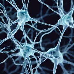
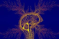
Post-Covid Neurological Symptoms

After contracting Covid-19 in August 2020, I developed a post-Covid syndrome with gradually increasing neurological manifestations. It took several months to turn into something that could be called a serious neurological condition without an evident cause.

I had a relatively mild form of Covid-19. I never had any chronic diseases that I was aware of, and I was a happy and energetic man in my late thirties. However, Covid changed that abruptly. Shortly after I was discharged from the hospital, I began to experience shortness of breath. It wasn’t happening all the time but was noticeable during physical loads and periodically at night when I was trying to get some sleep. During these episodes, I measured my oxygen saturation and it always was 99%. So, no reason to worry, right?

After the shortness of breath, I developed a form of a sore throat that was aggravated during the episodes of shortness of breath. I did not draw a link between the two at first, but consequent observations proved they were linked somehow. I was also fatigued and the fatigue lingered. I had an intellectually demanding job but could not work full time anymore. Five hours out of 8 were somewhat achievable but anything longer than that was problematic. I developed mild hypothermia as well. Instead of a normal temperature of 97.9 °F (36.6 °C) I always had 95.9 °F (35.5 °C). Finally, I also experienced intermittent episodes of slight tinnitus in the left ear.

I truly hoped these symptoms would be improved over time, but that did not happen. Moreover, in December 2020, four months after recovering from an initial infection, I began to experience what I can only describe as short and intermittent “halts” of consciousness. These episodes happened during work, especially when I was concentrating on a challenging task. The “halts” were like 1-2 seconds of fear that I was losing control over my body and was going to die. They happened every few minutes. If I relaxed a bit and moved away from the challenging task, the “halt” symptom weakened and disappeared.

In January 2021, I started to feel awkward. It was an unexplainable doom and gloom feeling of imminent death in the near future that was haunting me everywhere. I began to feel burning sensations in my right foot and right hand and my sore throat was becoming worse.

New Onset Panic Attacks

Meanwhile, I remained a big fan of walking and walked up to 8 miles (13 km) a day. After one such walk in February 2021, I experienced the first panic attack in my life. My heart rate went crazy and the shortness of breath reached its new maximum by turning into suffocation. I was thinking I was going to die and called the emergency.

I took every blood test I could imagine and visited every doctor I could reach. I went to a cardiologist, neurologist, endocrinologist, and phlebologist. Every test was negative. I was declared healthy except for a few seemingly unrelated things. I had a mild form of T2DM with A1C of 6.7 and insulin resistance with HOMA-IR of 7.8. My low-density lipoprotein levels were slightly elevated. An interesting observation is that I took blood tests right before the acute phase of Covid, and I had no T2DM back then. During a talk with an experienced endocrinologist, I was told that those relatively moderate levels of glucose in my blood could not wreak such havoc alone. It might be something else, but nobody knew what it was. A neurologist concluded that it was just severe depression and suggested taking some antidepressants, which I did without any positive results.

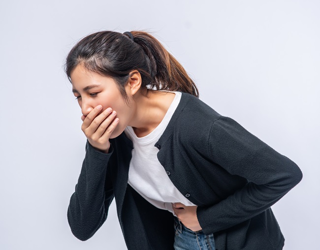
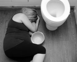
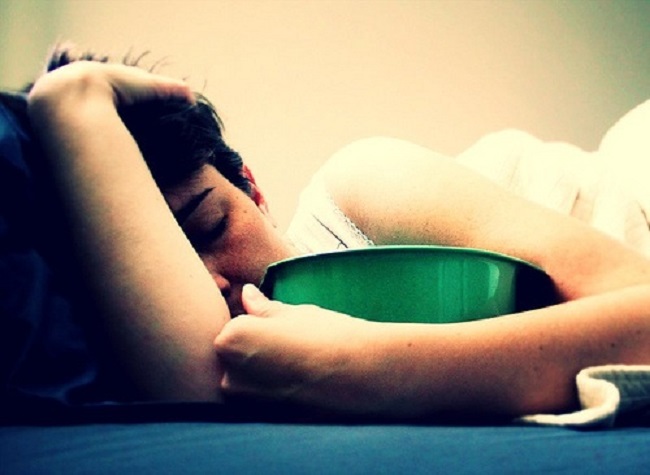
Meanwhile, my panic attacks intensified. The sore throat gradually transformed into dysphagia. The burning sensation in my right foot and hand took over the whole right side of my body. I started to lose the ability to speak fluently, and I started to have problems with my gait and street navigation.

At the height of a panic attack, I felt that my intrinsic biological processes were stopping and that one day it may have an ultimate ending. I started to develop a condition called intestinal ataxia. My right hand now had neurological edema. My peripheral neuropathy worsened significantly and became seriously painful. Everything went downhill very quickly, in a matter of weeks and days. At the beginning of April 2021, I faced an immediate risk of becoming an invalid and thought I might die.

The bitter part of the story is that nobody was going to help me. All the doctors I had met were helpless.

Strange Observations

I started to observe strange things while I was suffering from the condition. First, my symptoms slightly improved after consuming specific foods. For instance, it happened every time I ate a burger with real beef. The positive effect persisted for 2 or 3 days and then vanished. The same improvement I’ve experienced from consuming a glass of milk, but the effect was way shorter lasting just one day. Second, the condition was greatly improved on days when I consumed NSAIDs such as Aspirin or Ibuprofen for an unrelated reason. Third, I felt almost healthy on the rare days when I ate those specific foods and consumed NSAIDs together by pure coincidence. Unfortunately, I did not draw any specific conclusions back then. Such correlations just looked a bit weird to me as I never experienced anything like that in my whole prior life.

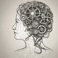
Discovering Poor Brain Glucose Metabolism

While the condition was gradually worsening, I measured my blood glucose levels several times a day. One day I decided to conduct an experiment to find out how blood glucose levels are affected by walking. I started by measuring my base glucose level in the morning which then was 126 mg/dL (7 mmol/L). Then, without eating anything, I decided to take an 8-mile (13 km) walk. To my amusement, the glucose level fell to 81 mg/DL (4.5 mmol/L) at the end of the route. Everything worked as expected, it seemed that I was not that insulin resistant after all.

But what about mental and intellectual activities? It would be a nice experiment to conduct too. The next morning, without eating anything, I went to my job with a glucometer and measured the base glucose level which was 117 mg/dL (6.5 mmol/L) at the time. Then I started to work taking the most challenging tasks. After 3 hours of intensive intellectual work, I started to experience the aforementioned “halts” of consciousness. Time to measure the glucose level. It was 115 mg/dL (6.4 mmol/L). Wait, what? How is that possible after all these intellectual activities? Yes, there is a process called gluconeogenesis that could raise the glucose level but still, I did not expect such a high value after such a massive cognitive load on an empty stomach. Clearly, something was going wrong with my brain as it had significant problems with glucose utilization. This was the crucial moment. A simple scientific experiment allowed me to see the light at the end of the tunnel. The cells of my nervous system were unable to consume the usual levels of glucose as they were insulin resistant, and I just proved that with my measurements.

Was I Deficient In Thiamine?

A quick web search of such a metabolic condition resulted in thiamine deficiency (vitamin B1) and beriberi as possible culprits. I knew about beriberi, but it never bugged my mind to link it with my own condition. I always thought it could be only caused by extreme malnutrition, which was not the case with me.

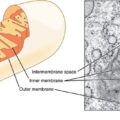
Going deeper on that route, I found the work of Hans Krebs, who described the process of cellular respiration and received the Nobel Prize for it in 1953. I was amused. Not only did it explain everything I had experienced, but now I had some actionable plan to try and improve my health.

If the usual levels of glucose cannot be consumed by the cells due to their insulin resistance, does it mean that by artificially raising glucose concentrations in the blood we can stop a metabolic panic attack? I conducted the experiment on several panic attacks of mine and received a positive answer. Yes, a metabolic panic attack can be stopped or at least significantly decreased by consuming 15 g of sugar. I immediately started to practice that to save my cells from further damage whenever a panic attack was mounting. That knowledge improved my condition a bit and gave me some time to find the appropriate therapeutic dose of thiamine.

Would thiamine help to reverse insulin resistance? I went to the pharmacy and bought Benfotiamine. It was hard to find, but luckily it was provisioned in two pharmacies in my hometown, so I did not have to wait for too long. Being in serious neurological suffering and pain, I immediately consumed 150 mg of Benfotiamine. Instinctively, I expected some kind of reaction, so I consumed the dose gradually. No reactions developed, but I immediately felt better in just 15 minutes after taking the pill. After that experience, I understood that I will be able to survive.

Given this experience and research, I wondered if the cellular damage would be reversed completely. It was an open question. Benfotiamine made me feel better, but I still experienced polyneuropathy and shortness of breath. The panic attacks went away completely, however.

Are Vitamins the Answer to Post-Covid Symptoms?

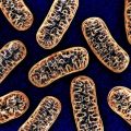
The new knowledge explained a lot of strange things that were happening with my body. Remember I talked about strange unexplained positive effects of consuming meat and milk? Now it became clear why: those foods contain nutrients including a rich set of vitamins and minerals. Vitamins help the mitochondria to process the substrates (glucose and lipids) to produce the energy (ATP) that powers up the cells.

It also explained the strange positive effects of consuming NSAIDs. As it turned out, this is relatively well known too. The mitochondrial dysfunction causes an auto-immune reaction of the body to its own metabolically subpar cells. This inflammation does more damage than good in that situation: the vessels’ endothelium gets damaged, leading to various blood flow problems including micro-clotting.  This further aggravates the tissue hypoxia and makes the mitochondrial dysfunction even more severe in the affected areas, leading to even more inflammation. This is a self-fueling pathological process with a positive feedback loop.

An acquired mitochondrial dysfunction can be reversed to some degree by using supplementary vitamins, co-factors, minerals, and antioxidants. Thiamine takes one of the instrumental roles in this process as it catalyzes the reactions at the very start of the oxidative phosphorylation pathway, but thiamine alone may not do much unless a full spectrum of nutrients is supplied. Glucose is not the only fuel a mitochondrion can consume, and other pathways need attention as well.

Combining all these pieces and using the protocol for treating beriberi as the basis, I came up with an experimental therapy, which I first tested on myself. It consisted of B1, B3, B7, B2, multivitamin, magnesium, potassium, CoQ10, alpha-lipoic acid, resveratrol, L-carnitine, zinc, cuprum, and aspirin in certain forms and proportions. Going beyond medicals, it also included dietary corrections (ketogenic non-vegetarian diet, no tea, no coffee, and no alcohol) and mild physical activities (walking).

To my surprise and amusement, it gave the desired results. I was able to get rid of the panic attacks, hypertension, tachycardia, neurological edema, tinnitus, insulin resistance, dysphagia, cognitive impairment, fatigue, and neuropathy. It took some time and effort. One part of that was my own body that needed the time to adapt and heal, another part was numerous therapy refinements.

At the beginning of the therapy, I took frequent blood tests to ensure the right therapeutic direction. All my outstanding markers were gradually normalizing, proving that I was on the right track.

At the time, I was not aware of Drs. Lonsdale and Chandler Marrs’ work or Elliot Overton’s videos. You can imagine the level of my sheer astonishment when I compared my humble findings to theirs. Now, almost two years after the disease’s inception, I can call myself a healthy man, again.

We Need Your Help

More people than ever are reading Hormones Matter, a testament to the need for independent voices in health and medicine. We are not funded and accept limited advertising. Unlike many health sites, we don’t force you to purchase a subscription. We believe health information should be open to all. If you read Hormones Matter, and like it, please help support it. Contribute now.

Yes, I would like to support Hormones Matter.

Photo by Marjan Blan | @marjanblan on Unsplash.

This story was published originally on August 11, 2022. 

Medication and Vaccine Adverse Reactions and the Orexin – Hypocretin Neurons

11657 views

A paper published in Science Translational Medicine, provides preliminary evidence that the H1N1 Flu Vaccine Pandemrix can evoke immune system mediated damage to the orexin – hypocretin neurons and induce narcolepsy in individuals with a particular genetic variant. The orexin – hypocretin neurons were only recently discovered in the mid 1990s, by two separate research groups, hence, the two names for the same molecule. For this paper, we’ll be utilizing the orexin nomenclature.

Initially, the orexin neurons were thought to be involved only in feeding behavior, as damage elicited hypophagia in animals. Soon it was learned that more severe damage to the orexin neurons induced narcolepsy and the orexin system became a key focus in narcolepsy related research. With time, however, it became quite clear that these neurons were involved in regulating a myriad of hormone and neurotransmitter systems and their consequent behaviors. Narcolepsy or rather the ability to sustain wakefulness, is but one of the many functions regulated by the orexin system.

In a previous paper, I touched briefly on the possibility that the orexin neurons might be damaged and have diminished functionality in individuals suffering from post Gardasil side effects. In particular, I suspected these neurons were indicated in post-Gardasil hypersomnia, a derivative of narcolepsy. That may be only the tip of the iceberg. As I soon learned, the hypocretin/orexin neurons are brain energy sensors and may be involved in array of post medication or vaccine adverse reactions. Indeed, they may be central to the ensuing state of sickness behaviors that emanate once an organism becomes overwhelmed.

The Orexin – Hypocretin Basics

Orexin nuclei are located in the lateral hypothalamus, the section of the hypothalamus that is most known for regulating feeding, arousal and motivation. The hypothalamus is the master regulator for all hormone systems and hormone related activity including feeding, sleeping, reproduction, fight, flight, energy usage – basically every aspect of human and animal survival. It sits at the interface between the central nervous system functioning and the endocrine system functioning.

From the lateral hypothalamus, orexin neurons project across the entire brain with its two receptors (OXA and OXB) differentially distributed throughout the central nervous system and even in the body, including in the kidney, adrenals, thyroid, testis, ovaries and small intestine. The orexin neurons also modulate local networks of adjacent neurons within the hypothalamus that in turn influence a myriad of behaviors.

The most densely innervated brain regions include the thalamus, the locus coeruleus, dorsal raphe nucleus, accounting for the hormone’s role in arousal, feeding and energy management. At the most basic level, release of the orexin induces wakefulness. When orexin neurons are turned on and firing appropriately, arousal is maintained. When orexin neurons are turned off, diminished or dysfunctional, melatonin, the sleep promoting hormone, is turned on. The two work in concert to manage wakefulness and sleep.

Orexin receptors are also located in the amygdala, the ventral tegmental area (VTA) and throughout the limbic system, accounting for its role in emotion and the reward system. Orexin directly activates dopamine in the VTA. The VTA is the reward, addiction, and in many ways, the pleasure center of the brain. All drugs of addiction, all pleasurable activities, activate dopamine in the VTA. Through the release of dopamine, here and elsewhere, orexin modulates the motivation to sustain pleasurable activities. When orexin is diminished, not only does dopamine diminish, but the motivation to sustain behaviors decreases and dysphoria increases.

That’s not all. Orexin influences the release of many other neurotransmitters and hormones, several of which are co-located on the orexin neurons themselves. For example, the neuropeptide dynorphin is co-located on orexin neurons. Dynorphin is an endogenous opioid involved in the perception of pain and analgesia. It has dual actions that can both elicit analgesia or pain depending upon dose and length of exposure. Stress activates dynorphin. Dynorphin then inhibits orexin firing by as much as 50%. Illness is a stressor, a vaccine is a stressor, either could activate dynorphin and inhibit orexin. After the initial activation of dynorphin, and the ensuing decrease in orexin, the presence of chronic stressors and chronic pain could begin a continuous feedback loop of diminished arousal, and increasing pain.

Other Neurochemical Connections

  • Consistent with orexin’s role in arousal, orexin neurons contain glutamate vesicles. Glutamate is the brain’s primary excitatory neurotransmitter. Drugs that increase glutamate, also increase orexin. Drugs that block glutamate, via its NMDA receptor, decrease orexin. Common migraine medications block glutamate and thereby may also diminish orexin.
  • Serotonin and norepinephrine decrease hypocretin/orexin firing (suggesting if one is concerned with hypersomnia, anti-depressants might not be a good option).
  • As one might expect, orexin neurons are inhibited by GABAα agonists – sedatives. From a women’s health perspective, consider that cycling hormones would also affect orexin neurons through the GABAα pathway. Progesterone is a GABAα agonist – a sedative, while DHEA and its sulfated partner DHEAS are GABAα antagonists, anxiolytics that block GABAα, reduce sedation, and thereby increase anxiety and wakefulness. There may be a cyclical nature to orexin firing that has yet to be investigated.
  • The hypocretin/orexin neurons also influence galanin, a GI and CNS hormone that seems to inhibit the activity of a variety of other neurons in those regions.

These are but a few of the brain systems that the orexin neurons touch in some way or another. Damage to this system would have serious health consequences by initiating a cascade of biochemical changes within the brain and body. Many of which, we have yet to fully understand.

How Might the Orexin Neurons Become Inhibited?

Quite easily, apparently. In addition to the orexin’s vast interconnected pathways with a myriad of neurotransmitters and neuropeptides, the orexin neurons act as energy and activity sensors with some unique intracellular mechanics that make them especially sensitive to the changing dynamics of the extracellular milieu. Disruptions in ATP, glucose and temperature, elicit reactions in orexin functioning.

Orexin neurons require as much as 5-6X the amount of intracellular ATP to maintain firing, and to maintain a state of wakefulness or arousal. This extreme sensitivity to reduced ATP makes the orexin neurons uniquely positioned to sense and monitor brain energy resources, early, before ATP levels become critical in other areas of the brain. The orexin neurons cease firing when ATP stores become low, thereby allowing the reallocation energy, perhaps to those cells required for survival, breathing and heart rate. As Hans Selye observed many decades ago, one of the first, and indeed, most consistent of the sickness behaviors, no matter the disease, is lethargy, fatigue and sleepiness. Orexin is at the center of this behavior.

Orexin neurons react to extracellular glucose levels, though perhaps not as one might expect. When extracellular glucose levels are high, orexin neurons stop firing via what is called an inward rectifying potassium (K+) channel that is ATP dependent. That means that when extracellular glucose is high, intracellular ATP is allocated to open K+ channels and flood the cell with the inhibitory K+ ions. K+ hyperpolarizes the cell, prohibiting it from firing. This mechanism reminds me of Dr. Peter Attia’s talk about the nature of Type 2 Diabetes and our approach to treatment. He proposes that the body’s metabolic response – the conservation of energy – to Type 2 Diabetes is not something aberrant but is exactly as it should be with a disease state. We’re just not treating the correct disease state.

Another way we can shut down the orexin neurons is via increased temperature. The orexin neurons are very sensitive thermosensors. Increased temperatures shut down orexin firing via the inward K+ flow. Again, this is consistent with sickness behaviors and the reallocation of resources.

Orexin – Hypocretin Neurons in Migraine and Seizures

Diminished orexin has been linked to migraine and seizure activity. With migraines specifically, orexin may contribute to the early warning, hours to days, of impending cortical disruption via changes in feeding and sleep patterns that often precede migraine onset. Orexin may also be linked to the pre-migraine aura mediated by changes in brain electrical activity that prelude the migraine pain itself by minutes, called cortical spreading depression or more appropriately, cortical spreading depolarization – the massive spreading change in ion balance of the neurons. Initially the wave is excitatory, neurons are firing, but that is soon followed by a period of neural silence. Finally, orexin is also connected to the vasodilation of the trigeminal nerve, the nerve responsible for migraine pain. These findings have led some to call orexin a migraine generator.

Diminished cerebral spinal concentrations of oxerin have been found in patients generalized tonic-clonic seizures. Conversely, in rodent studies, injections of orexin elicit seizure activity. Despite the somewhat contradictory findings in seizure activity versus migraine activity, it is likely that the orexin system is involved both disease processes.

Pulling it all Together: Orexins Monitor and Mark Disruptions in Brain Homeostasis

Here’s where it gets really interesting. Although some have argued orexin, particularly diminished orexin functioning, is the cause and culprit of disruptions in brain homeostasis, leading to narcolepsy, excessive sleepiness, migraine, seizures and other diseases, I think this system represents merely a marker of a disease process. I think the orexin system is the stopgap, the final barrier of disrupted cellular energetics, of mitochondrial function. Mitochondrial ATP is the key.

When we consider orexin’s role in migraine, in particular, we see clearly how environmental changes (diet, stress, illness, medication/toxin exposure) can lead to changes in the extracellular milieu where orexins reside. The orexin sensors adjust to these changes, mostly by reducing neural firing in attempt to counteract damages. The reduction in orexin then elicits the premonitory phases of the impending brain disruptions, sleep and hypophagia – the sickness behaviors. If it progresses, the massive waves of electrical disruption ensue, and migraine, perhaps even seizures are evoked. When the extracellular environment become chronically disrupted, so too does the diminishment of orexin activity, thereby initiating a perpetuating loop of dysregulated brain activity. We can hypothesize that similar progressions exist with disease processes marked by aberrant electrical activity, such as epilepsy.

We know that mitochondrial dysfunction is often generated by genetic polymorphisms and can predispose individuals to an array of seemingly unrelated conditions like migraine and fibromyalgia, dysautonomias and cognitive deficits. At the root of the dysfunction is a error of some sort in mitochondrial energy processing – ATP.

What has become increasingly clear, is that the production of cellular energy, can be disrupted environmentally, by diet, illness and exposures, if co-factors necessary for the production ATP like thiamine are diminished. It is via diminished ATP production, that I think some medications and vaccines evoke adverse reactions in some individuals. The orexin system, because it is so exquisitely sensitive to changes in cellular energy, is our warning system; first by subtle changes in neurochemistry, then by changes in arousal and feeding behavior, and finally, by an all-out reallocation of resources – excessive sleeping. If ATP remains deficient chronically, and an individual is so disposed, then the cortical misfiring we see in migraine and seizure ensues, along with autonomic dysregulation and the syndromes associated therewith. It is not the orexin – hypocretin system that is at root of many of these diseases, but rather, the causes are deeper yet and reside with mitochondrial health.

We Need Your Help

More people than ever are reading Hormones Matter, a testament to the need for independent voices in health and medicine. We are not funded and accept limited advertising. Unlike many health sites, we don’t force you to purchase a subscription. We believe health information should be open to all. If you read Hormones Matter, like it, please help support it. Contribute now.

Yes, I would like to support Hormones Matter. 

Image created using Canva AI.

This article was published originally on January 29, 2014.

Tank Estradiol and Lose Metabolic Flexibility: Pitfalls of Lupron and Oophorectomy

13953 views

Over the last several weeks, I have been looking at the role of estradiol in mitochondrial health. In the first post Hormones, Hysterectomy and the Aging Brain, we learned that estradiol depletion wreaks havoc on brain mitochondria turning them into misshapen donuts and blobs. Digging a little deeper, the next post (Lupron, Estradiol and the Mitochondria) pondered the connection between estradiol-depleting drugs such as Lupron, other Lupron-like drugs, and the devastating side effects that often follow suit. Could Lupron-mediated mitochondrial damage be at the root of these side effects? Quite possibly?  A question that remains is how. In this post, I will be digging even deeper into the role of estradiol in mitochondrial functioning, especially its role in something called metabolic flexibility.

A note of caution, while I focus on estradiol, the mitochondria, and what happens to health when we remove estradiol pharmaceutically via Lupron or surgically via oophorectomy, it is important to remember that estradiol is not the only hormone synthesized in the ovaries nor are the ovaries the only hormone-producing tissues. Moreover, the chemical castration induced by Lupron and other medications or via ovary removal disrupts and diminishes the synthesis of a myriad of hormones. Estradiol is simply where most of the research is focused, and so, it is where I too must focus, at least for the time being.

Steroid Hormones and Metabolic Flexibility: A Critical Factor in Post Lupron and Post Oophorectomy Ill Health

Steroid hormones regulate metabolic flexibility at the level of the mitochondria. Estradiol, the most frequently studied among the steroid hormones, plays a pivotal role in determining how food fuel is converted into cellular fuel or ATP.  When we eliminate estradiol with medications such as Lupron and other GnRH agonists or antagonists, or when we remove a woman’s ovaries, depleting her primary source for estrogen synthesis, metabolic flexibility diminishes significantly.  With the lack of metabolic flexibility comes several health issues, some noticeable, like weight gain, and others less noticeable, at least initially, like cardiac and neurodegenerative diseases. A common component of each of these conditions is mitochondrial dysfunction. Mitochondrial dysfunction can be initiated and accumulated via a number of mechanisms and over time, so estradiol is not the only variable, but it is a key factor that is often ignored.

Mitochondria

Mitochondria are the cellular powerhouses that consume oxygen and transform the foods we eat into a currency that cells can use (ATP) to perform all of the intricate tasks needed for survival and health. Mitochondria are also the site of steroidogenesis (steroid synthesis), immune signaling, and all sorts of other functions that determine cellular life and death. When you think about it, how well the mitochondria perform these tasks affects health at every level of organismal physiology. Without the appropriate amount of mitochondrial energy/ ATP, cell function becomes deranged, and ultimately, grinds to a halt. When that happens, disease is imminent. Indeed, genetic perturbations of mitochondrial function are some of the most devastating diseases known to medicine.

One has to wonder, what happens when we perturb mitochondrial function from the outside in – via toxicant exposure or by eliminating critical hormones or other co-factors such as nutrients that are necessary to mitochondrial operations? Worse yet, what if an individual with unrecognized genetic defects in mitochondrial functioning faces additional mitotoxicant exposures; what then? Complex, multi-system disease – that’s what. I would argue that mitochondrial dysfunction represents the final common pathway, a convergence point, connecting an array of seemingly disparate disease processes. Mitochondrial metabolism, and specifically, metabolic flexibility, may be at the heart of the derangement, with estradiol, and likely other hormones, in the driver’s seat.

Metabolic Flexibility: Adapt and Survive

When we think of stress and flexibility in general terms, it is easy to recognize that the more flexible one is in his/her behaviors or coping mechanisms, the easier it is for one to respond to, and survive stressors. Flexibility means that options exist for when everything hits the fan. Imagine if there were no options or if you had to respond to each and every stressful event in your life using exactly the same behaviors or response patterns. You would not get very far. The same holds true for cell behavior, and more specifically, mitochondrial behavior. The mitochondria need options to respond to the differing needs of the cells that they supply with energy. If those options become limited in any way, the mitochondria become less effective. They produce less energy, scavenge fewer oxidants (toxicants), and when stressors present, cannot easily adapt. In fact, the more inflexible the mitochondria are forced to become, the less likely they, and the cells, tissues, organs, and organism within which they reside, will survive. Estradiol is integral to mitochondrial flexibility. Remove the estradiol and the mitochondria become less metabolically flexible and less able to respond to the demands of a changing environment.

Estradiol Equals Increased Mitochondrial Efficiency and Decreased ROS

Estradiol maintains metabolic flexibility via two important mechanisms: increased mitochondrial efficiency and ROS management. With the former, estradiol regulates metabolic flexibility by altering the expression of genes that control the enzymes within the fuel conversion pathways. It is a complex algorithm of responses, with some proteins upregulated and others downregulated. The net result, however, favors increased efficiency in ATP production by maximizing metabolic flexibility or adaptability to the environment.

With the latter, estradiol, along with progesterone, manage the clean-up tasks inherent to any energy production process. In effect, estradiol manages ROS both on the front end and the back end of mitochondrial ATP production. On the front end, increased metabolic efficiency and flexibility equals fewer ROS byproducts. On the backend, estradiol cleans up the byproducts of processing -ROS – and tempers the damage these byproducts can cause.

Estradiol, Pyruvate, and ATP

Of particular interest to our work here at Hormones Matter, estradiol upregulates a set of enzymes called the pyruvate dehydrogenase complex, PDC. The PDC, responsible for converting glucose into pyruvate, is the first step in the long process that nets multiple units of mitochondrial ATP. The PDC is key to carbohydrate metabolism and more recently has been linked to fatty acid metabolism, making this enzyme complex central to mitochondrial energy production. Diminished PDC derails mitochondrial functioning, producing serious diseases. Children born with genetic pyruvate dehydrogenase deficiency suffer serious neurological consequences and rarely live to adulthood.

Importantly, the PDC (like all of the enzymes within these cascades) is highly dependent upon nutrient co-factors to function properly. Thiamine and magnesium, are critical to the PDC complex. Since PDC function demands thiamine, children and adults with thiamine deficiency also suffer significant ill-health, ranging from fatigue and muscle pain, to disturbed cognitive function, disrupted autonomic function affecting multiple organs, psychosis, and even death if not identified. Thiamine deficiency is most well known as a disease associated with chronic alcoholism but has recently begun re-emerging in non-alcoholic populations relative to medication and vaccine reactions.  Many medications and environmental variables deplete thiamine and magnesium, diminishing mitochondrial function significantly, by way of pyruvate.

Along with nutrient co-factors, estradiol is critical for pyruvate. Estradiol upregulates the expression of the enzymes that make up the PDC (in the brain). If estradiol is reduced or blocked, mitochondrial ATP production will take a hit. If estradiol is blocked in an already nutrient-depleted woman, the first step in mitochondrial fuel conversion would take a double hit. One can imagine the consequences.

In light of the direct role that thiamine, magnesium, and other nutrients play in the cascade of reactions required to produce ATP, can we maximize mitochondrial functioning with nutrients to compensate for the mitochondrial damage or deficiencies likely to occur post oophorectomy or as a result of GnRH agonist or antagonist drugs, like Lupron? I can find no research on the subject, but it is certainly a topic to explore given the millions of women already suffering from the mitochondrial damage induced by Lupron and/or pre-menopausal ovary removal. Even without the necessary research, correcting nutrient deficiencies and dietary issues should be undertaken for general health.

Another question in need of exploration, if we maximize mitochondrial functioning, does that then increase steroidogenesis in other endocrine glands? A section of the adrenal glands called the zona reticularus, for example, produces a complement of hormones similar to those of the ovaries. In postmenopausal women androgens, precursors for estradiol, produced by the adrenals account for a large percentage of total estradiol production. Could we take advantage of that to help stabilize circulating hormones?

Finally, beyond the nutrient requirements for mitochondrial ATP production, enzymes throughout the body, even those involved in post-mitochondrial steroid metabolism, require nutrient co-factors to function properly. Could we maximize those enzymes for more efficient steroid metabolism to net sufficient estradiol to maintain mitochondrial function?

What about Natural Declines in Estradiol?

It is not clear how menstrual cycle changes in estradiol affect mitochondrial functioning or how the postpartum decline in pregnancy hormones affects mitochondria. One would suspect there are compensatory reactions to prevent damage, but this has not been investigated. In natural menopause, however, researchers have noted that some form of compensation occurs as estradiol declines and, at least for a time, and in rodents, mitochondria maintain efficient production of ATP. In contrast, no such changes are noted with premature menopause or oophorectomy.

Also not investigated sufficiently, is the impact of chronic synthetic estrogen exposure on mitochondrial functioning. In other words, what are the effects of oral contraceptives, HRT, and the growing list of environmental endocrine disruptors, on mitochondrial ATP production? Since these compounds bind to estrogen receptors and displace the endogenous estrogens like estradiol, some evidence suggests endogenous production of estradiol is reduced. Do the mitochondria respond also by downregulating estrogen receptors or by some other mechanism?  Short-term, animal research suggests that supplementing 17B estradiol post oophorectomy reduces mitochondrial damage. In research in humans, where synthetic estrogens are used, results are less clear and longer-term studies do not exist beyond the broad brush strokes of epidemiology.

Metabolic Flexibility and Tissue Type

One of the more interesting aspects of estradiol’s role in metabolic flexibility is that it is site or tissue-specific and may point to novel therapeutic opportunities. Since different cell types, in different parts of the body, prefer different fuels for power to survive, when we eliminate estradiol from the equation, mitochondria from different tissues or organs respond differently to the lack of flexibility. Perhaps, we can utilize the information about fuel requirements to design diets that compensate for diminished metabolic flexibility.

Heart Cells. I’ve written about this research previously, not fully understanding the implications. Estradiol allows cardiomyocytes (heart cells) to switch from their preferred fuel of fatty acids to glucose during stressors such as heart attacks (and theoretically during any stressor like exercise). That ability to switch fuel types is protective and allows the cells to survive and heal. It may explain why women are more susceptible to heart damage post-menopause when endogenous estradiol declines. This may also point to a pathway for post oophorectomy and post Lupron declines in normal heart function.

Brain Health. Declining estradiol affects brain mitochondria differently. As I noted in a previous post, without estradiol, brain mitochondria become progressively less functional and misshapen. These structural changes impair mitochondrial ATP production. Unlike the heart, however, the brain prefers glucose as its primary fuel source. Estradiol appears to enhance glucose uptake from the periphery and across the blood-brain barrier. When estradiol is absent, brain glucose uptake diminishes significantly (in rodent studies), leaving the brain perpetually starved for glucose.

We know from brain cancer research, that with declining brain glucose, secondary fuels can kick in, but only when the mitochondria have sufficient flexibility to switch. For example, mitochondrial fuel flexibility is critical to battling brain tumors. Under conditions of stress and when brain glucose concentrations are low, healthy mitochondria can readily transition to ketone bodies for energy, at least in vivo. The transition from glucose to ketone bodies is believed to be an evolutionary adaptation to food deprivation allowing the survival of healthy cells during severe shifts in the nutritional environment. Estradiol appears to be key in maintaining that flexibility.

Weight Gain and Fat Accumulation. Post-menopausal, post-hysterectomy, and oophorectomy weight gain are well established research findings. Anecdotal complaints of Lupron weight gain are also common. These findings may be related to derangements in metabolic flexibility mediated by the relationship between estradiol and mitochondrial functioning. The increased lipid or fat accumulation in skeletal muscle though associated with impaired insulin-stimulated glucose metabolism may be related to the reduced capacity to adjust to a changing fuel environment. More specifically, weight gain may represent a declining ability to utilize fats effectively as a mitochondrial fuel source, possibly via a derangement in a mitochondrial channel responsible for shuttling fats and cholesterol into the mitochondria for processing. When the mitochondria become less flexible, a channel called the TSPO, shuts down, disallowing fats that would normally be shuttled into the mitochondria and processed for ATP (and steroid hormones), from entering. Instead, they are stored peripherally in adipocytes. I wrote about this in detail here: It’s All about the Diet: Obesity and Mitochondrial Dysfunction. It is possible in estradiol-depleted women that TSPO downregulation is a compensatory reaction to diminished metabolic flexibility.

It is also conceivable that the lack of brain glucose, as discussed above, leads to overeating and, more specifically, cravings for sugary foods. This would be a logical compensatory reaction to bring more fuel to the brain; one likely meant only for the short term and that when held chronically begins the cascade of other metabolic reactions known as obesity, diabetes, and heart disease. Perhaps, just as fat storage becomes a survival mechanism when mitochondria can longer process it effectively, the craving for sugar in estradiol-deprived women is also a survival mechanism.

Finally, adipocytes can synthesize estradiol. It is conceivable that in response to declining estradiol concentrations, the body stores fat to produce more estradiol.

Final Thoughts

Central to mitochondrial dysfunction, whether by genetic predisposition or environmental influence, is the inability to efficiently produce ATP (the fuel that all cells need to survive) and to effectively manage the by-products of fuel production and other toxicants. Estradiol plays a huge role in both of these processes. Eliminate estradiol and mitochondrial functioning becomes less efficient and less flexible initiating cascades of chronic and life-altering conditions. This suggests the ready application of medications like Lupron that deplete estradiol or the prophylactic removal of women’s ovaries is misguided at best, and dangerous at worst.

We Need Your Help

More people than ever are reading Hormones Matter, a testament to the need for independent voices in health and medicine. We are not funded and accept limited advertising. Unlike many health sites, we don’t force you to purchase a subscription. We believe health information should be open to all. If you read Hormones Matter, and like it, please help support it. Contribute now.

Yes, I would like to support Hormones Matter. 

Image by Triggermouse from Pixabay.

This post was published originally on Hormones Matter on February 11, 2015. 

Beriberi: The Great Imitator

33103 views

Because of some unusual clinical experiences as a pediatrician, I have published a number of articles in the medical press on thiamine, also known as vitamin B1. Deficiency of this vitamin is the primary cause of the disease called beriberi. It took many years before the simple explanation for this incredibly complex disease became known. A group of scientists from Japan called the “Vitamin B research committee of Japan” wrote and published the Review of Japanese Literature on Beriberi and Thiamine, in 1965. It was translated into English subsequently to pass the information about beriberi to people in the West who were considered to be ignorant of this disease. A book published in 1965 on a medical subject that few recall may be regarded in the modern world as being out of date and of historical interest only, however, it has been said that “Those who do not learn history are doomed to repeat it”. And repeat it, we are.

Beriberi is one of the nutritional diseases that is regarded as being conquered. It is rarely considered as a cause of disease in any well-developed country, including America. In what follows, are extractions from this book that are pertinent to many of today’s chronic health issues. It appears that thiamine deficiency is making a comeback but it is rarely considered as a possibility.

The History of Beriberi and Thiamine Deficiency

Beriberi has existed in Japan from antiquity and records can be found in documents as early as 808. Between 1603 and 1867, city inhabitants began to eat white rice (polished by a mill). The act of taking the rice to a mill reflected an improved affluence since white rice looked better on the table and people were demonstrating that they could afford the mill. Now we know that thiamine and the other B vitamins are found in the cusp around the rice grain. The grain consists of starch that is metabolized as glucose and the vitamins essential to the process are in the cusp. The number of cases of beriberi in Japan reached its peak in the 1920s, after which the declining incidence was remarkable. This is when the true cause of the disease was found. Epidemics of the disease broke out in the summer months, an important point to be noted later in this article.

Early Thiamine Research

Before I go on, I want to mention an extremely important experiment that was carried out in 1936. Sir Rudolf Peters showed that there was no difference in the metabolic responses of thiamine deficient pigeon brain cells, compared with cells that were thiamine sufficient, until glucose (sugar) was added. Peters called the failure of the thiamine deficient cells to respond to the input of glucose the catatorulin effect. The reason I mention this historical experiment is because we now know that the clinical effects of thiamine deficiency can be precipitated by ingesting sugar, although these effects are insidious, usually relatively minor in character and can remain on and off for months. The symptoms, as recorded in experimental thiamine deficiency in human subjects, are often diagnosed as psychosomatic. Treated purely symptomatically and the underlying dietary cause neglected, the clinical course gives rise to much more serious symptoms that are then diagnosed as various types of chronic brain disease.

  • Thiamine Deficiency Related Mortality. The mortality in beriberi is extremely low. In Japan the total number of deaths decreased from 26,797 in 1923 to only 447 in 1959 after the discovery of its true cause.
  • Thiamine Deficiency Related Morbidity. This is another story. It describes the number of people living and suffering with the disease. In spite of the newly acquired knowledge concerning its cause, during August and September 1951, of 375 patients attending a clinic in Tokyo, 29% had at least two of the major beriberi signs. The importance of the summer months will be mentioned later.

Are the Clinical Effects Relevant Today?

The book records a thiamine deficiency experiment in four healthy male adults. Note that this was an experiment, not a natural occurrence of beriberi. The two are different in detail. Deficiency of the other B vitamins is involved in beriberi but thiamine deficiency dominates the picture. In the second week of the experiment, the subjects described general malaise, and a “heavy feeling” in the legs. In the third week of the experiment they complained of palpitations of the heart. Examination revealed either a slow or fast heart rate, a high systolic and low diastolic blood pressure, and an increase in some of the white blood cells. In the fourth week there was a decrease in appetite, nausea, vomiting and weight loss. Symptoms were rapidly abolished with restoration of thiamine. These are common symptoms that confront the modern physician. It is most probable that they would be diagnosed as a simple infection such as a virus and of course, they could be.

Subjective Symptoms of Naturally Occurring Beriberi

The early symptoms include general malaise, loss of strength in knee joints, “pins and needles” in arms and legs, palpitation of the heart, a sense of tightness in the chest and a “full” feeling in the upper abdomen. These are complaints heard by doctors today and are often referred to as psychosomatic, particularly when the laboratory tests are normal. Nausea and vomiting are invariably ascribed to other causes.

General Objective Symptoms of Beriberi

The mental state is not affected in the early stages of beriberi. The patient may look relatively well. The disease in Japan was more likely in a robust manual laborer. Some edema or swelling of the tissues is present also in the early stages but may be only slight and found only on the shin. Tenderness in the calf muscles may be elicited by gripping the calf muscle, but such a test is probably unlikely in a modern clinic.

In later stages, fluid is found in the pleural cavity, surrounding the heart in the pericardium and in the abdomen. Fluid in body cavities is usually ascribed to other “more modern” causes and beriberi is not likely to be considered. There may be low grade fever, usually giving rise to a search for an infection. We are all aware that such symptoms come from other causes, but a diet history might suggest that beriberi is a possibility in the differential diagnosis.

Beriberi and the Cardiovascular System

In the early stages of beriberi the patient will have palpitations of the heart on physical or mental exertion. In later stages, palpitations and breathlessness will occur even at rest. X-ray examination shows the heart to be enlarged and changes in the electrocardiogram are those seen with other heart diseases. Findings like this in the modern world would almost certainly be diagnosed as “viral myocardiopathy”.

Beriberi and the Nervous System

Polyneuritis and paralysis of nerves to the arms and legs occur in the early stages of beriberi and there are major changes in sensation including touch, pain and temperature perception. Loss of sensation in the index finger and thumb dominates the sensory loss and may easily be mistaken for carpal tunnel syndrome. “Pins and needles”, numbness or a burning sensation in the legs and toes may be experienced.

In the modern world, this would be studied by a test known as electromyography and probably attributed to other causes. A 39 year old woman is described in the book. She had lassitude (severe fatigue) and had difficulty in walking because of dizziness and shaking, common symptoms seen today by neurologists.

Beriberi and the Autonomic Nervous System

We have two nervous systems. One is called voluntary and is directed by the thinking brain that enables willpower. The autonomic system is controlled by the non-thinking lower part of the brain and is automatic. This part of the brain is peculiarly sensitive to thiamine deficiency, so dysautonomia (dys meaning abnormal and autonomia referring to the autonomic system) is the major presentation of beriberi in its early stages, interfering with our ability for continuous adaptation to the environment. Since it is automatic, body functions are normally carried out without our having to think about them.

There are two branches to the system: one is called sympathetic and the other one is called parasympathetic. The sympathetic branch is triggered by any form of physical or mental stress and prepares us for action to manage response to the stress. Sensing danger, this system activates the fight-or-flight reflex. The parasympathetic branch organizes the functions of the body at rest. As one branch is activated, the other is withdrawn, representing the Yin and Yang (extreme opposites) of adaptation.

Beriberi is characterized in its early stages by dysautonomia, appearing as postural orthostatic tachycardia syndrome (POTS). This well documented modern disease cannot be distinguished from beriberi except by appropriate laboratory testing for thiamine deficiency. Blood thiamine levels are usually normal in the mild to moderate deficiency state.

Examples of Dysfunction in Beriberi

The calf muscle often cramps with physical exercise. There is loss of the deep tendon reflexes in the legs. There is diminished visual acuity. Part of the eye is known as the papilla and pallor occurs in its lateral half. If this is detected by an eye doctor and the patient has neurological symptoms, a diagnosis of multiple sclerosis would certainly be entertained.

Optic neuritis is common in beriberi. Loss of sensation is greater on the front of the body, follows no specific nerve distribution and is indistinct, suggestive of “neurosis” in the modern world.

Foot and wrist drop, loss of sensation to vibration (commonly tested with a tuning fork) and stumbling on walking are all examples of symptoms that would be most likely ascribed to other causes.

Breathlessness with or without exertion would probably be ascribed to congestive heart failure of unknown cause or perhaps associated with high blood pressure, even though they might have a common cause that goes unrecognized.

The symptoms of this disease can be precipitated for the first time when some form of stress is applied to the body. This can be a simple infection such as a cold, a mild head injury, exposure to sunlight or even an inoculation, important points to consider when unexpected complications arise after a mild incident of this nature. Note the reference to sunlight and the outbreaks of beriberi in the summer months. We now know that ultraviolet light is stressful to the human body. Exposure to sunlight, even though it provides us with vitamin D as part of its beneficence, is for the fit individual. Tanning of the skin is a natural defense mechanism that exhibits the state of health.

Is Thiamine Deficiency Common in America?

My direct answer to this question is that it is indeed extremely common. There is good reason for it because sugar ingestion is so extreme and ubiquitous within the population as a whole. It is the reason that I mentioned the experiment of Rudolph Peters. Ingestion of sugar is causing widespread beriberi, masking as psychosomatic disease and dysautonomia. The symptoms and physical findings vary according to the stage of the disease. For example, a low or a high acid in the stomach can occur at different times as the effects of the disease advance. Both are associated with gastroesophageal reflux and heartburn, suggesting that the acid content is only part of the picture.
A low blood sugar can cause the symptoms of hypoglycemia, a relatively common condition. A high blood sugar can be mistaken for diabetes, both seen in varying stages of the disease.

It is extremely easy to detect thiamine deficiency by doing a test on red blood cells. Unfortunately this test is either incomplete or not performed at all by any laboratory known to me.

The lower part of the human brain that controls the autonomic nervous system is exquisitely sensitive to thiamine deficiency. It produces the same effect as a mild deprivation of oxygen. Because this is dangerous and life-threatening, the control mechanisms become much more reactive, often firing the fight-or-flight reflex that in the modern world is diagnosed as panic attacks. Oxidative stress (a deficiency or an excess of oxygen affecting cells, particularly those of the lower brain) is occurring in children and adults. It is responsible for many common conditions, including jaundice in the newborn, sudden infancy death, recurrent ear infections, tonsillitis, sinusitis, asthma, attention deficit disorder (ADD), hyperactivity, and even autism. Each of these conditions has been reported in the medical literature as related to oxidative stress. So many different diseases occurring from the same common cause is offensive to the present medical model. This model regards each of these phenomena as a separate disease entity with a specific cause for each.

Without the correct balance of glucose, oxygen and thiamine, the mitochondria (the engines of the cell) that are responsible for producing the energy of cellular function, cannot realize their potential. Because the lower brain computes our adaptation, it can be said that people with this kind of dysautonomia are maladapted to the environment. For example they cannot adjust to outside temperature, shivering and going blue when it is hot and sweating when it is cold.

So, yes, beriberi and thiamine deficiency have re-emerged. And yes, we have forgotten history and appear doomed to repeat it. When supplemental thiamine and magnesium can be so therapeutic, it is high time that the situation should be addressed more clearly by the medical profession.

We Need Your Help

More people than ever are reading Hormones Matter, a testament to the need for independent voices in health and medicine. We are not funded and accept limited advertising. Unlike many health sites, we don’t force you to purchase a subscription. We believe health information should be open to all. If you read Hormones Matter, like it, please help support it. Contribute now.

Yes, I would like to support Hormones Matter.

This article was published originally on November 4, 2015. 

Thyroid Hormones, Mitochondrial Functioning, and Hair Loss

28521 views

Hair loss is a common symptom of thyroid disease. In our research, hair loss, changes in color or luster, and skin changes are regularly reported as one of the first symptoms noticed in an emerging non-allergenic adverse reaction to a medication or vaccine. These symptoms often coincide with unexplained fatigue and muscle pain. Given that many patients who develop the more chronic, multi-symptom medication or vaccine reactions also develop thyroid disease and frequently exhibit signs of mitochondrial damage, I wondered if somehow the hair and skin changes could be early warning signs of diminished mitochondrial functioning. I also wondered if all of these variables were connected. It turns out that not only are they connected, but incredibly interdependent.

What are Mitochondria?

Recall from high school biology, the mitochondria are those bean-shaped organelles inside cells that are responsible for cellular respiration or energy production. Through a variety of pathways, the mitochondria provide fuel for cell survival. In addition to cellular energy production, mitochondria control cell apoptosis (death), calcium, copper, and iron homeostasis, and steroidogenesis. In essence, mitochondria perform the key tasks associated with cell survival, and indeed, human survival. Damage the mitochondria and cellular dysfunction or death will occur. Damage sufficient numbers of mitochondrion and chronic, multi-symptom illness arises.

How to Damage Mitochondria

Mitochondria are remarkably resilient given the proper nutrients, but without those nutrients, they can be highly susceptible to damage. Mitochondrial damage can be inherited via mutations in maternal DNA (mtDNA) or nuclear DNA and present at birth or remain latent until triggered later in life, as in the case of mitochondrial endocrinopathies. Mitochondria are also susceptible to epigenetic changes, which can be heritable and acquired and remain latent until triggered.  Finally, mitochondrial impairment can derive from pharmaceutical or environmental exposures and nutrient or cofactor deficits. The sheer number of mechanisms that can influence mitochondrial functioning and heritability make diagnosing and predicting mitochondrial dysfunction difficult at best, particularly acquired or functional mitochondriopathies that are not evident from genetic or epigenetic testing. It is precisely those acquired mitochondriopathies, particularly those seemingly triggered by pharmaceutical reactions, that we are most interested in here at Hormones Matter. Indeed, acquired mitochondrial damage represents a nascent and emerging field in medicine, particularly in toxicology, as many drugs and vaccines damage mitochondrial functioning both directly and indirectly.

What Mitochondrial Damage Looks Like

Mitochondrial damage presents in a highly diverse, multi-organ, multi-symptom manner. On the surface, patients with mitochondrial dysfunction will appear to have multiple, unconnected diagnoses, from gastrointestinal distress to cognitive deficits, from cardiac arrhythmias to multiple sclerosis-like symptoms, and everything in between and beyond. According to Dr. Richard Boles, an expert on mitochondrial dysfunction:

“Mitochondrial dysfunction doesn’t really cause anything, what it does is predisposes towards seemingly everything. It’s one of many risk factors in multifactorial disease. It can predispose towards epilepsy, chronic fatigue, and even autism, but it doesn’t do it alone. It does it in combination with other factors, which is why in a family with a single mutation going through the family, everyone in the family is affected in a different way. Because it predisposes for disease throughout the entire system.”

This is partially because the human body contains over a billion mitochondria which are essential to cellular functioning in every cell of the body. Where the dysfunction emerges is dependent upon where the impaired mitochondria reside, by what mechanism the mitochondria are damaged, and how intervening variables, such as overall health, nutrition, and environment come into play. Given the mitochondrion’s role in energy production, highly energy-dependent tissues such as the brain, the heart, the liver, and even muscles, are most susceptible to direct mitochondrial damage. And considering the mitochondrion’s role in cellular energetics, fatigue is almost always present with mitochondrial dysfunction.

Hormone Synthesis and Mitochondrial Functioning

Adding yet another layer of complexity, mitochondria also control steroid production in the adrenal glands, ovaries, testes, and thyroid. Any impairment of mitochondrial functioning can have a significant influence on hormone production and regulation. Since hormones, like the mitochondria, also impact all facets of biological homeostasis, energy, and metabolism, damage to endocrine mitochondria can represent a double-hit and begin a cascade of endocrine ill-effects that are difficult to control. This is particularly true of the thyroid gland.

Thyroid Hormones and Mitochondrial Functioning

The cells within the thyroid gland are dependent upon proper mitochondrial functioning to maintain health and proper mitochondrial functioning is dependent upon thyroid hormones to manage cellular energy production. This reciprocal and interdependent relationship makes the thyroid especially susceptible to a mitochondrial spiral. Both thyroid and mitochondrial damage have been observed in our medication and vaccine adverse reaction populations.

Thyroid hormones regulate mitochondrial functioning. Triiodothyronine (T3) in particular is considered one of the major regulators of mitochondrial activity stimulating mitochondrial biogenesis (the birth of new mitochondria) both directly (genomic), indirectly (non-genomic), and epigenetically.

T3 is responsible for increasing cellular heat production and oxygen consumption, core activities of mitochondrial metabolism. In hypothyroid states, heat and oxygen are reduced, whereas, in hyperthyroid states, the two are increased. Here the intracellular patterns of heat and energy production correspond to the clinical symptoms of hypo- and hyperthyroid states. Other thyroid hormones along  the hypothalamus – pituitary – thyroid axis (HPT) and the other iodothyronines within the thyroid hormone metabolic pathway influence mitochondrial functioning. Remove or reduce the presence of the thyroid hormones and mitochondria produce less energy and eventually die. With them, the cells in which they reside die too. Conversely, as mitochondria within the thyroid become less efficient, smaller concentrations of thyroid hormones are produced.  With reduced thyroid hormones, mitochondrial efficiency continues to decline and so on, and so on.

Hair Follicles: Mini – HPTs

German researchers recently identified multiple mechanisms by which human hair follicles are responsive to thyroid hormones. Their research showed that human skin and hair follicles possess an equivalent peripheral HPT axis with all of the corresponding hormones such as the central HPT. It turns out that hair follicle mitochondria are differentially responsive to each of the thyroid hormones along that axis and are responsive to other iodothyronines not typically considered bioactive, such as diiodothyronine (T2).

An interesting finding, related specifically to the hair follicle, and perhaps other mitochondria, thyroid hormones were protective against reactive oxygen species (ROS) production via multiple mechanisms. ROS, also called free radicals or oxidants, are natural by-products of oxygen (energy) metabolism important to a number of basic cell and life processes, like signaling and the defense against pathogens, but ROS levels must be kept in strict balance. Too much or not enough ROS and health goes awry. In the case of adverse fluoroquinolone reactions, increased ROS production is implicated.  According to the hair-follicle study, thyroid hormones protect against ROS production and regulate the enzymes that scavenge for and eliminate free radicals – our own internal antioxidants. If this function is conserved throughout the body, it provides one more reason to investigate and appropriately manage thyroid damage in medication adverse reactions.

Hair Loss and Mitochondrial Damage

Skin and hair follicles are dense with mitochondria and highly regulated by thyroid hormones such that the mechanism for hair loss in some individuals can be attributed to either diminished thyroid hormones and/or damaged mitochondria. Since the relationship between thyroid hormones and mitochondria is reciprocal, it is difficult to tell which impairment comes first. However, given what we know about hair growth cycles and what we know about thyroid hormones and mitochondrial functioning, it is possible to speculate and backdate a chemical insult precipitating sudden and unexplained hair loss. For more incipient reactions, it is a bit more difficult. Regardless, however, it appears that unexplained hair loss is a sign of poor mitochondrial functioning.

Hair growth occurs in phases. The anagen phase is the growth cycle where hair follicles grow about 1 cm per day for 28 days. This growth phase lasts for 2-7 years. The exact time frame is genetically, or more specifically, epigenetically determined by factors associated with the health of the maternal grandmother. After the anagen phase, the hair follicles reach a transitional, quiescent period lasting approximately 2-3 weeks. This is then followed by the telogen phase where hair begins to fall out. At any given time, up to 90% of hair follicles are in the anagen or growth phase while the remaining follicles are either catagen (10-14%) or telogen phases (1-2%).

Chemo Induced Hair Loss: Answers in the Mitochondria?

With chemotherapy, hair loss begins 2-4 weeks after treatment begins and although multiple mechanisms have been investigated, none have been able to explain or treat effectively chemo-induced hair loss. I would suspect that given the time frame, the toxic insult of chemotherapy, the role of mitochondria in hair growth, and the connection to thyroid damage, that chemo-induced alopecia is representative of mitochondrial damage. The ability to maintain hair growth during chemo may be related to supporting mitochondrial and/or thyroid health.

In the case of other presumed less toxic or at least less directly toxic chemical insults such as medication or vaccine adverse reactions, the initial loss of hair that begins either in the weeks preceding the full onslaught of symptoms or coincident with those symptoms, marks a decline in mitochondrial functioning and likely an impending decline in thyroid functioning.

Hair Loss: A Reallocation of Mitochondrial Resources

Considering, that hair generation is an energy (read mitochondrial) intense process, sudden hair loss could be an early marker that mitochondrial resources are limited and being reallocated towards more critical operations like brain and heart functioning. When the components for proper mitochondrial functioning are absent, be it the thyroid hormones or the co-factors necessary for cellular energy (ATP) production, the first wave of resource allocation might be to cease non-essential activities. The non-essential activities would include hair growth (and wakefulness in general – read Medication and Vaccine Adverse Reactions and the Orexin – Hypocretin Neurons). Sudden or unexplained hair loss could indicate mitochondrial impairment. Backdate the hair loss 2-4 weeks and an illness, a medication, vaccine, or environmental exposure could be the culprit. Whatever the cause, the thyroid and mitochondrial health should be considered and treatment initiated accordingly because if the disease process continues, the symptoms will expand beyond the hair, potentially to every tissue and organ in the body. Concurrently, investigate and amend nutritional status. Mitochondrial functioning is critically dependent on proper nutrients. Deficits in important nutrients, like thiamine, can have severe repercussions.

Feed your thyroid. Feed your mitochondria.

We Need Your Help

More people than ever are reading Hormones Matter, a testament to the need for independent voices in health and medicine. We are not funded and accept limited advertising. Unlike many health sites, we don’t force you to purchase a subscription. We believe health information should be open to all. If you read Hormones Matter, and like it, please help support it. Contribute now.

Yes, I would like to support Hormones Matter.

This article was first posted on Hormones Matter in May of 2014.

Cyclic Vomiting Syndrome and Mitochondrial Dysfunction: Research and Treatments

13783 views

Cyclic vomiting syndrome (CVS) is a debilitating disease characterized by episodes of severe nausea and persistent vomiting interspersed with periods of wellness. CVS affects about 2 percent of school-aged children, and also affects adults, although in adults it is often not recognized. Getting a diagnosis can be challenging, and sometimes takes a long time. Episodes of CVS can be extremely debilitating, and are sometimes difficult to treat and require hospitalization.

My daughter has suffered from this disease for 10 years, since she was 2 years old (see her story here). Her episodes were somewhat predictable when she was younger, but have changed and become less predictable, and more difficult to manage with medication, as she gets older. Although we try to avoid triggers such as stress and fatigue, being a pre-teen girl, she likes to have sleepovers with her friends and stay up chatting all night.  Unlike other pre-teen girls, however, she suffers the aftereffects of the sleepovers sometimes by vomiting for 24 hours or more.

What Causes Cyclic Vomiting Syndrome?

Although it has long been thought to be related to migraines, many sources state that the cause of cyclic vomiting syndrome is not known. Mechanisms that may be involved include episodic dysautonomia (malfunction of the autonomic nervous system that can result in a variety of symptoms), mitochondrial DNA mutations that cause deficits in cellular energy production, and heightened stress response that causes vomiting. However, there is mounting evidence for the role of mitochondrial dysfunction in the pathogenesis of this disease, a fact that is not often understood by the average practicing gastroenterologist. The connection to mitochondrial dysfunction has important implications for effective treatment of cyclic vomiting syndrome.

Mitochondrial Dysfunction, Cyclic Vomiting and Other Conditions

Mitochondria are small organelles within the cell responsible for energy production and other critical functions. Because of these crucial functions, Dr. Richard Boles, Director of the Metabolic and Mitochondrial Disorders Clinic at Children’s Hospital Los Angeles, explains that “30 years or so ago, many scientists couldn’t believe that mitochondrial disease could exist, because how does the organism survive?” However, mitochondrial dysfunction plays a role in many diseases, including CVS, and according to Dr. Boles:

“these are partial defects. Mitochondrial dysfunction doesn’t really cause anything, what it does is predisposes towards seemingly everything. It’s one of many risk factors in multifactorial disease. It can predispose towards epilepsy, chronic fatigue, and even autism, but it doesn’t do it alone. It does it in combination with other factors, which is why in a family with a single mutation going through the family, everyone in the family is affected in a different way. Because it predisposes for disease throughout the entire system.”

DNA mutations that affect mitochondrial function can occur in the DNA that is found in the nucleus of the cell (genomic DNA), or they can occur in the DNA that is found within the mitochondria themselves. Mitochondrial DNA is inherited differently than nuclear DNA. Most people are familiar with the inheritance of nuclear DNA, in which we have two copies of every gene, and we inherit one copy from each of our parents. However, mitochondrial DNA is inherited exclusively through the mother; therefore, mutations that affect the mitochondrial DNA can be traced through the maternal lineage of a family.

A possible relationship between cyclic vomiting syndrome and mitochondrial dysfunction was suggested by the finding that in some families, CVS was maternally inherited. Mitochondrial DNA mutations and deletions have been reported in patients with CVS, and disease manifestations of mitochondrial dysfunction have been found in the maternal relatives of patients with CVS. In other words, conditions such as migraines, irritable bowel syndrome, depression, and hypothyroidism, are often found in the maternal relatives of patients with CVS.

Mitochondrial DNA mutations don’t cause CVS directly, in the way that a DNA mutation causes cystic fibrosis, for example. In some patients, mitochondrial dysfunction plays a greater role in the causation of their disease, and in other patients, it may be less of a factor. Dr. Boles explains: “In some cases it’s a clear mitochondrial disorder, they have multiple other manifestations and it drives the disease. However, in most patients, it is one of many factors in disease pathogenesis.” Patients with classical mitochondrial disorders have disease manifestations such as muscle weakness, neurological problems, autism, developmental delays, gastrointestinal disorders, and autonomic dysfunction. Some patients with CVS have these other disease manifestations, and some have only CVS symptoms.

Treatment for Cyclic Vomiting Syndrome and Mitochondrial Dysfunction

As with many diseases, understanding as least some of the cause of CVS has allowed for the development of treatments tailored towards fixing the root cause. Co-enzyme Q10 and L-carnitine are two dietary supplements that have been used to treat a wide variety of conditions.  Both supplements may be able to assist the mitochondria with energy production and thus, help compensate for mitochondrial dysfunction. A retrospective chart review study found that using these two supplements, along with a dietary protocol of fasting avoidance (having three meals and three snacks per day), was able to decrease the occurrence of, or completely resolve, the CVS episodes in some patients. In those patients who didn’t respond to treatment with supplements alone, the addition of amitriptyline or cyproheptadine, two medications that have been used for prevention of CVS episodes, helped to resolve or decrease the episodes. Treatment with the cofactors alone was well tolerated with no side effects, and treatment with cofactors plus amitriptyline or cyproheptadine was tolerated by most patients. Therefore effective treatment for prevention of CVS episodes does exist, although it may not be widely employed by most gastroenterologists.

My daughter is currently trying to treat her CVS with the combination of co-enzyme Q10 and L-carnitine. So far she hasn’t experienced any side-effects, and over the next few months we will see if she experiences a decrease or even a complete cessation of her episodes. My hope for her is that she won’t have to choose between missing out on a fun night with her friends, and being able to be functional for the rest of the weekend. Maybe she can be like every other teenager and go to a sleepover, and just be grumpy the next day, instead of spending the next day vomiting and lying on the bathroom floor in pain.

Dr. Richard Boles, MD:  Dr. Boles completed medical school at UCLA, a pediatric residency at Harbor-UCLA, and a genetics fellowship at Yale. He is board certified in Pediatrics, Clinical Genetics and Clinical Biochemical Genetics. His current positions include Associate Professor of Pediatrics at the Keck School of Medicine at USC, an attending physician in Medical Genetics at Children’s Hospital Los Angeles, and Medical Director of Courtagen Life Sciences. Dr. Boles practices the “bedside to bench to bedside” model of a physician-scientist, combining an active clinical practice in metabolic and mitochondrial disorders with clinical diagnostics (DNA testing) and research. Dr. Boles’ clinical and research focus is on polymorphisms (common genetic changes) in the DNA of genes involved in energy metabolism, and their effects on the development of common functional disorders. Examples include migraine, depression, cyclic vomiting syndrome, complex regional pain syndrome, autism and SIDS. He has 50 published papers on mitochondrial disease.

Postscript: Using this advice, we were able to manage my daughter’s vomiting. Here is the follow-up story.

We Need Your Help

More people than ever are reading Hormones Matter, a testament to the need for independent voices in health and medicine. We are not funded and accept limited advertising. Unlike many health sites, we don’t force you to purchase a subscription. We believe health information should be open to all. If you read Hormones Matter, like it, please help support it. Contribute now.

Yes, I would like to support Hormones Matter.

Image by jcomp on Freepik

This article was first published on January 28, 2014. 

Hyperemesis Gravidarum – Severe Morning Sickness: Are Mitochondria Involved?

15019 views

Hyperemesis gravidarum, more commonly known as severe morning sickness, is the type of intractable vomiting that lasts well beyond morning and well after the first trimester. It affects up to 2% percent of all pregnant women and often leads to serious maternal and fetal health complications, including mortality. Although many theories abound, hormone changes and psychosocial stressors among them, the research is extremely limited and, more often than not, steeped in tried and not-so-true aphorisms of the blatantly obvious. Of course hormones play a role and of course stress is involved but neither are requisite to evoke the continuous vomiting experienced by some women.

As a result of our fealty to the obvious, we have no idea what causes the vomiting or how to treat it; leaving women to suffer and their physicians and midwives few tools to alleviate the vomiting. Nevertheless, there are clues from other disciplines and other diseases processes that if we piece them together correctly might point us towards a cause, and more importantly, new treatment options.

If you have followed my work here on Hormones Matter, you’ll know that I spend a lot of time understanding pharmaceutically and environmentally induced damage to the mitochondria. Over the years, I have come to realize that every illness involves the mitochondria in some manner or another. In some instances mitochondrial impairment precipitates illness. In others, it is a consequence of the illness and, in yet other cases, the disease processes involved are a gobbled mess with mitochondrial cascades initially meant to be protective promoting a sort of self-perpetuating damage that is difficult to unwind much less assign fundamental causation. No matter the origins of mitochondrial distress, however, it is my belief that if we look to the mitochondria first we can solve a great number of previously unsolvable disease processes, including hyperemesis.

Outside the Box with Hyperemesis Gravidarum

Not known for coloring within the lines, I often look for clues about disease processes outside the given discipline. So disregarding most of the hyperemesis research, I looked for other ways into this condition. Specifically, I wondered if mitochondrial disorders that cause vomiting independent of pregnancy, like Cyclic Vomiting Syndrome (covered here) or pregnancy complications that fell outside of the hyperemesis classification, but caused severe vomiting nonetheless, such as Acute Fatty Liver of Pregnancy (AFLP), would provide clues and treatment opportunities for severe morning sickness. Indeed, they did.

In both of these disease processes (and a few others), severe, ‘unexplained’ nausea and vomiting are present and, more importantly, share mitochondrial components in the form of deficient fatty acid oxidation. It appears that with Cyclic Vomiting Syndrome (CVS) and AFLP, a critical component of mitochondrial energy production is impaired within the liver (and likely elsewhere) that hinders the liver’s capacity to metabolize fatty acids and detoxify metabolic waste products effectively. When hepatic mitochondria are defunct, liver function is compromised leading to the nausea and vomiting. We get deficits in mitochondrial bioenergetics (made worse by the increased energy demands of pregnancy), but also, a buildup of toxins (energy starved mitochondria cannot clear waste products effectively), and an accumulation of unprocessed fatty acids, all leading to the body’s only mode of clearance, vomiting.

Mitochondrial Fatty Acid Metabolism

The mitochondria take nutrients from food, consume oxygen, and convert those nutrients into a fuel source (adenosine triphosphate ATP) that the cells use to function (learn more). There are three primary mitochondrial fuel pathways (and a whole bunch of secondary and tertiary pathways), one for carbohydrates, one for proteins, and the other for fats, disrupt one or more and all sorts of problems arise. Disrupt these pathways in the liver, the organ responsible not only for toxic waste removal but also for glycogen and fatty acid processing and storage, and the problems become exponentially worse. In the case of the severe morning sickness of pregnancy, I suspect that the mitochondrial beta oxidation pathway, the route for turning fatty acids into ATP, is disrupted.

How to Damage Mitochondria: Let Me Count the Ways

Mitochondrial function can be disturbed by a number mechanisms. Sometimes there are heritable genetic mutations, but not always. Heritable genetic mutations are called primary mitochondrial disorders and occur in up to 1 in every 200 individuals. Fortunately, not all mutations result in illness, but when they do, the results are often devastating.

More frequently, researchers are seeing what are called secondary, acquired, or functional mitochondrial damage evoked lifestyle variables. Epigenetic injuries, sometimes from generations past, have become increasingly common routes to disease. Epigenetic injuries do not induce mutations per se, but rather, aberrantly turn on or turn off gene activity that then influences mitochondrial function. Epigenetic activation or deactivation occurs relative to environmental influence, exposure to toxicants, stressors and/or other variables.

Among the least well recognized secondary mitochondrial injuries are those that are purely environmental; cumulative dietary and lifestyle exposures that damage multiple aspects of mitochondrial functioning. Many environmental and pharmaceutical chemicals evoke mitochondrial damage by leaching critical nutrients needed for mitochondrial energy production and other mitochondrial and cellular functions, but they also damage the structural or functional integrity of these organelles. The cumulative damage of everyday exposures when combined with genetic, epigenetic and/or poor dietary choices, render many individuals susceptible to mitochondrial illnesses. I suspect many of the idiopathic pregnancy complications, like hyperemesis, have their roots in mitochondrial dysfunction.

Although most of this paper, and indeed, most of the popular press focuses on mitochondrial bioenergetics, we must keep in mind that the mitochondria regulate a number of other important and endlessly reciprocal cellular functions, namely: steroidogenesis, immune signaling and cell death. Disturbances in mitochondrial bioenergetics, thus, would be expected impair hormone regulation, induce uncontrolled inflammation (chronic inflammatory and autoinflammatory diseases) and initiate tissue and organ injury. Individuals with mitochondrial issues would be expected to have a broad range of subtle and not-so-subtle health issues; many of which are endemic and epidemic in Western cultures.

Clues for Hepatic Mitochondrial Dysfunction in Hyperemesis Gravidarum

Backing up a bit, let’s connect some dots from the AFLP research. From the research on AFLP, we know that a mutation in the mitochondrial enzyme responsible for processing an important mitochondrial transporter evokes some, but not all of the cases of this disease process. Notably, in some women with hyperemesis, the fetus carries the mutation and evokes the vomiting, while mom is simply a heterozygous carrier.

The mutation (L-3-hydroxyacyl-CoA dehydrogenase deficiency – LCHAD) involves an enzyme (carnitine palmitoyltransferase I – CPT I) responsible for synthesizing the protein that acts as key transporter for fatty acids across the mitochondrial membrane. The protein involved is called carnitine.

When a fetus carries the CPT I mutation, the fetus’ inability to metabolize fatty acids and the associated bi-products are kicked back into maternal circulation effectively overriding the mom’s capacity to process these compounds. The increased load on the mom’s liver induces the vomiting, leading, in some cases, to the compensatory reaction of fat deposits within the liver cells – AFLP.  Since AFLP is relatively rare, developing in only 7-10 per every 100,000 pregnancies, is not present in all hyperemesis cases (50% of women with severe vomiting show some liver damage), and the fetal mutation is even rarer, we can deduce that neither AFLP nor the mutations that impair fetal fatty acid metabolism account for the totality of hyperemesis cases or even the morning sickness of early pregnancy.

Nevertheless, this research provides several important clues about hyperemesis. First, given the right set of circumstances, e.g. pregnancy or another high intensity stressor, carriers of a particular mutation may become symptomatic. We often view heterozygous carriers as being asymptomatic or less symptomatic than their homozygous counterparts. This may not be true. We may be simply viewing the symptom status incorrectly. Secondly, mitochondrial fatty acid metabolism is likely impaired and in some manner related to carnitine. Thirdly, maternal hyperemesis may not be a primary mitochondrial disorder in the classical sense (those definitions are changing, however). Even though there are a number of possible genetic mutations involved with the carnitine pathway, most are either severe enough to be identified during infancy (save except CPT II, which may remain latent until adolescence or early adulthood) and/or present differently (with muscular weakness and cardiomyopathy), and therefore preclude them from our differential. For all intents and purposes, hyperemesis presents during pregnancy, mostly in women with no known fatty acid oxidation or carnitine-related mutations, suggesting non-genetic mechanisms at play. In other words, I think we’re looking for functional mitochondrial disturbances in fatty acid metabolism related to carnitine.

What is Carnitine?

Carnitine is an essential micronutrient derived from the amino acid lysine with the help of methionine (an essential amino acid derived from diet). It is highly expressed in liver, testes and kidney. Dietary carnitine from meats, dairy and other sources yield carnitine. (L-carnitine is biologically active isomer. The research nomenclature varies considerably. For consistency, the word carnitine will be used throughout except when speaking of supplementation, where L-carnitine is more appropriate.) Carnitine is then shuttled off to skeletal and cardiac muscle where fatty acids are used as a primary fuel source. Although it is believed that endogenous carnitine homeostasis is maintained to some extent despite dietary contributions, there are number of conditions that override the internal synthesis of carnitine. These include genetic mutations that limit carnitine synthesis, difficulties with nutrient absorption (leaky gut or bacterial imbalances), kidney dysfunction which limits carnitine re-absorption, pharmacological inhibition of carnitine transporters, and nutrient deficiencies that disrupt any of the many enzymes involved in carnitine biosynthesis or metabolism.

In addition to its direct role in fatty acid metabolism, carnitine is also involved in glucose metabolism (the other major source of mitochondrial ATP) via its potentiating role in the pyruvate dehydrogenase complex, its modulation of  acyl-coenzyme A (CoA) and the storage of acylcarnitine. So when we disrupt carnitine availability, by whatever mechanism, not only is fatty acid metabolism derailed, but the other primary pathways for mitochondrial energy production are negatively impacted, as are the storage and clearance pathways.

Carnitine, Fertility and Pregnancy

We know very little about carnitine during pregnancy except that it generally declines. Below is a review the literature.

In women undergoing in vitro fertilization, higher maternal carnitine concentrations are associated markedly improved fertilization rates and overall better outcomes. Competent fatty acid oxidation is required for oocyte and embryonic development.

During pregnancy maternal carnitine concentrations diminish significantly. Indeed, at delivery, plasma carnitine concentrations have been reported 50% lower than in non-pregnant women. Researchers don’t know why carnitine decreases so much during pregnancy. There is some indication that carnitine concentrations are inversely related to iron status. Iron is needed for carnitine biosynthesis and so the increased demands for iron during pregnancy, if not met, may negatively impact carnitine synthesis. Since carnitine crosses the placental barrier, maternal carnitine deficiency would lead to fetal carnitine deficiency. The research, however, is all but nonexistent.

From animal research, we know that supplementing with L-carnitine, maintains carnitine concentrations across the pregnancy and improves a number of variables associated with reproductie function. Supplementation with L-carnitine also appears to offset liver damage and improve liver function in a mouse model of acetaminophen induced liver toxicity. Similar to the human IVF research mentioned above, L-carnitine supplementation improves oocyte development while increasing overall fatty acid oxidation capabilities.

Carnitine Deficiency with Nutrient Depletion

Population data for carnitine deficiency are unknown but nutrient deficiencies in general are postulated to be non-existent in the developed world, except with poverty. This assumption is erroneous and dangerous in the land of nutrient stripped processed foods. What little data exist for different nutrients, show that a significant portion of the Western population is deficient in one or more nutrients. Nutrient deficiencies impact enzyme function and the mitochondria’s ability to produce ATP and perform other critical functions. Carnitine synthesis alone requires five different enzymes, each with their own nutrient demands. This is in addition carnitine’s requirement for lysine and methionine. Given such demands, it is entirely conceivable, and in fact likely, that Western women come to pregnancy deficient, either marginally or grossly, in any one of the many nutrients involved in the carnitine pathway. Here are just a few.

Possible Nutritional Culprits in Functional Carnitine Defiency

Endogenous carnitine synthesis requires methionine. Methione concentrations in foods have steadily decreased (by as much as 60%) in parallel with the increase in glysophate (Roundup) used in conventional agricultural practices. Methionine synthesis also requires vitamin B12a nutrient deficiency common with the Western diet and exacerbated by many medications.

One of the only accepted treatments said to reduce the nausea in hyper-emetic women is vitamin B6 supplementation. Vitamin B6 is involved in carnitine synthesis. It is also an important anti-inflammatory, especially in the central nervous system.

The other nutrients required to maintain active enzymes for carnitine synthesis include: iron, niacin (B3) and vitamin C.

Finally, with pregnancy in general, but especially, with pregnancies involving severe nausea and vomiting, the risk of nutritional deficits is exacerbated as the intake of nutrients diminishes. Not only would we expect carnitine depletion but deficits in many of the other vitamins and minerals required by the mitochondria to produce ATP either via fatty acid metabolism or via glucose metabolism. The vomiting itself depletes nutrient stores, and thus, becomes self-propagating; fewer nutrients > more vomiting, more vomiting  > fewer nutrients.

Connecting the Dots: Potential Treatment Options for Hyperemesis Gravidarum

Thus far, the clues point to some sort of functional, epigenetic, or even an unrecognized, but latent, genetic derailment of fatty acid metabolism involving carnitine. The deficit in carnitine then precipites the severe morning sickness of pregnancy known as hyperemesis gravidarum. The nausea and vomiting worsen nutrient deficiencies and continue the cascade. If this is true, and I think it is, then the question becomes, can we support the carnitine system and mitochondrial function in general, to alleviate or completely eliminate the vomiting. I think we can.

I mentioned cyclic vomiting syndrome in the early sections of this post but haven’t spent any time on the topic. It is from the cyclic vomiting research that we find our treatment options. Specifically, Dr. Richard Boles has successfully treated pediatric patients who have cyclic vomiting syndrome with L-carnitine and Co-Enzyme Q10 (CoQ10), as have others. Indeed, we have personal experience with Dr. Boles’ work, as the daughter of one our writers had treatment refractory cyclic vomiting syndrome; that is, until the L-carnitine and coQ10 eliminated the constant vomiting. Cyclic vomiting syndrome is believed to be a mitochondrial disorder falling under a category of disorders called dysautonomias. And though a specific mitochondrial genotype has not been linked to CVS, Dr. Boles’ clinical data shows a clear association with mitochondrial fatty acid oxidation (L-carnitine supplementation) and the electron transport function (coQ10 supplementation).

Other Bits and Pieces

Fatty acid and carbohydrate metabolism within the mitochondria are closely tied to each other, with multiple interleaving levels of reciprocity. Both pathways demand nutrients to power their enzymes. A nutrient that is particularly high on food chain for mitochondrial function, is vitamin B1 or thiamine. We’ve written about thiamine deficiency repeatedly, as it seems to be leached from the mitochondria by a number of medications and vaccines and is implicated in a wide variety of adverse medication reactions. As a core nutrient in the pyruvate dehydrogenase enzymes, thiamine is critical for ATP production. Thiamine is also critical for fatty acid metabolism. A borderline thiamine deficiency would impair fatty acid metabolism and is linked to hyperemesis related liver damage and Wernicke’s Encephalopathy. Thiamine deficiency also impairs brainstem control of vomiting, thereby exacerbating the already difficult-to-control pregnancy hyperemesis. Thiamine supplementation should also be considered for hyperemesis gravidarum. Our own Dr. Lonsdale tells us that he has used thiamine in clinical practice to reduce cyclic vomiting in pediatric patients. The research on hyperemesis gravidarum, however, is extremely limited, focusing solely on the use of thiamine to curb the effects of hyperemesis-induced Werknicke’s syndrome.

Final Thoughts

Although there is little direct evidence linking a functional carnitine deficiency in pregnancy to hyperemesis gravidarum, there are a enough indirect data to suggest this may be a mechanism worth investigating. If this work bears fruit, L-carnitine, CoQ10, thiamine, vitamin B6 and likely other nutrients may be all that are needed to alleviate the nausea and vomiting across pregnancy.

Please note, I am not a medical doctor and this should not be construed as medical advice. Please speak to your healthcare practitioner before beginning any treatment protocol.

If there are any physicians or midwives who have used L-carnitine in patients with hyperemesis, please comment below.

We Need Your Help

More people than ever are reading Hormones Matter, a testament to the need for independent voices in health and medicine. We are not funded and accept limited advertising. Unlike many health sites, we don’t force you to purchase a subscription. We believe health information should be open to all. If you read Hormones Matter, like it, please help support it. Contribute now.

Yes, I would like to support Hormones Matter.

This post was published originally on Hormones Matter on July 22, 2015.

Thoughts on Inflammation, Vaccines and Modern Medicine

6556 views

One of the core components of an HPV vaccine adverse reaction inevitably includes some degree of seemingly unexplainable but observable brain inflammation and white matter disintegration. The brain inflammation falls under a number of different names and diagnoses, some are regionally specific, cerebellar anomalies for example, while others demarcate a more diffuse injury including, acute disseminated encephalomyelitis (ADEM), myalgic encephalomyelitis (ME), sometimes known as chronic fatigue, multiple sclerotic (MS) type lesions and, the newest and perhaps more prescient among them, a set of conditions designated as Autoimmune/Inflammatory Syndrome Induced by Adjuvants or ASIA that denote chronic inflammation both centrally and peripherally relative to vaccine adjuvant exposure.

Is the Brain Immune Privileged?

Despite the observance of brain inflammation in many post HPV vaccine victims, many practitioners, and indeed, the FDA and CDC, seem loathe to recognize that an aluminum lipopolysaccharide adjuvanted virus vector might induce a neuro-inflammatory response, leaving patients with little recourse post injury. The difficulties attributing brain inflammation to a vaccine reaction stem from a long held belief that the blood brain barrier is stalwart in its protection against peripheral trespassers.  The brain has long been considered, ‘immune privileged’ having little to no communication with peripheral immune function. Indeed, the perceived impenetrableness of the blood brain barrier is so extensive that brain-body separation might as well be complete, with a brain in bottle and a decapitated body.

Logically, we know this cannot be true. There must be crosstalk between the immune systems of brain/central nervous system and that of the body. How else could we survive if the two modalities were segregated so completely? It turns out, that logic may be prevailing. A decade of research suggests that the long held notion brain immune – privilege is completely and utterly incorrect. Indeed, the immune system not only guides early neurodevelopment (and so mom’s immune function matters) but communicates and affects brain morphological changes chronically. Likewise, signals from the brain continuously influence peripheral immune function.

The immune system appears to influence the nervous system during typical functioning and in disease. Chronic infection or severe illness may disrupt the balance of normal neural–immune cross-talk resulting in permanent structural changes in the brain during development, and/or contributing to pathology later in life. The diversity, promiscuity, and redundancy of “immune” signaling molecules allow for a complex coordination of activities and precise signaling pathways, fundamental to both the immune and nervous systems. 

It should not be surprising then, that nutrient status and toxicant exposures in the periphery, in the body, affect central nervous system function and are capable of inducing brain inflammation and vice versa. And yet, it is; perhaps even more so than any of us realize.

Re -Thinking Brain Inflammation

When one reads through the definitions, research and case reports of ADEM, ME, MS or other instances of brain inflammation, the notion that biochemical lesions in the periphery are linked to observed neuro-inflammatory reactions is far from center stage. Nevertheless, if we can accept the premise what happens in and to the body does not stay in the body, then we can begin to re-frame our approach to brain inflammation. Specifically, we can look at inflammation more globally and ask not only what triggers inflammation, but allows inflammation to persist chronically, regardless of its location. If there is an on-going peripheral inflammatory response, is it not prudent to suspect that a similar response might be occurring within the central nervous system, even if our imaging tools are not yet capable of visualizing the inflammation; even if it is too premature to observe demyelination, neuronal, axonal swelling or other telltale signs of chronic brain inflammation?  I think it is.

Vaccine Adjuvants: A Pathway to Brain Inflammation

With the HPV vaccine, and indeed, any vaccine, the deactivated viral vectors come with a cocktail of additional chemical toxicants and a metal adjuvant to boost the recipient’s immune response, as measured by the increase in post vaccine inflammatory markers. It is believed that without these adjuvants (and data back this up), the recipient’s immune response is insufficiently activated to merit ‘protection’ against the virus. The strength or size of the immune response is then equated with success and protection.

By this equation, an excessive immune response that continues chronically and is eventually labeled ‘autoimmune’ as innate systems begin to fail, is in some way not a failure or side effect, but an example of extreme success; the larger the immune response, the stronger the vaccine. And so, skewed as this observation may seem, within the current vaccine-paradigm there can be no ‘side-effects’, not really. By design, there should be inflammation, even brain inflammation; the more the better. Also by design, metal, lipid soluble, adjuvants cross the blood brain barrier and directly induce brain inflammation. To say vaccines don’t or somehow couldn’t induce brain inflammation is ignorant, if not, utterly negligent, and quite simply, defies logic. Again, for prudence and safety, shouldn’t we assume that an inflammatory reaction in the body might also ignite some concordant reaction in the central nervous system?

Why Aren’t We All Vaccine Injured?

What becomes apparent though, is even with exposure to the most toxic brew of vaccines, not all who receive vaccines are injured, at least observably. (I would argue, however, even those who appear healthy post vaccine, had we the tools to observe brain inflammation more accurately, would show a central inflammatory response, at least acutely, and likely, progressively). So what distinguishes those individuals who seem fine post vaccine, particularly post HPV vaccine, from those who are injured severely and sometimes mortally?

More and more, I think that the fundamental differences between vaccine reactors and non-reactors rest in microbial and mitochondrial health. Indeed, all vaccines, medications, and environmental toxicants damage mitochondria, often via multiple mechanisms, while altering microbial balance. Whether an individual can withstand those mitochondrial insults depends largely upon a balance struck among three variables: 1) heritable mitochondrial dysfunction, genetic and epigenetic; 2) the frequency and severity of toxicant exposures across the lifetime; and 3) nutrient status. Those variables then, through the mitochondria, influence the degree and chronicity of inflammation post vaccine. With the HPV vaccine in particular, the timing of the vaccine, just as puberty approaches and hormone systems come online, may confer additional and unrecognized risks to future reproductive health.

Mitochondria and Microbiota

The mitochondria, as we’ve written about on numerous occasions, control not only cellular energy, but cell life and death. Every cell in the body, including neurons in the brain, require healthy mitochondria to function appropriately. Healthy mitochondria are inextricably tied to nutrient concentrations, which demand not only dietary considerations but balanced gut microbiota. Gut bacteria synthesize essential nutrients from scratch and absorb and metabolize dietary nutrients that feed the mitochondria. Indeed, from an evolutionary perspective, mitochondria evolved from microbiota and formed the symbiotic relationship that regulate organismal health. Disturb gut bacteria and not only do we get an increase in pathogenic infections and chronic inflammation, but also, a consequent decrease in nutrient availability. This too can, by itself, damage mitochondria.

When the mitochondria are damaged, either by lack nutrients and/or toxicant exposure, they trigger cascades of biochemical reactions aimed at conserving energy and saving the cell for as long as reasonably possible. When survival is no longer possible, mitochondrial sequestration, and eventually, death ensue, often via necrosis rather than the more tightly regulated apoptosis. Where the mitochondria die, cells die, tissue dies and organ function becomes impaired. I should note, as steroid hormone production is a key function of mitochondria, hormone dysregulation, ovarian damage and reduced reproductive capacity may be specific marker of mitochondrial damage in young women.

Mitochondria and Inflammation

Mitochondria regulate immune system activation and inflammation and so inflammation is a sign of mitochondrial damage, even brain inflammation. Per a leading researcher in mitochondrial signaling:

The cell danger response (CDR) is the evolutionarily conserved metabolic response that protects cells and hosts from harm. It is triggered by encounters with chemical, physical, or biological threats that exceed the cellular capacity for homeostasis. The resulting metabolic mismatch between available resources and functional capacity produces a cascade of changes in cellular electron flow, oxygen consumption, redox, membrane fluidity, lipid dynamics, bioenergetics, carbon and sulfur resource allocation, protein folding and aggregation, vitamin availability, metal homeostasis, indole, pterin, 1-carbon and polyamine metabolism, and polymer formation.

The first wave of danger signals consists of the release of metabolic intermediates like ATP and ADP, Krebs cycle intermediates, oxygen, and reactive oxygen species (ROS), and is sustained by purinergic signaling.

After the danger has been eliminated or neutralized, a choreographed sequence of anti-inflammatory and regenerative pathways is activated to reverse the CDR and to heal.

When the CDR persists abnormally, whole body metabolism and the gut microbiome are disturbed, the collective performance of multiple organ systems is impaired, behavior is changed, and chronic disease results.

Reducing Inflammation

Instinctively, we think reducing inflammation pharmacologically, by blocking one of the many inflammatory pathways, is the preferred route of treatment. However, this may only add to the mitochondrial damage, further alter the balance of gut microbiota and ensure increased immune activation, while doing nothing to restore mitochondrial and microbial health. In emergent and acute cases, this may be warranted, where an immediate, albeit temporary, reduction in inflammation is required. The risk, however, is that short term gains in reduced inflammation are overridden by additional mitochondrial damage and increased risk of chronic and/or progressive inflammation. The whole process risks becoming a medical game of whack-a-mole; a boon to pharmaceutical sales, but devastating to those who live with the pain of a long-standing inflammatory condition.

In light of the the fact that damaged mitochondria activate inflammatory pathways and that vaccines, medications and environmental toxicants induce mitochondrial damage, perhaps we ought to begin looking at restoring gut microbial health and overall mitochondrial functioning. And as an aside, perhaps we ought to look at persistent inflammation not as an autoinflammatory reaction, but for what is it, an indication of on-going mitochondrial dysfunction.

We Need Your Help

Hormones Matter needs funding now. Our research funding was cut recently and because of our commitment to independent health research and journalism unbiased by commercial interests we allow minimal advertising on the site. That means all funding must come from you, our readers. Don’t let Hormones Matter die.

Yes, I’d like to support Hormones Matter.

Image by Pavlo from Pixabay.

This post was published originally on Hormones Matter on September 22, 2014.

1 2 3