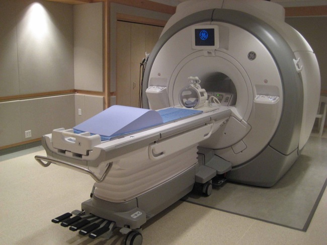

As the use of brain imaging increases in routine clinical care, clinicians and researchers are confronted with incidental anomalies appearing on the scans. One such anomaly is the white matter hyperintensity. A white matter hyperintensity indicates increased signal intensity that the MRI has picked up – basically a spot on the MRI image. Remember brain white matter is formed by the myelin protected axons of neurons. This is where electrical messaging takes place across the brain and spinal cord. A white matter hyperintensity means that something is going on with these axons. But what and why?
As more and more of these hyperintensities appeared, researchers began to identify the causes of the increased signal. They were lesions in the axons. The question then became, what would cause such lesions in otherwise healthy individuals. The obvious suspects included stroke, age-related damage, Alzheimer’s disease and even migraine. The not so obvious reasons for the lesions included cardiovascular and thyroid disease.
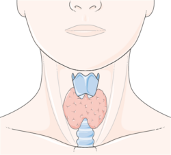
In a recent study, high levels of homocysteine (a protein associated with cardiovascular disease and stroke), chronic migraine with frequent attacks (~20 years and >5 migraines per month) and subclinical hypo- and hyperthyroidism were significantly associated with white matter lesions in people as young as 40 years old. There was no difference in the incidence of lesions in men versus women or in smokers versus non-smokers. Although, smoking increased the frequency of migraines and so indirectly may lead to these lesions.
The association among elevated homocysteine, chronic migraine and thyroid disease share a potentially similar though untested etiology – one commonly disrupted in other conditions that affect women – mutations in the MTHFR gene. I can’t help but think there are some connections to be made here.
How are Homocysteine, MTHFR and White Matter Lesions Connected?
Homocysteine levels are controlled by the B vitamins, which is in turn controlled by the MTHFR gene. Deficiencies in Vitamins B6, B9 or B12, either by nutritional deficiencies or via a MTHFR mutation that impairs metabolism, elicits high homocysteine levels. (A great video explaining this common mutation can be seen here).
The impaired metabolism of Vitamin B, leads to demyelination of the axons – the white matter lesions observed by the MRI as white matter hyperintensities. It is important to note that low vitamin B levels (high homocysteine) are associated with demyelinating diseases such as multiple sclerosis, Alzheimer’s and Parkinson’s Disease. MTHFR mutations have been identified in migraine sufferers as have the white matter lesions.
As we learned last week, pre-menopausal hysterectomy, leading to brain iron accumulation is also associated with white matter damage. And as luck would have it, iron levels may be regulated in part by homocysteine as well. Hypothyroid patients also exhibit elevated homocysteine levels whereas hyperthyroid patients have low homocysteine. Could the underlying cause of the cause of the thyroid, migraine, brain lesion trifecta be impaired homocysteine metabolism – an MTHFR mutation? Interesting possibility.
We Need Your Help
More people than ever are reading Hormones Matter, a testament to the need for independent voices in health and medicine. We are not funded and accept limited advertising. Unlike many health sites, we don’t force you to purchase a subscription. We believe health information should be open to all. If you read Hormones Matter, like it, please help support it. Contribute now.
Yes, I would like to support Hormones Matter.
This post was published originally on March 21, 2013.


Thank you for this information! I am wondering if you might every have heard of subacute combined degeneration being associated with thiamine deficiency. I underwent an MRI for cervical disc issues in July, and incidental changes of SCD were noted. No etiology has been found. My labs showed a high B12, normal MMA, homocysteine of 11, normal copper, and normal zinc, ruling out the usual culprits for SCD. I am MTHFR compound heterozygous. Levels of iodine and molybdenum are quite low, although thyroid panel studies are otherwise normal. I am a physician, and find myself at a loss, since multiple internists and neurologists have said I am a medical mystery. Thank you very much for your help!
Thanks for this article! I have white matter lesions and have had lots of MRIs and last year a lumbar puncture, which I reacted badly to. I have Hashimoto’s disease and a few years ago, developed a tremor in my right leg. The neurologist thought I had cerebrovascular disease, although I have none of the risk factors and was recommending statins to me. My case was reviewed by other neurologists and they concluded I didn’t have cerebrovascular disease, but that there was demyelination although this hadn’t spread. Personally, I thought a lack of B vitamins was the cause and I periodically take these individually (I react to biotin so can’t take that). Unfortunately the NHS doesn’t test homocysteine.
Now struggling with low iron levels so I’m wondering about taking B vitamins again soon
Try thiamine — vitamin B1. Thiamine, mitochondria and the thyroid have a tightly reciprocal relationship. Thiamine deficiency is often implicated in thyroid disease and in demylination. We have lots of articles on thiamine on the website, suspect you’ll recognize your symptoms.
I am compound heterozygous for MTHFR,
I have had migraines since the age of 20,
I have hypothyroidism,
and I have incidental areas of white matter on my brain MRIs (which my neurosurgeon told me not to worry about, saying they were probably caused by migraines).
However, I do NOT have high homocysteine.
Apparently people who are compound heterozygous for MTHFR often do not have homocysteine problems.
Apparently, it’s generally the people who only have *C677T* MTHFR mutations (heterozygous or homozygous on C677T only) who have problems with homocysteine.
(I’ve read this in internet sources, and also this is what a hematologist told me after testing me for MTHFR mutations at a surgeon’s request, prior to a surgery that I had. All the surgeon was concerned about was whether I had any issues with homocysteine or not, and the hematologist said that I do not.)