autism

ASD, Seizures, and Eosinophilic Esophagitis: Could They Be Thiamine Related?

2977 views

My 18 year old son has ASD and has had a seizure disorder since he was 6 years old. He has tried virtually all anti-epileptic drugs. Either the side effects were unbearable, they made his seizures worse, or had no effect on his seizures. He was diagnosed with Eosinophilic Esophagitis. He is underweight and of short stature, and always has been. Mitochondrial tests show that complex II is working at 26% capacity. He is also autistic. He has tested positive for folate receptor antibody.

Over the years he has done several rounds of antibiotics, including Flagyl, which I have since learned that it significantly depletes the body of thiamine. He has also taken several rounds of Diflucan, Azithromycin, Vancomycin, Augmentin, Amox for various issues including candida, clostridia, gram negative gut bacteria, etc.

He is currently on Lamictal and just started Briviact for seizures. The Briviact causes anger and aggression issues. He currently deals with OCD tendencies. He was recently found to have bone density of 2.8 standard deviations below normal. This falls in the range of osteoporosis, but he has not been diagnosed with it because of his age.

He eats fresh and a lot of dried fruit, meats, raw and cooked greens, white rice, lots of cooked veggies, eggs. He also takes Lipothiamine 100 mg/day, Magnesium 550 mg, a multi-vitamin, calcium, vitamin D, and K, all at the direction of his doctors.

Childbirth and Infancy

M was born on July 9th 2005 7lbs 9oz. He was full-term. I had high blood pressure at 41 weeks and labor was induced. He would not drop into position and he became distressed and so was delivered via cesarean while I was under general anesthesia.

He spent 4 days in the NICU because he aspirated meconium and would not latch to feed. While in the NICU, he was administered antibiotics. He was formula-fed, not breast-fed.

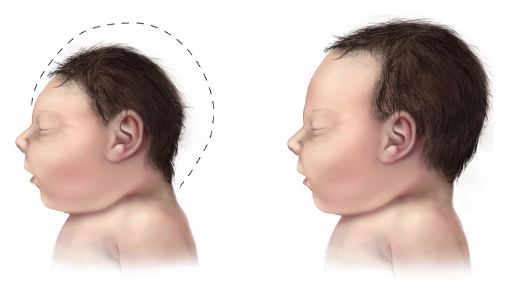
As an infant, the large size of his head was somewhat of a concern for the pediatrician. He was administered vaccinations according to the CDC guidelines for the first 12 months. He had infantile spasms off and on. He spiked a fever for every vaccination. Tylenol was administered. He received 3 doses of flu vaccine, accidentally, within 3 months.

He did not sleep well, and still doesn’t.

Initially, he was very precocious. As an infant, he would put puzzles together that were for much older children. He would complete sorting activities that were well beyond his age range. He did not babble and eye-contact was fleeting.

After his 18 month vaccination, he lost just about everything within 2 weeks. After these vaccinations, he couldn’t do his puzzles, bring food to his mouth, smile, couldn’t stand to be read to when he previously loved to be read to. He also developed a sensitivity to light and sound and cried a lot.

At 24 months, he was diagnosed with profound autism.

PANDAS/PANS and Eosinophilic Esophagitis

At age 10 years, he abruptly lost skills again and it was thought he had PANDAS/PANS as he had several strep infections treated with antibiotics. He did a several month long courses of Augmentin or Azithromycin to treat PANDAS/PANS. He had a severe trauma at age 11. He was horrifically abused by a school employee.

He has always been of short-stature nearing 5th percentile for height, and slightly overweight for his age, until age 14 when he started having symptoms of Eosinophilic Esophagitis. He was diagnosed with EoE at 15 and has struggled to keep his weight high enough as he dealt with the intense pain, fatigue, and esophagus issues with this condition. He is currently taking Dupixent for his Eosinophilic Esophagitis as the PPI and Budesonide slurry were not addressing the issues. So far Dupixent is allowing him to eat. His diet remains very restricted due to having so many trigger foods and he has almost no appetite.

He eats a lot of dried and fresh fruit. He loves greens, raw and cooked. He also eats meat, white rice noodles.  He eats mostly an organic diet. He does occasionally enjoy candy.

Seizures

He developed seizures at age 6. These were controlled for a while on Depakote, but the side effects of Depakote were too much for him and so we had to stop. His seizures are now not controlled. He has 1-2 tonic-clonic seizures per week, plus several staring spells all throughout the day. Recent EEG showed abnormal spikes and discharges in the frontal and temporal lobes. It indicated his seizures involved many places on his brain. Brain surgery was being considered for seizures at this time, but ruled out as an option due to the nature of his seizures.

He has failed several other seizure meds including Vimpat, Zonegran, Aptiom, Topamax, Onfi, and others. He is currently on Lamotrigine and Epidiolex for his seizures. He also takes trazadone and gabapentin for sleep, although these do not consistently help him sleep. He is so consumed by fatigue and can hardly get out of bed even to walk across the room. With tons of encouragement he can do brief periods of school work. The meds cause him to lose focus and become frustrated. He seems to almost always be lost in a fog and unable to participate in basic conversations without losing focus or becoming too exhausted to continue. Each seizure will cause him to be in bed for 2-3 days. He has fallen many times going into a seizure and is now afraid to leave the safety of his bedroom. He will come out, but rarely.

He has intermittent issues with nystagmus. He had a bad case of COVID 2 years ago, which caused clusters of seizures and constant nystagmus.

He has an exaggerated startle response.

Despite It All

M is a sweet young man. He is brilliant. He loves animals. He tells everyone he sees that he is so happy to see them. He is working with a local legislator on how to improve rights for non-speaking people, especially in the court room. He is completing all of his high school courses at home with straight A’s and he is a published poet.

He does not speak, but he communicates by pointing to letters on an alphabet board. This is a skill that took him years to learn. He communicates at an age-appropriate level or higher. He is working, slowly, toward a standard high school diploma.

Postscript

Based upon what I have learned from this website, I discussed thiamine with our physician. It turns out, she heard Dr. Lonsdale speak years ago. She recommended 50mg of Lipothiamine. The entire time he was taking it, he had no seizures. I was not sure that it was thiamine or the meds until we ran out for about a week. The seizures returned, but as soon as we resumed the Lipothiamine, they disappeared again. He has been taking it again and now it has been 2 weeks without seizures. I don’t want to get my hopes up, but it could definitely be a piece of the puzzle. Are there others out there with similar experiences?

We Need Your Help

More people than ever are reading Hormones Matter, a testament to the need for independent voices in health and medicine. We are not funded and accept limited advertising. Unlike many health sites, we don’t force you to purchase a subscription. We believe health information should be open to all. If you read Hormones Matter, like it, please help support it. Contribute now.

Yes, I would like to support Hormones Matter. 

Image by Gerd Altmann from Pixabay.

Healing Our Daughter, Healing Ourselves

8603 views

Our Little Tadpole

Abby is our little tadpole. Sensitive to seemingly everything, she morphed into something she should never have been. Many people openly stare quietly, thankful their kids aren’t like her and go about their business without much thought. Or worse were those who only stood by saying “God, somebody DO something!” when our daughter was screaming in pain in public as if WE weren’t there. There is much to be learned from them, if we only had eyes to see. We’ve made mistakes in our journey with our daughter, but the 10 specialists we’ve taken her to in the last nine years have provided numerous test results with little to no answers. The last allergist I took her and told me to “stay off the internet” as he was handing me free pharmaceutical samples as I was walking out the door. They made it as far as the trash can in the restroom. And so began our journey of doing something different, looking for answers outside of what we’ve been told. In our journey to heal our youngest daughter, we are healing ourselves too as we all seem to be dealing with very similar issues of low stomach acid, connective tissue dysfunction, and nutritional deficiencies/dependencies. I will start with her story first, since it is because of her that we’re figuring ourselves out.

The Early Red Flags: Hypermobility, Digestive Issues, Speech Problems and Intense Reactivity

Abby was adopted from China in September 2010 at the age of 10 months. Her birth and family history is unknown. She was found in a very poor district at three weeks old, underweight and jaundiced. She also had a congenital heart defect called atrial septal defect, which later closed up on its own. She was kept in ICU for a period of time. She was bottle fed until 10 months, then introduced to gluten and dairy. Orphanage caregivers reported she had a much heartier appetite than other babies (an early red flag that we all missed).

When we adopted her, we found her to be a very happy and social baby, who transitioned well. Her eye contact was always good. We did notice, however, her hips, elbows, knees, and fingers were hypermobile. In hindsight, we recognized that she played differently with infant toys. More red flags.

  • In October of 2010, she had her first post-adoption doctor’s appointment.
  • In December 2010, she was walking on her own. She tested positive for TB exposure and put on Isonaizid. She tested negative for lead. She became ill and was in pain for 10 days with diarrhea 3x day. We switched to Rifampin for the next 6 months. She was on Isonaizid and/or Rifampin from December 2010 through July 2011. Anti-tuberculosis antibiotics required monthly liver checks. She was vaccinated during this time as well, a mistake we came to regret. Our once happy child now became stoic and would remain so for nearly a year, but things had changed.
  • In spring of 2011, she experienced continuous and severe congestion along with loose stools.
  • In July we stopped Rifampin. She was a good eater, often ravenous.
  • In spring of 2012 we began speech therapy, 3x per week, but progress was slow. After removing gluten she spoke her first three word sentence.
  • July 2012 her hearing was checked. “Possibly mild frequency decrements. Minor issues”.
  • July 2013, probiotics stopped her diarrhea, unless she was exposed to a problematic food.
  • In 2014, we found she was a MTHFR 677ct double mutation. We strongly suspect she has a CBS mutation due to very strong reactions to various things: ALA, NAC, Epsom salts, CLO, methylated B12/Folate. Though over time, some of these we have been able to get in her in small amounts.
  • Over the next few years, and a multitude of negative tests, all we were able to determine was that her B12 levels were consistently high, even when not supplementing much, as were her B6 levels (though iodine brought her B6 levels down into the normal range). Creatinine was low, and a few amino acids were only slightly elevated. Prostaglandin F2 were extremely high and liver enzymes were elevated.

Despite all of this, Abby is a very happy, socially engaged and intelligent girl. Her speech has always been intermittent, ranging from very slurred to full complete clear “normal” sentences. It switches at random. Her former teacher of 5 years doesn’t think it is ASD, but in truth, it doesn’t really matter. Our kids are often labeled for the convenience of others.

Altered Pain Sensitivity

Abby appears to have a high pain threshold, except stomach pain. Since she was young, she has preferred to be barefoot and wore few clothes, even in winter. Over time this has changed and she has grown more “normal” in her body’s adjustment to temperatures.

The only observable nervous system affect was that she tightens/clenches her fingers when very excited. Excitement seems to trigger degranulation in her unstable mast cells. She had been extremely reluctant to draw, write, or color when young. She’s doing all this now, not as age-appropriate, but gaining.

She has had many problems with probiotics in the past; often creating an immediate OCD/stuffing whatever she could get her hands on behavior, under furniture, peeling birch tree bark for hours, etc.  Her brain was almost immediately affected.

Severe Reactions to Triggering Substances

Trying to solve her medical problems has been difficult. She has had so many reactions in the past when trying various vitamins/minerals and supplements recommended by her physicians. The results were always mixed and reactions could be extreme. We often, and still do, dose her vitamins and minerals separately, mixed together. Many reactions were not to the main ingredient, but to the binders or fillers added to the supplement. Some treatments would spur a short snippet of normal speech but only 1-2x then nothing more.

Below are the symptoms that we have been navigating.

  • Dry, itchy skin. Rashes, hives, angioedema, large welts from some foods/chemicals and insect bites.  Her skin feels like it’s on fire and she tore at her clothing after a small amount of Pure Vegetable Glycerin (99.9% pure) was applied. In 2015, her skin peeled off her arm, wrist to shoulder in a 3” wide band of deeply reddened dry/cracked skin, after eating non-organic strawberries. It looked like a third-degree burn, minus the blisters. Epsom salts, baking soda, Vick’s Vapor Rub, various other skin oils like jojoba, or almond oil, all caused painful reactions.
  • Severe abdominal pain. She experiences severe abdominal distress and pain after ingestion of various foods or charcoal-grilled food. She may also develop constipation/diarrhea, headaches/migraines. Probiotics often dramatically changed her behavior within an hour to severe OCD. Fruit-based digestive enzymes would cause facial rashes and behavior changes. Pancreatic enzymes caused much less speech, very quiet per her teachers.
  • Urinary. She was unable to urinate 9+ hours after ingesting cough syrup on two occasions. She was not dehydrated either time. She formerly had urinary incontinence on occasions and enuresis. The enuresis resolved with the addition of vitamin K2 MK7.
  • Insomnia. Occasionally she would develop insomnia, often after ingesting or exposure to an offending food or chemical. Tap water seems to be particularly problematic.
  • Behavioral. She has experienced severe OCD, irritability, extreme aggression/anger, hyperactivity.
  • Heart and Lungs. She develops a rapid heartbeat at rest and persistent coughing for 6+ hours following ingestion of a trigger.
  • Head and nose. Congestion, puffiness/eyes, headaches/migraines (based on focused tearing behavior).
  • Speech Problems. Her ability to speak various greatly relative to exposures. It goes from single words to full clear “normal” sentences. With gummy vitamins, recommended by her doctor, she developed a very notable and immediate regression in speech when she was four years old.  The day before she took the vitamins, she had clearly-spoken emerging speech, i.e., “I eat” “I do” “I wash”.  Immediately after giving her the vitamins, she walked about the entire day just saying “mmmmmm” over and over. Unsure of the cause, I was thinking dyes, rancid hydrogenated oil, or some such.  I would not make the sugar connection for a few more years.
  • Severe pain after exposures. She had a strong reaction to Cassia cinnamon. In class, she and other children were making Christmas ornaments with lots of Cassia cinnamon. Although none was ingested, her teacher said she was inhaling it and handling it for hours. Near pickup time, the teacher said she was not feeling well, began to be irritable, like her head hurt. As we were walking out of the building, she went down fast onto the ground and began writhing in pain (not sure if head or gut related). Teacher held her head to keep her from hitting it on the pavement, while I ran to get my Lavender essential oil rollerball. Applied it, and within a few minutes she was fine and got into the car. No further incident. Ceylon cinnamon causes no problems. Cassia can affect B1 levels, or so I read.

Our Journey to Healing Began With Vitamin K and Thiamine

In October 2018, we learned about thiamine and suspected that many of her problems may have been the results of a longstanding thiamine deficiency. We began in August slowly increasing Thiamine HCL. She began to improve at school, but results were inconsistent. We then moved to Benfotiamine for a while and results seemed better, but still inconsistent. By October, she was taking Sulbutiamine and we worked our way up slowly to 200 mg. Organic Acid Test (Great Plains) showed her lactic acid levels came down with the addition of the high dose of thiamine.

Nighttime enuresis persisted several years beyond toilet training. In 2016, we added approximately 700 mcg of vitamin K (MK7) working up slowly to this dose and her nighttime accidents completely stopped. The addition of vitamin K (MK4), reduced her food intolerances and allowed her to eat a broader diet, but that form of the vitamin did not stop the enuresis, the MK7 form did. We have since lowered her doses and now she just takes a D3/K2 liquid form with no return of the enuresis and food tolerances seem good, though we monitor her diet closely.

We use a variety of homeopathic remedies to treat reactions, illnesses, and injuries and reduce chemical exposures at home. Once her lactic acid levels came down into the normal range with the thiamine, we were able to add probiotics without negative reactions.

Her diet is mostly organic, grass-fed beef, organic chicken, wild-caught fish, cage-free eggs, local raw honey, coconut and olive oil, ghee, no GMOs. MTHFR mutations seems to be sensitive to gluten and dairy, but I wonder if that’s because of our need for the TTFD form of thiamine. She has been sugar free since July 2018.

My Big Takeaway: Healing Requires Resolving Nutrient Deficiencies Dependencies

EDS and ASD both share very similar nutritional deficiencies and/or dependencies. I wonder how much of autism isn’t simply the undiagnosed trio of EDS/MCAD/POTS. As most genetic testing is beyond the reach of most family budgets, it is difficult to know. It seems like it would be worth looking into one’s broad family history. A friend once told me that the foods we crave the most can be our biggest problems.

Years ago when Abby was in preschool, her teacher had me in for a conference. She showed me her notebook, which sadly only had a few scribbly lines in it. She slowly closed the book and moved it to one side. She looked me straight in the eyes and said “this isn’t autism”. Her son was on the spectrum. She said “Abby is smart, very, very smart. I think she’s gifted”. I looked at her dumbfounded, asking “then why?”.  She said, “I don’t know what’s going on, but she knows… she knows!” She proceeded to tell me something Abby did that proved to her unquestioningly her assessment. Giftedness and learning disabilities seem to share many commonalities.

We sort of figured some things out in reverse. For example, the MK4 form of vitamin K2 allowed for more food tolerances, and the MK7 stopped her enuresis. Bacteria in the gut (bacillus subtilus) produces K2, but then too much lactic acid was a problem because her thiamine was low and the CBS mutation seeming caused trouble as well. K2 seems to be very important in the distribution of calcium in the body.

We often see admonitions to heal the gut on the internet; so many opinions and recommendations. As Abby’s case suggests, it is far more complicated than simply taking a probiotic. It is also highly individual. Our daughter’s journey may not be applicable to someone else, but perhaps something can be gleaned.

We continue to avoid triggers, eat and live clean, heal the gut, use holistic remedies, play and laugh a lot. Thankfully, her reactions are now infrequent and fairly mild, but it was long road to get to this point. Her appetite is now normal with no real cravings or hunger extremes. We use vitamins/minerals, fish oil, and probiotics less cautiously now. She is gaining speech rapidly. She may still not be typical, but she is a far cry from what she had morphed into and much more normal than even a year ago.

We’ve been fortunate to avoid prescription drugs overall and use natural remedies, diet, and vitamins and minerals to affect change. We are avoiding further vaccinations, as our belief is her body has had enough and can’t deal with the stress at this time. Overall many people’s demeanor changes rapidly when mentioning alternative approaches to western medicine. If outside the norm, we may even be deemed a quack, but since we’ve been able to heal various family members of numerous ailments, if we’re seen as strange, so be it.  We can heal our bodies, probably not 100%, but often without prescription drugs.

Perhaps even the most complicated puzzles among us are not as hard to put together after all. We are still healing and our journey is not over. We tell ourselves and our kids to eat less junk because a nutrient-dense diet is helpful to everyone, but it seems that it is even more vital to those who suffer both the blessings and curses of a good brain.

Our brightest lights are ever so vulnerable.

We Need Your Help

More people than ever are reading Hormones Matter, a testament to the need for independent voices in health and medicine. We are not funded and accept limited advertising. Unlike many health sites, we don’t force you to purchase a subscription. We believe health information should be open to all. If you read Hormones Matter, like it, please help support it. Contribute now.

Yes, I would like to support Hormones Matter.

This story was published originally on August 19, 2020. 

Food Aversions in Children with Autism

5779 views

Dietary Aversions and Preferences in Children with Autism

“They are a super picky eater. They won’t eat vegetables.”

These are some of the most common words I hear from the parents of my autistic patients. Of course, we all know that eating our veggies is healthy, but for a child on the spectrum what the bacteria would do with the vegetable fiber can have a massive impact on how their immune system and brain work. There are a few reasons they might dislike certain types of foods.

  • Altered gut bacteria. They may have too many bacteria in the small intestine due to poor bowel motility which then could cause pain and bloating.
  • Parietal lobe issues. It is also possible that a child on the spectrum could have their parietal lobes affected. This could lead to an aversion to the texture as well as the taste of certain foods. Since there is typically altered sensory perception with a child on the spectrum, including on the lips and tongue, this will impact how they perceive food textures and the taste of food itself.
  • The foods they like may be exorphins. Certain foods like breads with gluten, for example, can activate opioid receptor sites. That is, when the child, eats these foods, he or she gets a boost of nature’s endogenous painkillers. This makes the child addicted to the very foods that have the potential to be the most problematic. When a parent asks me what foods their child should avoid, I typically ask them what foods they seem addicted to and we have some of the answers.

Pieces to the Puzzle: Immune System and Microbiome Crosstalk

Although the research is ongoing, it is becoming more apparent that either the child with autism or his/her mother suffer from a pattern of immune activation that can lead to disrupted gut barrier. This, in turn, contributes to an alteration not only in the gut bacteria, but also, in how the immune system in the brain respond to immune insults. Since these bacteria are responsible for metabolizing the food we eat and extracting from those foods essential nutrients, that then are used for building neurotransmitters, and modulating immune function, the alteration in gut bacteria can lead to an inflammatory cascade and cycle, which then affects the proper development of the nervous system. As the diet becomes narrower, it brings further nutrient depletion, causes more of a microbial imbalance, and leads to a pro-inflammatory environment. Although most people with chronic disease have some form of inflammatory response, researchers have found that when a subset of immune cells called TH17 are hyperactive, either in the mom during pregnancy, or the child themselves, we see an inflammatory response so imbalanced that a neurodevelopmental disease is more likely to develop.

A Few More Pieces: Bacteria, Fiber, and Immune Modulation

When bacteria eat different types of fiber from dark leafy green vegetables or grains, they make compounds called short-chain fatty acids (SCFA). These SCFAs activate several parts of the enteric nervous system, which then communicate with the central nervous system. Specifically, one SCFA called butyrate seems to particularly important. Butyrate is a primary energy source for cells in the large intestine used to ‘fix the gut’, but it also has a great impact on the immune system and healing the gut lining. It does this by multiple mechanism, but mainly by reducing bad bacteria that can be driving the immune issues in the first place. Butyrate helps turn off pro-inflammatory signals as well as turn on anti-inflammatory signals and bring balance back to the immune system by helping with something called T-regulatory cells. T regulatory cells do exactly what the name suggests, they regulate the immune response. This is especially important given that the brain and GI tract are in constant communication, not only via the vagal nerve, but the sympathetic nerve fibers send signals directly to the brain. Given that the microbiome and inflammatory cytokines are an important part of this relay system, their involvement with something like Autism should not be ignored.

For those of us that have been doing this enough, it is blatantly apparent that there is immune system involvement, with some articles showing upwards of 70% of children on the spectrum have an autoimmune disease process going on in their brain. Hence, to help modulate the altered immune response, the bacterial by-products of fermenting fibers can go a long way in helping bring balance to the immune response, gut lining, and nervous system.

Returning to the picky eater problem in children with autism, it appears that because of altered bacterial balance and possibly also sensory issues, they may be drawn to the very foods that sustain an inflammatory response, and conversely, naturally avoid those foods that might dampen that response and rebalance the gut microbiome. The concept of oral tolerance is where the immune system is told to ignore foods that are being consumed so they don’t contribute to an inflammatory response. This can be a large catch-22 in the sense that the less diverse a child’s diet, typically the lower their oral tolerance of foods, so unfortunately, the immune system and microbiome get painted into a corner that can be difficult to get out of.

A Picture Emerges

Although these are only a few pieces of the puzzle, giving the gut and immune cells of a child with autism what they need can really help. Eventually, the goal is to have these kids lose their taste aversion and start eating veggies and other healthier foods. To accomplish this, we have to address the root causes of their food aversions. This means addressing proper magnesium levels of the brain and re-balancing gut microbiota. Once we see how all the pieces are connecting, we can paint the clearest picture possible. Since each child is unique, each picture is unique as well. The good news is, once a path to healing is identified in a child, the effect can snowball in the sense that the fewer food aversions a child has, the better the gut and immune system work and so the better the brain works, which means the more foods they will eat and enjoy. That is, the food/health/immunity cycle becomes more favorable.

We Need Your Help

More people than ever are reading Hormones Matter, a testament to the need for independent voices in health and medicine. We are not funded and accept limited advertising. Unlike many health sites, we don’t force you to purchase a subscription. We believe health information should be open to all. If you read Hormones Matter, like it, please help support it. Contribute now.

Yes, I would like to support Hormones Matter. 

Image by StockSnap from Pixabay.

This article was published originally on April 8, 2019. 

Energy Deficiency and ASD

6028 views

I was a pediatrician at Cleveland Clinic from 1962 to 1982 and the clinical research, much of which I have published, was done during those years. I can distinctly remember that a patient with autism was considered to be unusually rare at that time. It was a habit of the pediatric staff to meet regularly with the residents in order to discuss clinical cases, particularly those that were uncommon or rare. I remember a case of autism coming up for one of those conferences because of its rarity. No longer rare, there are many ways in which a child can present symptoms, so the disease is now referred to as autistic spectrum disorder (ASD). This may imply that a lot of different disorders are conceived as being within a spectrum, each with a different cause, or it may simply refer to the wide variation of symptoms.

I looked up the statistics on the Internet and found that ASD today affects one in 68 children (1% of the childhood population). Parents who have a child in the spectrum have a 2% to 18% chance of having a second child affected, implying a genetic risk. I took early retirement from Cleveland Clinic and joined a private practice specializing in nutrient therapy. I became active in the group of physicians who are members of the American College for Advancement in Medicine (ACAM, acam@acam.org). A very big fraction of my patient population during those years was many children with ASD. My clinical experience between 1962 and 1982 had strongly suggested that thiamin(e) deficiency was common in the United States, in spite of the general medical opinion that it never occurred. During those years I had worked with an associate, a doctor who worked in the laboratory. Because of my clinical need, he had researched the literature for ways and means of accurately measuring thiamine deficiency. He had initiated a blood test known as transketolase activity and I need to explain this briefly.

Understanding How Thiamine Works and How to Test for Deficiency

Transketolase is the name of an enzyme whose activity is completely dependent on the presence of thiamine and magnesium, both known as cofactors to the enzyme. There are a series of biochemical reactions in which this enzyme plays an integral part. Known as the hexose monophosphate shunt, this series of reactions occurs in red blood cells. By taking some blood from a patient, the activity of this enzyme can be measured in the laboratory and is reported as transketolase activity (TKA). What we discovered was that the TKA could be quite normal when the symptoms of the patient strongly suggested thiamine deficiency. In order to prove that the patient was deficient, there was a second part to the test. Thiamine pyrophosphate (TPP), the biologically active form of thiamine in the body, had to be added to the reaction container and the transketolase activity measured again. If the TKA accelerated, it showed that the enzyme was lacking the necessary cofactor, in spite of it being in the normal range of activity with the first analysis. It was reported out as a percentage increase of activity as compared with the baseline (thiamine pyrophosphate effect or TPPE).

My friend in the laboratory had done a series of control tests on people that were purportedly healthy. His results compared favorably with reports of the test in the literature and these healthy people could have a TPPE of up to 18%. Anything over 18% for the TPPE guaranteed deficiency of thiamine or magnesium, so both of them would have to be supplemented for the benefit of the patient. Unfortunately, laboratories in prestigious institutions presently only do the TKA and assume that the patient does not have thiamine deficiency if the TKA is in the normal range. From my experience, I am aware that they are missing the majority of patients who have this deficiency. The TKA is not a valid indicator of deficiency by itself. Having given considerable thought to this, I concluded that if the enzyme is equipped with an adequate supply of thiamine there should be no acceleration in its activity after the addition of TPP. The only strictly normal TPPE would be zero (i.e. no TKA acceleration). The percentage acceleration of TKA would reveal a gradual deficiency compatible with no clinical significance until a certain increase in TPPE was recorded as being symptomatic of deficiency. Therefore a TPPE of, say, 15%  might be more clinically significant in one individual, whereas 20% might be less significant in another individual. The use of the test requires the clinical experience and adequacy of knowledge of the observer.

Clinical and Laboratory Experience

Without thiamine and magnesium as cofactors, the several enzymes dependent on them begin to become less and less efficient. One of these enzymes is essential to energy synthesis and so thiamine deficiency, because it powers pretty well every cell in the body, can literally cause any disease, since energy is integral to all functions of the body. Depending on the cellular distribution of the deficiency, the clinical expression will vary and there is no typical repetitive clinical expression (phenotype) for thiamine deficiency. However, the brain and heart being the most metabolically active organs are therefore much more susceptible to this deficiency. Thus, brain and heart symptoms are the commonest forms of clinical expression. Thus, it can be assumed that any gross deviation of behavior is evidence of electro-chemically driven changes in brain metabolism related to energy availability.

Case Evidence

Many of the children that I saw with ASD had an increase in their TPPE. Most of them with an accelerated TPPE had a TKA that was in the low normal range. When they were treated with thiamine, many of them responded clinically and their TPPE decreased into the acceptable range, so the evidence was published . The case histories of a mother and her two children were reported . The mother was a recovered alcoholic and alcohol is frequently responsible for causing thiamine deficiency. Some alcoholics react to sugar, particularly if there is a family history of alcoholism that suggests genetic risk. She and both of her children had symptoms that were typical of autistic spectrum disorder, but also they all were recognized to experience the clinical effects of dysautonomia. A description of beriberi clearly indicates that thiamine deficiency disrupts the normal functions of the autonomic nervous system (dysautonomia). All of them had intermittently abnormal transketolase studies indicating abnormal thiamine pyrophosphate homeostasis. Both children had unusual concentrations of arsenic in the urine. All of them had symptomatic improvement with diet restriction and supplementary vitamin therapy, but they quickly relapsed after ingestion of sugar, milk, or wheat. All three individuals became aware that their symptoms were related to their dietary indiscretion, but were quite unable to resist their ingestion of the “foods” that they all knew to be responsible for their relapse. The presence of arsenic in the urine was in keeping with our experience in finding the presence of heavy metals in ASD. My colleagues and I were so impressed with thiamine deficiency as a major cause of ASD that we conducted a pilot study, using a derivative of thiamin known as thiamin tetrahydrofurfuryl disulfide (TTFD). Eight of 10 children showed clinical and biochemical improvement .

The negative relationship of the brain with sugar is mindful of a case that I reported in 2016 and whose story appears on this website. A 14-year-old boy was strongly addicted to sugar and throughout the first eight years of his life his repetitive symptoms had been classified elsewhere as psychosomatic in nature. At the age of eight years it had been discovered that he had the relatively recently described disease known as eosinophilic esophagitis. He also had the classical clinical picture of dysautonomia. It was hypothesized that some form of genetic risk, coupled with thiamine deficiency from out-of-control sugar ingestion, was the responsible combination. The vagus nerve, whose course runs from the brain to the spleen, (and beyond) presides over the control of inflammation. The medical literature has reported food allergy as the cause of eosinophilic esophagitis. The vagus nerve requires acetylcholine as its neurotransmitter, a chemical that is derived from the citric acid cycle (CAC), the cellular “machine” that synthesizes energy. In turn, the nerve is dependent on thiamine that is essential for the entry of glucose into the CAC. Lacking the suppressive function of the vagus on inflammation resulted in a failure to suppress it in the esophagus. Thus, it was conjectured that thiamine deficiency was the primary underlying cause of the esophagitis.

Insufficient Energy and ASD: The Three Circles of Health

Mitochondria are very sensitive to environmental stressors such as toxicants, medication, immune activation, and metabolic disturbances suggesting that mitochondria are involved as a cause of ASD.

Figure 1. Three Circles of Health


From what has been written above, it must become clear to the reader that I am proposing a very different approach to the cause of ASD and disease in general. It is based on the use of Boolean algebra as represented in Figure 1. This is a statistical method of measuring the influence of variables by representing them as overlapping circles. The individual effect of one circle is represented by its area and its relationship with the other two circles by the area within the overlap. All three circles also overlap, representing the concept that disease might be 100% from one circle alone, by the combination of two defective circles, or by the variable combination of all three.

Genetics

The construction of the human body is based on a code in the form of DNA and represents a “blueprint”. Known as the genome, perfection in the code would create a perfect human body. However, we know that DNA has imperfections, but in most cases a human individual can get along quite well in spite of the imperfections.  Although an abnormal gene may be the single cause of disease, the late onset of most genetically determined conditions indicates that an additional factor is required to cause disease expression. This emphasizes the importance of the quality of nutrition in preserving health. For example, type 1 diabetes has a genetic risk, but is not usually expressed for many years, so another factor is necessary. That is why the disease is stress related, making its clinical appearance following something as simple as an infection, a divorce or an injury. Type 2 diabetes often has a genetic risk where either diet or stress, or both, may be initiators.

Epigenetics: Environment and Nutrition

Epigenetics is a relatively new science that seeks to explore how malnutrition and poor lifestyle can have a negative effect on our genes. There are several genetically determined diseases known as inborn errors of metabolism that make their appearance at birth. If any one of them is not recognized in the newborn infant, the result is often a mentally retarded individual. Some of them, perhaps the commonest being phenylketonuria (PKU) require a special diet initiated at birth in order to prevent the mental retardation. That is why in every state in the United States, a special laboratory has been set up to screen a blood test from every newborn infant, in spite of the rarity of the diseases. Sometimes, an infant is born with a gene that creates a risk for generating the disease, but it does not appear unless a secondary factor such as an infection or an injury (stressor) is experienced. There is a disease known as maple syrup urine disease (because the urine smells exactly like maple syrup) that can appear immediately at birth spontaneously or will only appear when the child is hit by some infection or trauma. There are some cases of this disease that respond to megadoses of the vitamin (thiamine/magnesium) that is normally necessary for the mechanism whose failure produces the disease. This has significant importance because a head injury or an infection might be blamed as the sole cause of a clinical problem, whereas the truth could be that the stress factor has initiated a metabolically determined disease previously unsuspected. If such a similar disease responds to the necessary associated vitamin, it can be said that the patient was successfully treated epigenetically.

Stress

In my view, this word is used too carelessly. It is most frequently used to describe the result of stress by saying that someone is “all stressed out”. So let us be clear that stress is some kind of force that a person has to meet and to which an adaptive response is required. It may be a mental force such as a divorce, a business deadline, or a physical assault such as an infection, or an injury. The three circles of health uses the philosophy proposed by Selye. This famous researcher used many forms of physical and mental injury (stress) on rats and studied the biological effects by examining their blood and tissues. He came to the conclusion that each animal went through a process of resistance that he called the General Adaptation Syndrome (GAS). The laboratory results that occurred if the GAS failed to restore health were strikingly similar to those registered in sick people and he proposed the idea that human diseases were the diseases of failure to resist or adapt to stress. What was completely revolutionary was that the GAS required some form of energy to power the machinery that enabled the animal to adapt.

In Selye’s time the biochemical mechanisms of energy synthesis were not well known. Today, these mechanisms are understood and our lives are spent in meeting the daily stresses to which we have to adapt. Our failure to meet the GAS may well be of course that the form of stress is overwhelming, such as a car collision or a virulent infection. However, what we meet on a daily basis is a mental or physical form of stress to which we have to adapt or from which we have to heal. The central factor is the mobilization of sufficient energy to meet the demands of defense. Therefore, the three circles of health predict that the cause of human disease agrees with the concept of the “diseases of adaptation” proposed by Selye.

Energy and the Ability to Adapt

Assuming that the adaptive machinery in the body is genetically adequate, all that is required is sufficient energy to drive it. The symptoms generated during the process of adaptation simply represent the locality of the energy deficiency and constitute a warning by their perception in the brain. They have to be interpreted for their underlying meaning rather than accepting them as the effects of a named disease such as Alzheimer’s or Parkinson’s. Since the brain is an electrochemical “machine”, the nature of the patient’s diet, as well as family history, are essential to beginning to unravel the problem. In the case of ASD, the mother’s diet during pregnancy is of vital importance. Occasionally, a seemingly irrelevant observation by a physician can be valid. I was riding in a car with a gentleman who had a group of infant’s shoes dangling behind the windscreen. I asked: ‘what was the significance?’ He responded by telling me that his first child died in infancy from a rare genetically determined disease. Subsequent children had survived and this was his method of keeping them in memory.

Nutrition as Medicine

When Homo sapiens arrived on the face of the earth, or even when his ancestors were present, the food was available and the general concept is that we were hunter gatherers. To find out what kind of food an animal should be taking, you have to look at the teeth. We have cutting teeth, canine teeth and grinding teeth, indicating that we are omnivores and can consume meat, vegetables and fruit. The necessary non-caloric nutrients (vitamins and essential minerals) were in the natural food. In the modern world, our food is far from natural and the high calorie content, particularly in the form of sweets, is overwhelming the cellular machinery that synthesizes energy. For this reason, many of the illnesses that haunt a physician’s office are merely a reflection of a mild to moderate energy deficiency in brain cells. If recognized for what they represent and treated with appropriate nutrition, the symptoms quickly disappear. If not, we can hypothesize that there may be permanent damage that is diagnosed as one of the neurodegenerative diseases.

The trouble with understanding this concept is that the distribution of the energy reducing mechanism, together with genetic risk, will vary from individual to individual. A child with a genetic risk, or, more commonly, a victim of poor maternal diet, is born a candidate for energy deficiency. That is why ASD can be the result of thiamine deficiency or any other mechanism that interrupts the flow of energy. However, the same deficiency can give rise to many other conditions presenting with an unpredictable array of symptoms. There is a great deal of evidence that food is burned (oxidized) in the cells of the body to create a form of chemical energy (ATP) that is transduced to electromagnetic energy. It is this energy that is used to drive physical and mental action. There is also evidence that a biological form of thiamine (thiamine triphosphate) is important in the electomagnetic transduction process, thus making thiamine uniquely indispensable. It is for this reason that I have spent many years emphasizing the life-giving properties of this extraordinary vitamin. I am not therefore surprised when I read that a constellation of symptoms called Parkinson’s disease has been reported to respond to treatment with thiamine.

We Need Your Help

More people than ever are reading Hormones Matter, a testament to the need for independent voices in health and medicine. We are not funded and accept limited advertising. Unlike many health sites, we don’t force you to purchase a subscription. We believe health information should be open to all. If you read Hormones Matter, like it, please help support it. Contribute now.

Yes, I would like to support Hormones Matter.

Autistic Spectrum Disorder, Mitochondria, and Nutrient Deficiencies

5459 views

Much has been written in the medical literature concerning the biochemical and potentially treatable nature of a group of conditions known as Autistic Spectrum Disorders (ASD). Then why are practicing physicians treating these children with medications rather than nutrients? The answer, of course, is that the disease model is being rapidly antiquated by a better knowledge of Mother Nature. This post discusses some of the important published information that indicates very strongly where the research should be.

Mitochondria

Hopefully, the reader of this post will be aware that mitochondria are the organelles that produce cellular energy. It has become clear that what the authors describe as a large subgroup of individuals with ASD demonstrate abnormalities in mitochondrial function and suffer gastrointestinal symptoms. These authors state that the majority of children with ASD do not have a primary genetic mutation. This raises the possibility that their mitochondrial disorder is acquired, or results from a combination of genetic risk interacting with a wide range of environmental triggers. “Spotty”energy deficiency in the brain may be the bottom line. Cellular distribution of the deficiency might vary from individual to individual, thus explaining the variety of symptoms. These researchers also reviewed the evidence that enteric bacteria play a part in this disease. The microbiome is the term used to describe the full complement of bacteria that live in the human intestine and play a huge part in maintaining health. The microbiome and its connection with disease was recently reviewed on Hormones Matter.

ASD and Malnutrition

One hundred and five children with ASD and 495 typically developing children of 6 to 9 years were compared in Valencia, Spain. The affected children failed to meet dietary recommendations for thiamine, riboflavin, vitamin C, and calcium. It was recognized that cultural patterns and environment may influence food intake. The majority of parents reported some concern regarding the feeding behavior of their ASD children in India. Relative to controls, the affected children had significantly lower daily intake of potassium, copper and folate although they did not differ in their calorie intake. We have pointed out in these posts that a high intake of empty calories with a normal blood concentration of thiamine is the equivalent of a normal calorie intake with dietary thiamine deficiency.

Autism rates in the United States are increasing at a rate of 15% a year. A recent study established the autism rates for each of the 50 states and calculated the percentage of infants who participate in the Women, Infants and Children program (WIC). The states with the highest WIC participation had significantly lower autism rates. Infants who were solely breast-fed, however, had diets that contained less thiamine, riboflavin, and vitamin D than the minimal daily requirements. The results suggest that autism may be nutritionally related to vitamin deficiency. However, the question remains as to whether the malnutrition was the effect of poor eating behavior resulting from brain changes due to autism, a typical “chicken and egg argument”. Which is cause and which is result?

An 11-year-old boy with autism developed liver dysfunction and became less responsive. His diet for several years was self-limited exclusively to a single “fast food”. He improved rapidly with the administration of thiamine but developed epileptic fits two weeks later that required the administration of vitamin B6. It has long been recognized that pharmacological doses of vitamin B6 can be used in some cases of seizure disorder, requiring the physician to be alert to the possibility.

Genetic Factors, Nutrition, and Autism

To illustrate that nutritional effects can be linked with genetic risk, we reported a recovered alcoholic mother who had two children with ASD. All three subjects had evidence of autonomic dysfunction that improved with dietary restriction but quickly relapsed after ingestion of sugar to which they were clearly addicted. Improvements and relapses were marked by an intermittently normal and abnormal laboratory test for thiamine deficiency. A genetic relationship between sugar consumption and substance abuse, including alcohol, has been demonstrated. I came across several cases where an individual with sugar addiction had a family history of alcoholism. In one instance a boy had developed autonomic dysfunction (Postural orthostatic tachycardia syndrome, POTS) following a vaccination with Gardasil. His test for thiamine deficiency was strongly positive. His father was known to have a classic case of brain thiamine deficiency (Wernicke encephalopathy) and a family history of alcoholism on his side of the family. This case suggests that the boy had a genetic risk for alcoholism from his father and that sugar ingestion might have increased that risk, creating a marginal, possibly mildly symptomatic or asymptomatic, state of thiamine deficiency before he received the vaccination. The question remains therefore whether his succumbing to POTS demanded a combination of all three factors, genetic risk, asymptomatic marginal thiamine deficiency with symptomatic precipitation by the “stress” imposed by the vaccination.

Oxidative Metabolism

The published research suggests that the primary cause of autism (and many other brain diseases) is an underlying defect in the energy requirement for the brain. Although we do not claim that thiamine deficiency is the only cause of this disease, it is strongly suspected that the cause in common is anything that interferes with energy metabolism, an idea that has already been published. Since thiamine deficiency appeared to be a common cause of mitochondrial dysfunction, we reviewed our medical records from ASD children that we had treated. Besides blood tests revealing thiamine deficiency, there was evidence of traces of heavy metal accumulation from hair analysis, suggesting that some children with ASD may have particular trouble excreting these heavy metals.

Genetic studies have not revealed dominant genetic errors common to all cases of ASD, although it is assumed to be a complex disorder due to mutations in hundreds of common variants. Insight has been published, offering evidence that perhaps many of these neurologically afflicted children could be successfully treated with micronutrients. Evidence has been published that, in infantile autism, the areas of brain damage that occur are the same as those resulting from alcohol abuse, well known to be associated with thiamine deficiency. It is of particular interest that concentrations in blood testing of inactive thiamine (thiamine and its monophosphate) were normal whereas the concentrations of active thiamine (thiamine pyrophosphate) in autistic children were significantly decreased.

Many people (including physicians) are not aware that thiamine ingested from the diet does not have any action in the body until it is “activated” (converted to its phosphate form). Failure in the body to activate it is of course equivalent to being diet deficient in the original vitamin. In 2002, I published a paper reporting that I had treated 10 children with ASD using a derivative of thiamine, now available as Lipothiamine. Although eight of 10 children had marked clinical improvement, it was only a pilot study to assess whether this substance had any value in treating this disease. I had intended to set up a major study in collusion with a number of university scientists but the particular circumstances we required were not permitted by the FDA and we were unable to go ahead. Two recent publications have provided further details concerning the role of thiamine in health and disease (Lonsdale, Marrs 2017. Lonsdale, 2018)

Conclusions

It seems to be clear that ASD is a group of disorders with overlapping symptoms represented by a mixture of genetic risk, response to the stresses of life, and poor nutrition. The symptomology may come from genetic risk alone, but it is argued that in most cases it is a variable mixture of all three factors.

We Need Your Help

More people than ever are reading Hormones Matter, a testament to the need for independent voices in health and medicine. We are not funded and accept limited advertising. Unlike many health sites, we don’t force you to purchase a subscription. We believe health information should be open to all. If you read Hormones Matter, like it, please help support it. Contribute now.

Yes, I would like to support Hormones Matter. 

Image by H. B. from Pixabay.

Sudden Infant Death Syndrome, Autism, and Maternal Thiamine Deficiency

5819 views

I have addressed this problem before on this website. However, in the Wall Street Journal on Tuesday, March 27: an article appeared entitled “A Study of Sudden Infant Deaths Divides Doctors“. It goes on to say “The finding on sudden and unexpected infant deaths is surprising, says Joel Bass, of the pediatrics department at Newton Wellesley Hospital in Newton Mass”, the first author of the study, who   said “that’s more than one newborn baby dying of SUID [sudden and unexpected infant death] a day”. Oddly enough, the article indicates that “among the possible causes, some researchers point to the promotion of certain hospital practices to encourage breast-feeding. But some doctors vehemently disagree with that theory”. Apparently, the theory alluded to the practice of encouraging skin-to-skin care between mother and infant during breast feeding. I had not read the article so I do not understand how this connection was made. What arrested my attention was the reference to breast-feeding, long encouraged by pediatricians as the best way to feed newborn infants. How could skin-to-skin contact be involved? However, an article was published in 2011 in which the author had carefully examined the nutrient content of breast milk in the United States (Shamberger, 2011). It reminded me of an extremely important article that was written in the 1940s.

Breast Milk Toxicity Syndrome

It may be remembered that Hong Kong at that time was a British protectorate. A medical officer of health was sent out from Britain to Hong Kong to investigate a relatively common occurrence of sudden death in breast-fed infants of Chinese mothers. This death commonly occurred between three and four months of age and happened in infants that, from their appearance, were considered to be the healthiest in the family. Fehily, the author, was able to show that the breast milk was deficient in vitamin B1 and it was well known by early researchers of beriberi that this form of infant death was virtually pathognomonic (indication of cause) of the infantile form of this disease. In fact it was well known by these early researchers that there was no other disease of infancy that behaved like this. Fehily herself was struck by the exact likeness to “cot death”, the term used in England for what we call Sudden Infant Death Syndrome in the United States today.

Autism Rates for Each State Connected to Maternal Nutrient Status

The paper published in 2011, referred to above, was in consideration of the cause of autism, not SIDS. The author had studied the rate of autism associated with nutrition. The paper stated that autism rates in the United States are increasing at a rate of 15% per year. The study abstract reported that the design of the study used nutritional epidemiology and an ecologic study design. In other words, he studied public health data and the nutrient content of breast milk. The objective was to try to link the possible cause of autism to nutrition by creating autism rates for the 50 states of America and comparing them with published measures of infant nutrition. These included the duration of exclusive breast-feeding and participation in the Women, Infants and Children (WIC) program. The results were impressive. The states with the highest WIC participation have significantly lower autism rates (P <0.02).

In contrast, there was a direct correlation with the increasing percentage of women exclusively breast-feeding from the years 2000 to 2004. Infants who were solely breast-fed had diets that contained less thiamine, riboflavin, and vitamin D than the minimal daily requirements. Although the author was studying the rates of autism and was not in the least interested in SIDS, his study supports the finding of vitamin deficiency in breast milk, in turn supporting the possible relationship between breast-feeding and SIDS. Of course, the modern medical model would find it objectionable to hypothesize a cause common to two diseases, although both autism and SIDS have a slight male dominance and are diseases that occur during rapid growth, particularly of the brain.

My Particular Interest in SIDS

Many years ago, because of clinical experience, I became interested in SIDS. At about that time, the idea of threatened SIDS had become an acceptable diagnosis, whereas previously it had been considered that the death was truly sudden and completely unpredictable. I can remember two parents who had brought their infant to see me. They had observed him in his crib when he had stopped breathing. When one of them picked him up he started breathing again. They took him to the nearest emergency room where he was pronounced completely fit and the situation was dismissed. The parents were so scared that one of them took turns to sit up all night to watch in case this thing should happen again.

This is exactly the clinical situation that I began to experience with other infants. A monitor had been invented that could be attached to an infant that would sound an alarm if either his heart slowed or his breathing stopped. It had become well-known that picking the infant up or giving him a little slap on the buttock would instantly return him to a normal state (SIDS is more common in male infants). To cut a long story short, my colleagues and I performed a lot of clinical research on these threatened SIDS babies. We had found that a machine known as brainstem auditory evoked potential (BAEP) gave abnormal readings . This strongly suggested that the mechanism was an electrochemical dysfunction in the brainstem. This is the part of the brain that connects with the spinal cord and it contains the controls for automatic breathing. Let me explain this a little.

Most of us are not aware that our essential breathing is controlled by centers in the brainstem when we are unconscious, as in sleep. The condition known as sleep apnea is a failure of this mechanism (apnea is a temporary cessation of breathing). It was during my library research that I discovered the 1944 paper by Fehily. It seemed only to be common sense to look at the possible association of thiamine deficiency as the underlying biochemical cause. We treated several infants with thiamine injections that seemed in every case to stop their episodes of apnea. I received a visit from a university researcher in Australia. His group had been studying SIDS and they had come to the same conclusion. I was so impressed by their work that I took sabbatical leave in Australia with Dr. Read who was the leader of the group. Abnormal brainstem responses in infants at risk of SIDS was reported at about the same time. The authors said that this suggested immature development of brainstem. A recent publication found an association between maternal alcohol consumption and SIDS. The relationship of thiamine deficiency in brain and alcohol consumption is well-known. It is not too difficult to imagine that the concentration of alcohol in the blood of the mother would have an exaggerated effect in the brain of the fetus, perhaps injecting a risk factor for the infant after birth.

Conclusion

Can a parent who has not experienced the sudden death of a precious infant at the age of 3 to 4 months even imagine the horror and the enormous stress imposed on such a parent? We now know that there are quite nonspecific symptoms that may appear in an infant who threatens SIDS. They are sometimes unusually irritable and a “runny nose” may be mistaken for a cold, but such symptoms may be so slight as to be ignored. I must emphasize that the lower part of the brain that organizes automatic body functions, including breathing, is peculiarly sensitive to thiamine deficiency. If the infant is breast-fed with thiamine deficient breast milk as the 2011 study suggests, studies that I have reported here would make sense. Even if only a few of the cases were due to this biochemical phenomenon, giving some thiamine on suspicion can do no harm.

A diet history from the mother might offer clues, particularly emphasizing whether there is ingestion of sugar in all its different forms. If I were a physician in charge of such a pregnancy, I would not hesitate to add thiamine supplementation, starting as early as three months of gestation, as advised in the book written by an American obstetrician/gynecologist by the name of John B Irwin, M.D. He had found that supplementary thiamine removed the complications of pregnancy such as toxemia and even prevented premature delivery. It might well provide a nutritional legacy for the infant. Of course, I am not suggesting that alcohol ingestion by the mother is THE cause of SIDS. However, I am suggesting that the present widespread use of unhealthy nutrition may well be at fault for both SIDS and autism. Genetic risk, coupled with some form of stress (e.g. mattress flame retardants, a cold virus) and vitamin deficiency breast-feeding, might be more or less important individually, or more than one of the three items collectively.

We Need Your Help

More people than ever are reading Hormones Matter, a testament to the need for independent voices in health and medicine. We are not funded and accept limited advertising. Unlike many health sites, we don’t force you to purchase a subscription. We believe health information should be open to all. If you read Hormones Matter, like it, please help support it. Contribute now.

Yes, I would like to support Hormones Matter. 

A Tale of Two Autisms: Folic Acid, Gene Silencing, and Estrogen

11960 views

In the current public discussion about autism, one could easily conclude that autism must be caused by either genetic mutations or vaccines. The media, besides portraying these positions as the only two possibilities, typically go even further by portraying them as mutually exclusive possibilities. This kind of distorted presentation crowds out discussion of other causation ideas.

Over the last 10 years I’ve developed my own theory of autism causation, a set of interconnecting biological mechanisms that, taken together, provide viable explanations for a substantial amount of autism data from multiple research areas. In summary, I am convinced that broad and indiscriminate folic acid supplementation practice has caused two concurrent and overlapping autism epidemics in certain susceptible subgroups of the population.

Part I: Early Pregnancy and Neural Tube Formation

The neural tube is the first well-defined structure of the fetal central nervous system, and it is formed by embryonic day 28. It was shown in 1980 that taking folic acid prior to conception and during the first few weeks of pregnancy prevented birth defects of the brain, spine, and spinal cord caused by the neural tube’s failure to close completely (called neural tube defects, or NTDs). In 1992, the U.S. Public Health Service recommended that all women capable of becoming pregnant consume 400 micrograms of folic acid per day, and in 1998, a folic acid food fortification program was implemented. It is estimated that about 1,300 NTD cases per year are avoided in the U.S. due to these measures.

More than 30 years after the initial groundbreaking NTD study, it was reported that prenatal folic acid supplementation was also associated with reduced risk of autism. One study found that folic acid reduced autism risk when it was taken during the first month of pregnancy, and another study confirmed folic acid’s early pregnancy protective effect, but found that it did not extend to mid-pregnancy. The results of these studies are fascinatingly counter-intuitive, given that both autism rates and prenatal folic acid uptake increased during the same time period. If folic acid does prevent autism, then it is puzzling that autism rates fail to track NTD rates; in other words, we would have expected to see a tremendous reduction in autism rates – certainly not the increase in autism rates that did occur – after the U.S. Public Health Service issued its recommendation and folic acid food fortification was implemented in the 1990s. This implies, at the very least, that folic acid’s relationship with autism is not as straightforward as it is with NTDs.

One extremely critical point is that most women in the U.S. are now supplementing with folic acid throughout pregnancy, despite the neural tube’s closure by embryonic day 28. Since there are plenty of substances – alcohol, rubella virus, and thalidomide, to name some better known examples – that have dramatically different effects on a fetus depending on when the exposure occurs, it’s not unreasonable to suppose that folic acid might have different potential effects on a fetus depending on the gestational period of exposure. Folic acid has extraordinary properties, and should not be dismissed as “just a vitamin.”

Part II: Mid Pregnancy and Neurogenesis

A Brief Summary of Normal Brain Development

In humans, neuron production begins on embryonic day 42, two weeks after neural tube closure, and is largely complete by mid pregnancy. Neurons form in a layer of tissue called the ventricular zone, and migrate away to form the brain’s structures and neural networks. The largest processing networks involve the neocortex, the layer of cells on the brain’s surface, and the subcortical nuclei, which lie underneath the neocortex and relay information to and from the neocortex. After migration, cortical neurons undergo extensive structural changes to form the pathways of the brain’s neural networks. They extend long fibers called axons to connect with other neurons, and grow arrays of fibers called dendrites for receipt of electrochemical information from other neurons.

Brain development depends upon complex cascades of molecular signaling involving many genes and gene products. The integrity of the entire process depends on the right elements appearing at the right times and in the right amounts, such that one genetic disruption can impact the entire downstream course of development.

Gene-disrupting mutations are not the only way that the molecular products of genes can be altered; disruptive epigenetic alteration of gene expression can also cause the normal cascade of developmental events to veer off course. Epigenetics is the molecular alteration of a gene’s expression without changing the underlying DNA sequence. These alterations to genes are usually heritable through cell division, although perhaps not from one generation to the next. The most widely understood way that epigenetic change can occur is via DNA methylation, where a methyl group forms a bond with cytosine in the DNA sequence, typically at a point in the sequence where cytosine is followed by guanine, called a CpG site. CpG islands are DNA regions with a high frequency of CpG sites; and hypermethylation of CpG islands, especially in promoter and enhancer regions, is highly correlated with silencing of the associated gene. In other words, too much methylation can render a gene just as nonfunctional as a disruptive mutation can.

Brain Development in Autism

A key factor in the widespread rejection of a causal link between vaccines and autism is the weight of evidence showing that autism is associated with a fundamental disruption in prenatal brain development.

Structural indicators of a disruption in brain development are very common in autism. These indicators include dysgeneses, heterotopias, altered cell morphologies, impairments in synapse formation and synapse plasticity, microcephaly, and macrocephaly. Studies have also found increased numbers of neurons in the prefrontal cortex and abnormal positioning of cortical projection neurons in subjects with autism, which suggests a disruption in neurogenesis between gestational weeks 7-20.

Researchers think that nearly one thousand genes may be involved in autism, with some of the most strongly autism-associated genes being those that are involved in neurodevelopment, synapse formation, and epigenetic functions. In fact, recent estimates suggest that 88% of high-risk autism genes are integral to early stages of brain development, regulating neurogenesis and early neuroblast differentiation, and that 80% of these genes also influence later stages of neurodevelopment such as synapse formation. It has been suggested that this strong association of autism with genes that impact multiple stages of brain development may indicate that autism occurs when the number and nature of gene-disrupting mutations involved in neurodevelopment reach a critical threshold of cumulative impairment in neural connectivity.

Beyond genetics, there is ample evidence that atypical epigenetic activity is also involved in autism. Post-mortem analyses show that tissues from certain regions of the brain are differentially methylated in autism, to the point that researchers have called these differences “profoundly distinctive” as compared to controls and have suggested that autism may be generally associated with a disruption to methylation-associated neurodevelopment. Specific genes found to be differentially methylated in autism include oxytocin receptor (OXTR), glutamate decarboxylase 1 (GAD1), Engrailed-2 (EN2), Reelin (RELN), and MECP2, all neurodevelopment-impacting genes, and the autism-associated genetic conditions of Angelman, Fragile X, and Rett Syndromes all involve disrupted epigenetic processes. Particularly fascinating results were obtained in a study of DNA methylation in peripheral tissue, where the subjects were three sets of monozygotic twins. In each twin set, one twin was diagnosed with autism and the other twin had milder autistic features that did not rise to the level of diagnosis. The twins with diagnosed autism had higher methylation levels than either their undiagnosed twins or their unaffected siblings, hinting at the presence of some environmental factor that increases both methylation activity and autism risk.

As noted above, hypermethylation is typically associated with gene silencing, and gene silencing can affect the entire downstream course of development. So what could cause a derailment of fetuses’ normal methylation activity, causing gene silencing during critical periods of brain development on a population-wide scale?

The answer to that question may lie in prenatal vitamins.

Bathing the Fetal Brain in Methyl Groups

The B vitamin folate is an extremely critical source of methyl group donors for DNA methylation, and folic acid is the synthetic version of folate. Standard obstetric protocol now recommends folic acid supplementation through the entire nine months of pregnancy, which means that the majority of pregnant women in the U.S. are consuming historically unprecedented amounts of methyl groups between gestational weeks 7 and 20, just when fetal genes are causing neurons to form, migrate, and differentiate.

It is known that dietary consumption of methyl groups impacts DNA methylation and gene expression. In animals, it is well established that administering folic acid to pregnant mice alters genetic expression in their offspring. In humans, researchers have found that maternal use of folic acid increases methylation in young children, whereas maternal low folate status is associated with both NTD and hypomethylation in the fetal brain. In sum, current research suggests that “in the rapid period of cell division occurring during fetal development, the level of methyl donors can have a significant impact on transcriptional activity that is maintained into adulthood.”

To explore possible effects of folic acid during mid-pregnancy, we can engage in a thought experiment based on the fairly noncontroversial idea that autism results from the cumulative effect of multiple genetic mutations impacting neurodevelopment. In particular, let’s consider a fetus with some genetic mutations that impact brain development, but with a cumulative effect still below the threshold of impaired neural connectivity that would cause autism. What happens if his mother takes folic acid throughout the neurogenesis period? And what if the fetus also has a common genetic impairment in folate metabolism, perhaps causing folic acid to accumulate in the womb? One possible progression of events might develop as follows:

  1. The mutated genes cause some errors in brain development.
  2. Because of the fetus’s impaired folate metabolism, excess folic acid accumulates and causes abnormally high methylation activity during brain development. This causes some unmutated genes to become hypermethylated, which renders these genes essentially non-functional. More developmental errors occur because of this gene shut-off.
  3. Downstream brain development is altered by both the mutations and the inactivated genes. Hypomethylation of certain downstream genes could be a possibility, if either the mutated genes or the inactivated genes functioned to create molecules involved in epigenetic processes.
  4. The autism threshold is reached as neural connectivity becomes severely impaired by both genetic and epigenetic malfunctioning. Perhaps neither the genetic mutations nor the hypermethylation would have been sufficient to cause autism in isolation, but in combination they do.

If the above sequence of events were to occur on a population-wide level, it might (a) cause an overall rise in autism rates in supplemented populations, and (b) lead to a finding that genetic mutations involving brain development are particularly common in autism, because the presence of these mutations in utero would put a fetus closer to the autism threshold than a fetus without such mutations.

In fact, children with autism do present with significantly more de novo, gene-disrupting mutations than their unaffected siblings.

Part III: Mid Pregnancy and Estrogen

In mammals, male fetuses go through a period of development where fetal testosterone enters the developing brain, converts to estradiol (an estrogen), and stimulates neuronal estrogen receptors. This period of development, called sexual differentiation, essentially masculinizes the male brain. In humans, it is thought that fetuses are sensitive to estrogen-sensitive brain growth in mid-gestation.

In 2011, I published a paper describing, I believe for the first time, the large overlap between estrogen-sensitive cognitive functions and functions impacted in autism, including anxiety, motor deficits, stereotyped and repetitive movements, epilepsy, hyperactivity and attention deficit, disrupted pain recognition, deranged serotonin system, atypical memory function pattern, and atypical visual-spatial information processing. If we speculate that some form of autism results from excessive estrogen-sensitive brain growth, then male fetuses, with their generally higher baseline level of such growth, would logically be more susceptible than females to this form of autism. This could explain the high ratio of boys to girls in autism.

In my article for Hormones Matter last year, I described autism-linked environmental factors that are also linked to estrogenic effects, including hypothyroidism, which is associated with breast cancer; high levels of fetal testosterone, which is relevant because high levels of testosterone would be expected to lead to increased estradiol synthesis; exposure to pyrethroid insecticides, which are known to be estrogenic; maternal usage of selective serotonin reuptake inhibitors (SSRIs), a type of antidepressant that has also been found to be estrogenic; and maternal vitamin D deficiency, a condition also associated with increased risk of breast cancer.

Folic acid is known to stimulate estrogen-sensitive growth in mammals when in the presence of estrogenic substances. It may therefore be the case that excessive quantities of folic acid in the womb during mid-pregnancy, coupled with an estrogenic exposure, is a second path to autism.

Part IV: Two Autisms

Notably, the SSRI and pyrethroid insecticide studies referenced above both found that third trimester exposures were associated with autism. This is well after most neurogenesis is complete. I would suggest that this may demonstrate two distinct risk periods for autism, perhaps even corresponding to two different, yet frequently overlapping, types of autism.

We can imagine that both male and female fetuses with neurodevelopmental genetic mutations might be at greatest risk for hypermethylation-induced autism during neurogenesis. We can also imagine that males with prenatal exposure to estrogenic substances might be at greatest risk for estrogen-induced autism in later pregnancy. The first group might be at greater risk of falling on the low functioning, severe end of the autism spectrum, given the fundamental alteration to brain architecture, whereas the latter group might be more likely to develop a type of high-functioning autism. Accordingly, we’d predict that the ratio of hypermethylation-induced cases to estrogen-induced cases would be markedly higher in females, such that females with autism would be predicted to generally present with more gene-disrupting de novo mutations than males with autism. In fact, this is consistently reported.

Common mutations causing impairment in folate metabolism would be predicted to increase risk for both hypermethylation-induced and estrogen-induced autism. We don’t typically test for these kinds of mutations in fetuses or pregnant women, however, so by the end of pregnancy some fetuses might be swimming in retained folic acid. Up until recently this was not thought possible, but in 2016, researchers from Johns Hopkins found that it was. Moreover, this study found that mothers who retained very large amounts of both folic acid and vitamin B12 in their bloodstreams shortly after birth were 17.6 times more likely to give birth to a child with autism, suggesting that maternal and/or fetal impairments in the folate cycle (which uses vitamin B12 as a co-factor) may be risk factors for autism in supplemented populations. This is very concerning, given that approximately 60% of the population has some degree of impairment in folate metabolism.

Part V: Vaccines

It is important to emphasize that the theory outlined in this article predicts vaccine reaction to be associated with autism.

As I discussed in my first article for Hormones Matter, the kinds of genetic mutations associated with impairment in folate metabolism are also associated with lowered glutathione levels, oxidative stress, and difficulties in fighting infections, resolving inflammation, and removing toxins like metals from the bloodstream. There would thus be an anticipated correlation between generalized detoxification disability and both types of autism. Moreover, eight studies have demonstrated that autism is associated with disrupted genetic expression (but not genetic mutations) with respect to pathways involving inflammation and immunity. This could perhaps indicate that these pathways are associated with autism only because they are vulnerable to hypermethylation via folic acid, not because they play a role in causation. In any case, the administration of vaccines to a child with disrupted inflammation and immunity pathways may have any number of unknown negative consequences – both in the womb and after birth.

Part VI: Conclusion

Folic acid appears to be critical to the earliest phase of human development, as we can see the effects of insufficient early methylation in terminated fetuses with NTDs, and we’ve learned that early supplementation appears to be protective against autism. After embryonic day 28, however, we’re no longer taking folic acid to prevent NTDs. The neural tube has either closed or failed to close by that point, and the fetus has moved onto new stages of development.

Taking folic acid after embryonic day 42, when neurons start forming, may unnecessarily expose developing fetuses to excessive amounts of methyl groups, in some cases disrupting the delicate genetic machinery that produces the molecular products necessary for normal brain growth. And taking folic acid into late pregnancy may aggravate any negative effects of environmental estrogenic exposures, in a world where environmental estrogens are increasing in quantity at an alarming rate. As a standard operating protocol, then, population-wide folic acid supplementation after neural tube formation seems likely to pose an unnecessary risk, especially to fetuses with extraordinarily common mutations that impact folate metabolism.

We Need Your Help

More people than ever are reading Hormones Matter, a testament to the need for independent voices in health and medicine. We are not funded and accept limited advertising. Unlike many health sites, we don’t force you to purchase a subscription. We believe health information should be open to all. If you read Hormones Matter, like it, please help support it. Contribute now.

Yes, I would like to support Hormones Matter.

Image credit: love_K_photos / Flickr; CC 2.0; tinting added.

Trehalose for Autism?

4832 views

There is nothing more heart wrenching than a suffering child, except perhaps the look of despair in his mother’s eyes. We want to stop the suffering of our children. We want the magic pill or substance that can, if not reverse, at least reduce, even if only for a little while, the severity of their illness. When some new treatment shows promise we jump head first, hoping against hope that this will be the one. “This will be treatment that will return my child to me,” we tell ourselves. Businesses understand this and they prey on us; sometimes intentionally and sometimes from a similar desperation, but they prey nonetheless. They sell us one product after another, each with more promise than the last. And we buy them, even when logic and instinct suggest otherwise.

Trehalose for autism is just such a product. Based upon just one preliminary study showing that trehalose diminished the amyloid plaques seen in Alzheimer’s disease in cell culture, trehalose was marketed as a treatment for autistic and special needs kids. Nearly every autism site I could find had an article boasting the benefits of trehalose. Although subsequent studies have been conducted, mostly in animal models, because trehalose is considered generally regarded as safe (GRAS – a premarket review requiring no testing) by the FDA, the safety of this additive was not contemplated in any significant manner. Not only is the lack of research troubling, but if we dig into the mechanisms of action of this commercial food additive, we find that trehalose is anything but the magic substance it is purported to be. I wrote about many of the mechanisms that make trehalose problematic for the general population here, but let us explore why it is even more worrisome for special needs kids.

Is Trehalose the Solution Parents Are Looking For?

In nature, trehalose is a sugar molecule synthesized and used by microorganisms, plants and insects to survive intense stressors, typically dehydration and lack of fuel. Humans cannot synthesize it but most can metabolize it, at least in small quantities. The synthesis of trehalose represents the organism’s last ditch effort to keep itself alive until fuel can be replenished. It is a backup fuel for when all else fails. In this regard, it seems somewhat logical that trehalose might prove beneficial to stressed cells. And some research confirms this. Trehalose, at least in the short term, appears to reduce what is called oxidative stress.

Oxidative stress is the mitochondrial response representing the release of increased free radicals – oxidants or reactive oxygen species (ROS). ROS are the natural byproducts of mitochondrial respiration, even healthy mitochondria. To compensate for these end-products of oxidation, the body has a host of endogenous antioxidants. Under normal and healthy circumstances, this process is more or less in balance. Where things go awry is when those natural processes cannot keep up and clear sufficient ROS. There, all sorts of compensatory reactions begin, many of which result in increased inflammation, reduced immunity, and inevitably, progressively more dysfunctional, deformed, and inefficient mitochondria. In children with Autism, and indeed, in most children or adults who have been ill and/or have chronic health issues, or mitochondrial issues, elevated ROS are present. ROS is not specific to any one disease process, but representative of mitochondrial distress in general. The higher the distress and dysfunction, the higher the ROS.

Considering the damage that increased ROS can cause, an entire market of commercial anti-oxidants has sprung up, promising all manner of restorative properties. And some probably work, but for the vast majority, they do not. That is because the problem lays not in the increased ROS, but in the cause of the increased ROS. At the fundamental level, elevated ROS represents a mismatch between energy availability and energy needs. Since the whole process of clearing ROS is itself energy dependent, the backend approach of increasing antioxidants, absent resolving frontend energy production, may evoke more damage than it prevents and some evidence suggests this is the case.

When we consider trehalose, we cannot dissociate it from its intended purpose in nature. It is a source of fuel that can be used by microorganisms to create energy. Its synthesis only occurs during times of intense stress, either involving excess energy demands, such as in insect flight, or insufficient fuel supply, like drought and dehydration. Trehalose keeps the organism alive until fuel can be found.

Modern humans are not lacking in fuel sources. So we have to ask, what is causing this need for a backup fuel? This is the paradox of the modern diet. Though we have an ample supply of food, we have become deficient in actual nutrients, the vitamins and minerals or micronutrients needed to fuel mitochondrial processes. Thiamine, in fact, is key among them both for the organisms that utilize trehalose and human mitochondria. It turns out, that thiamine is one of the key regulators of trehalose synthesis. That is, trehalose synthesis is only upregulated in the absence of thiamine, at least in yeast and bacteria, where much of the research has been conducted. If organisms only upregulate trehalose in times of severe stress – thiamine deficiency – might thiamine be a more appropriate supplement for special needs kids? Perhaps. It seems a bit more logical to me and indeed, there is quite a bit of research and clinical case work that supports this approach.

Perhaps as problematic, why would we want to use something not naturally available in human chemistry, something that exists only as a last resort fuel for single cell organisms, plants and some insects, to restore stressed human mitochondria? This to me sounds foolhardy at best. In the one clinical trial using trehalose in kids, a daily dose of 3000 mg was given to 12 kids, for 45 days; a relatively small dose considering that with its use in commercial food products even by conservative estimates we are eating at least 6-20 grams of trehalose per day (1-4 teaspoons), over extended periods of time. Five of the children dropped out because of gastrointestinal issues. The remaining 7 showed modest improvement in the Autism Treatment Evaluation Checklist (ATEC). Not the most stellar results. The GI distress was written off as benign, but let us reconsider it for a moment.

Trehalose’s function in nature is to prevent, or at least markedly postpone, single celled organisms from dying. We have to ask, where else might we find a plethora of these microorganisms? Yes, that’s right, the microbiota of the GI system. There, we have a delicate ecosystem of millions of bacteria, yeast and other microbes that regulate many critical functions, including metabolizing nutrients and signaling mitochondria. What do you suppose happens to that ecosystem when we provide a ready supply of trehalose, a backup fuel that prevents apoptosis for bacteria and yeast? Gastrointestinal distress is the least of our worries. Providing those microorganisms with trehalose ignites pathogenic virulence. Clostridium difficile (c diff), glabratalisteria and e coli, are but a few of the infections known to feed off and prosper with trehalose. Tuberculosis is another. In fact, I suspect, given its role in survival, all of the major pathogens are likely to thrive on added trehalose and do so when thiamine is absent. It may very well be the root of many of the recent outbreaks of intractable GI infections.

As if all of this were not troubling enough, ingested trehalose interrupts fatty metabolism, distressing mitochondrial energy processing by yet another mechanism. Again, I have to ask, why would we give this to kids to heal mitochondria, when 1) it is used by microorganisms to sustain life in times of thiamine deficiency, 2) it doesn’t heal the mitochondria, and 3) it introduces a significant risk for pathogenic virulence? This makes no sense at all.

We Need Your Help

More people than ever are reading Hormones Matter, a testament to the need for independent voices in health and medicine. We are not funded and accept limited advertising. Unlike many health sites, we don’t force you to purchase a subscription. We believe health information should be open to all. If you read Hormones Matter, like it, please help support it. Contribute now.

Yes, I would like to support Hormones Matter. 

Image by M W from Pixabay.