cerebellar ataxia

Thiamine Deficiency and Dependency Syndromes: Case Reports

27290 views

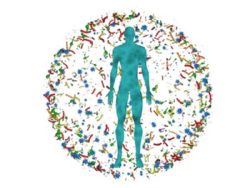
I have been studying thiamine metabolism since 1969 when I published the first case of thiamine dependency: Intermittent cerebellar ataxia associated with hyperpyruvic acidemia, hyperalaninemia, and hyperalaninuria. The case involved a 6-year old boy experiencing recurrent  episodes of cerebellar ataxia (a brain disease resulting in complete loss of a sense of balance). These episodes, occurring  intermittently, were naturally self-limiting without any treatment and were triggered by inoculation, mild head trauma, or a simple infection such as  a cold. In other words, his episodes of ataxia were repeatedly initiated by an environmental factor. I have called each of these variable factors  a “stressor”. Our studies showed that one of these stressors would unmask the true underlying latent thiamine dependency, falsely giving the impression that the stressor was the primary cause. This may be the principle of post vaccination disease in some cases. It may also be too easy to explain symptoms arising from trauma or infection as primary cause. These recurrent ataxic episodes were prevented from occurring by giving him mega-doses of a thiamine supplement.

Cerebellar Ataxia of Metabolic Origins?

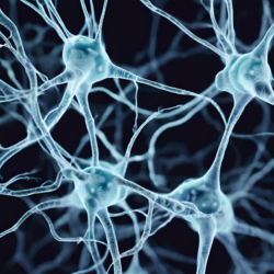
When ataxia, as in this child,  or other symptoms, occur intermittently, as they did in many other patients whom I would treat across my career, it is difficult to identify the true cause. The studies performed by neurologists, neurosurgeons and others inevitably would be  normal, causing diagnostic confusion. In other patients with less serious symptoms, they are considered to be somehow feigned or of psychological origin. Symptoms that appear and disappear in a seemingly random manner and are not supported by conventional laboratory data are often explained this way. Please be aware that ataxia should never be regarded as psychosomatic. The point is that less serious symptoms that cause deviant behavior may not be recognized as biochemical changes in the brain.

With the present medical model, it is difficult to understand and accept that a stress factor can initiate the symptoms of a metabolically caused disease that has been relatively innocuous or silent until the stress is imposed. Let me give you another example.

Loss of Consciousness, Edema, Joint Pain: Rheumatic Disease or Metabolic Disorder

Since I was working at a multi-specialty clinic I was sitting having lunch with an ear, nose, throat (ENT) surgeon who knew of my interest in sudden death in infants (Treatment of threatened SIDS with megadose thiamine hydrochloride). He had been called to put in a tracheostomy to a middle-aged woman who had suddenly stopped breathing. Unlikely as it sounds, he suggested that I should go and look at the situation unofficially.

In the hierarchy of specialization, a pediatrician is not supposed to know anything about adult conditions, so I was not welcome. Because the internists who were taking care of her were rheumatologists, it was considered to be some kind of rheumatic disease, because of aches and pains in joints and limbs. She had had periods of unconsciousness over many years and her body was profoundly swollen, the hallmark of beriberi. Without going into details I was able to prove that this was indeed beriberi.

When I approached the rheumatologist who was her primary physician, I could not convince her of what appeared to her as too bizarre to contemplate. Notwithstanding, I had the cooperation with the nurses who followed my directions.  When the patient was given injections of thiamine, she recovered consciousness and the gross body edema disappeared.

So fixed in the mind of many physicians is the concept that a vitamin related emergency simply does not occur, it was called “spontaneous remission” by my colleagues and “had nothing to do with vitamin therapy”. When I asked the rheumatologist whether we could conference the patient, she ignored the request. Well, this was not the end of the story.

Resolving One Deficiency Often Unmasks Another

After she started the injections of thiamine, with recovery of the nervous system, she began to develop a progressive anemia. It was considered by the internists to be internal bleeding and a thorough search produced only negative results.  So ingrained is the negative attitude to vitamin therapy, I was even in fear that I might be blamed for causing the anemia. In the meantime, I took a specimen of urine and found a substance in the urine that suggested a deficiency of folic acid. Readers will remember that folic acid is a member of the B group of vitamins, as is thiamine. A blood test proved that she was indeed deficient in folic acid. When this vitamin was given to her, the anemia rapidly disappeared. This, believe it or  not, still did not interest my colleagues.

She was discharged from the hospital, receiving supplements of thiamine and folic acid and her nervous system gradually improved. Some months later she developed a rash of a type that had been reported a few months previously as due to vitamin B12 deficiency. She was given an injection of vitamin B12 and over the next few days suffered slight fever and variable joint pains. These were symptoms with which she was familiar and had been responsible for the diagnosis of rheumatic disease.  This sometimes happens temporarily with vitamin therapy, but often enough that I refer to it as “paradox”, meaning that things seem to be worse before they get better. Note that this paradox is not the same as side effects from a drug. The symptoms that cause a patient to see a doctor are temporarily exacerbated. With our present model the patient concludes that this is side effects from the vitamin(s) being used. I had to learn that paradox was the best sign that improvement would follow with persistence. She then continued on the thiamine, folic acid and vitamin B12.

The Role of Lifestyle and Diet Disease Expression – Oft Ignored Stressors

The fact that this woman was a chronic beer drinker and smoker had been ignored.  They were, if you will, the “stressors” that were the dominant cause, perhaps impacting on genetic risk factors. The relationship between alcohol and thiamine deficiency is well known and so she had induced her own disease. Since there was a profound ignorance concerning vitamin deficiency diseases, the beriberi had been referred to by her internists as “rheumatic” in nature. This is because joint and limb pain, usually not recognized for what the pains represent, are often associated with compromised oxidative metabolism, either in the limb itself or in the brain where the pain is interpreted.

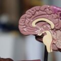
Defective oxidative metabolism caused in this patient’s case by thiamine deficiency, causes exaggerated brain perception. The brain induced a pain that gave the false impression that the disease originated in the joints and other parts of the body. Even if the origin of the pain is truly from a joint or muscle, defective oxidative metabolism in the brain will exaggerate the sense of pain perceived by the patient. Although this “phantom” pain is known as “hyperalgesia”, the mechanism is not well known as being due to compromised oxidation in the pain perception brain centers. Thiamine deficiency was responsible for the hyperalgesia experienced by the case of a patient with eosinophilic esophagitis that was posted recently on this website.

Beyond Thiamine: Multi-Nutrient Deficiencies

What interested me in the woman with beriberi was that folic acid deficiency was not revealed until her metabolism had been accelerated by the pharmacological use of thiamine. The folic acid deficiency then became clinically expressed as her metabolism “woke up”. It had been well known for some time that folic acid produced anemia would have to be treated with both folic acid and vitamin B12.

In the case of folic acid deficient Pernicious Anemia, if vitamin B12 was not given at the same time, the patient would develop a disease known as subacute combined degeneration of the spinal cord. Because I had forgotten this fact, I had neglected to give her vitamin B12 until it was finally expressed clinically in the form of a rash. Associating a skin rash with a vitamin deficiency is certainly not commonly accepted as a possible indicator of an underlying cause by physicians.

Vitamin Deficiency Versus Dependency

Returning to the case of the 6-year old boy discussed above, we learned over time that his health was dependent on high doses of thiamine to function. Believe it or not, this child required 600 mg of thiamine a day in order to prevent his episodes of illness. If he began to notice the beginning of an infection he would double the dose. The recommended daily allowance for thiamine is between one and 1.5 mg a day. Here, and in many other cases, huge doses of the vitamin are required in order to accomplish the physiologic effect. This represents what I call vitamin dependency.

Thiamine and magnesium, like many other vitamins, are known as cofactors to enzymes. An enzyme without its cofactor works inefficiently if it works at all. The “magic” of evolution has “invented” this cooperative action which is in itself under genetic control. In technical terms, the vitamin has to “bond” with the enzyme. If this bonding mechanism is genetically compromised, the concentration of the corresponding cofactor has to be increased enormously by supplementation in order to prevent the inevitable symptoms. You can see that this requires a clinical perspective tied to unusual biochemical knowledge. This is in complete contrast to what is usually regarded as vitamin deficiency, arising from insufficient concentrations in the diet.

What is perhaps not known sufficiently is that prolonged vitamin deficiency appears to affect this bonding mechanism. For example, it has long been known that to cure chronic beriberi, megadoses of thiamine are required for months. I have concluded that the megadoses of thiamine given by supplementation to a patient with long term symptoms arising from unrecognized deficiency appears to re-activate the inefficient enzyme. It is as though the enzyme has to be repeatedly exposed to megadoses of its cofactor to stimulate it and restore its lost function.

This may mean that even if the bonding mechanism is normal in chronic deficiency, enzyme function has simply decayed from lack of stimulation. This may explain why genetically determined dependency and long term dietary deficiency will produce the same clinical effect. The dosing of vitamins, if the clinical effects of deficiency are recognized, is not well understood in traditional western medicine. When insufficient doses are given and the symptoms fail to abate, the practitioner views it as evidence that supplements do not work.

Biochemical Diagnoses are Complex

I want the general public to begin to understand the principles that underlie the complexity of biochemical diagnosis. Perhaps a reader might find that a case like this is a reminder of a loved one whose illness was never understood after seeing many different specialists, all of whom were like the blind men and the elephant. Each was confined to his specialist status but none of them could see the overall big picture.

Reading these cases, you might easily come to the conclusion that they represent a rarity. Chronically unrecognized thiamine deficiency is common. Dependency is  not uncommon. It is not as rare as is presently thought. Believe me, cases like these are surprisingly common and are responsible for a great deal of diagnostic confusion.

Vitamins are essential to consumption of oxygen in all life processes. To go against the principles of diet dictated by Mother Nature is a risk to life and limb that is not worth the derived pleasure. When limb pain is experienced without an obvious trauma, it is difficult to accept that it is because of inefficient use of oxidation in the brain, but that is exactly what we found.

We Need Your Help

More people than ever are reading Hormones Matter, a testament to the need for independent voices in health and medicine. We are not funded and accept limited advertising. Unlike many health sites, we don’t force you to purchase a subscription. We believe health information should be open to all. If you read Hormones Matter, like it, please help support it. Contribute now.

Yes, I would like to support Hormones Matter.

Treating Intractable Insomnia and Cerebellar Ataxia With Thiamine

5512 views

Twenty years ago, I attempted suicide after years of alcohol and drug abuse. I almost succeeded, but by some miracle I survived. I suffered a cerebellar stroke and as result was left with severe cerebellar damage. The doctors said that I had lost almost 75% of my cerebellum. I couldn’t walk or talk, I could not swallow and was barely functioning, if you could call it that. At some point, I decided I would live and forced myself to improve. I worked the physical therapy and eventually began lifting weights. I wrote about my journey here. Despite my gains in strength and balance though, since the stroke I had suffered from severe and intractable insomnia. Although I was under the care of multiple doctors, none could offer any help beyond increasing this or that medication, none of which worked. Insomnia is common in individuals with cerebellar injury. Then I learned about thiamine.

Persistent Insomnia Treated with Thiamine

Around the time my initial article was published, I began supplementing my diet with thiamine. This vitamin may have been mentioned in passing by some of my physicians, but it was never prescribed or even really emphasized to any great degree. Neither was it ever touted by the medical community during my initial stroke recovery in 2003. I am fairly sure much of the reluctance to use thiamine could be attributed to the pervasive fear of it not benefitting my health.

Being entrenched in the world of mental health and alcoholism for about 25 years (sober 9), I am well aware of Wernicke-Korsakoff’s syndrome. I had heard about massive doses of thiamine being administered to others in medical detoxification facilities. I also have loved ones grappling with Parkinson’s Diseases. I quickly began to see how many of our neurological symptoms were similar, and that thiamine was slyly mentioned to help all of us.

In early 2021, it was strongly suggested that I gradually increase to a large dose of thiamine (not by doctors, mind you, but by others from this website). At the time, I was lucky if I slept 3 hours per night, which of course, exacerbated my ataxia symptoms. I was on so many medications that it raised red flags, but I was never warned about the negative neurological effects. And worse yet, nothing was working.

Once my initial dose of thiamine was entrenched in my system (50 mg of Thiamax), I began sleeping with greater ease. At first, this was approximately an hour, but I discovered that I also slept more soundly and had acquired greater rest during the night. As time progressed, my sleep patterns became increasingly regular.

Better Functioning With Improved Sleep

For several years, I have been making progress in all areas of my life. By August of 2022, thanks to the thiamine, I was off of all prescription medications. I was sleeping through the entire night, soundly. I was also able to complete difficult feats at the gym without falling asleep on a mat in the stretching area (usually resulting in me being taken home, so I didn’t sleep at the gym.) This is one of the reasons that I didn’t train publicly until recently. With my sleep problems, I wasn’t sure if I’d be overcome with an insatiable need to sleep mid-workout.

In addition to the improvements in sleep and the elimination of medication after thiamine came on board, I was able to lift a barbell by myself, while standing. This is no small feat for someone with ataxia. Remember, I was told I would not walk again, or function in any semblance perceived as normal. They said that all my neurological systems had crossed the threshold believed salvageable. In September of 2022, I competed in my first powerlifting meet, where I deadlifted 182 pounds and bench pressed 78 pounds. I am currently deadlifting 225 pounds (January 2023). While I am still ataxic and I still have struggles relative to my injury, I have improved so much since beginning the thiamine.

thiamine for insomnia and cerebellar ataxia
Me lifting 225lbs at the gym.

As I write this, right before the New Year of 2023, I am overcome with the amount of remarkable progress that has been made in my life since August. To deny that would fact would be denial of any amount of truth or reality in existence. I fail to see how anyone can deny the differences in my life since using regular thiamine.

We Need Your Help

More people than ever are reading Hormones Matter, a testament to the need for independent voices in health and medicine. We are not funded and accept limited advertising. Unlike many health sites, we don’t force you to purchase a subscription. We believe health information should be open to all. If you read Hormones Matter, and like it, please help support it. Contribute now.

Yes, I would like to support Hormones Matter. 

Photo by Jake Charles on Unsplash.

More Thoughts on Thiamine Deficiency

16146 views

Thiamine Dependency and Intermittent Ataxia

Readers of this blog will recognize that there are many posts concerned with thiamine deficiency and that is associated with a huge number of symptoms. I want to use this post to illustrate the difficulties experienced by a boy whose thiamine deficiency was proved. His case was written up in the medical literature because it was the first example of thiamine “dependency“. That meant that he was required to take massive doses of the vitamin in order to prevent intermittent recurrences of the brain disease known as cerebellar ataxia. Curiously, each episode was self-limiting, but as they recurred, each one left him with a little bit more brain effect. It was this case that forced me to devote myself to library study and change in medical practice throughout the remaining years before my retirement.

Although this boy’s problem was an example of a condition, thought to be extremely rare, it illustrated the kind of medical problems that occur as a result of dietary deficiency. In fact, I have come to the conclusion that prolonged dietary deficiency of this vitamin will make it difficult to restore health by its administration as a supplement. The enzymes in the body that require it seem to degenerate over time if the deficiency persists. Huge doses are required and it is not a simple vitamin replacement. We are using the vitamin as a drug in an attempt to coerce the enzymes back into a healthy state. We know from the history of beriberi, the traditional vitamin B1 deficiency disease, that large doses of the vitamin were required to restore health.

This case was reported in 1969 and the last time that I saw him was in the 70s. He had grown into a handsome boy with a marvelous personality. For example, he cut the grass for the neighbors without charging them and his customers were delighted with him on his paper route. This escape from a potentially lethal disease by the use of what might be called a megadose of vitamin B1 was incredibly impressive to me, making me wonder whether a healthy dose would benefit everyone. Is the modern diet so artificial that vitamin deficiency is common, in spite of their enrichment by the food industry?

I recorded the fact that the family had visited Florida. While they were in a store that was cooler than the outside temperature, it was without air conditioning and John became suddenly unconscious and was taken to an emergency room where his examination proved to be quite normal. On the following day, he went from a 95° temperature into a store with an estimated air-conditioned temperature of 60 to 65°. He immediately experienced difficulty in breathing and produced a kind of strident asthma that was self-limiting. He was again taken to the emergency room where his examination once again proved to be quite normal. The diagnosis given at the time was that it was due to “nerves”. A similar episode occurred on the way home when the air-conditioning was put on in the car. Note that an environmental temperature difference was enough for him to lose consciousness on one occasion and produce asthmatic breathing in another similar situation. He had never been known to experience asthmatic breathing previously. One can readily see that he would be a complete mystery to the doctors in the emergency rooms to which he was taken.

My interpretation would be as follows: as repeatedly pointed out in various posts on this website, his brain had an energy problem from the poor association of thiamine with its energy enabling enzymes and the stress of a sudden change in temperature, requiring an energy surge to adapt, could not be met. The energy deficiency affected the functions of the brain, causing syncope in one example and asthma in another. The diversity of this response illustrates the fact that different parts of the brain can be affected by the overall deficiency, perhaps even on a day to day basis. It is not surprising that such episodes are a diagnostic problem for ER physicians or any physician for that matter.

Syncope and Sudden Death

A very common incident occurring in dietary deficient people is a sudden fainting attack, known as a syncope. They invariably wind up in the ER where it is usually written off as being due to “nerves”. It is impossible to understand this without knowing the chemistry involved. It reminds me of two siblings, a boy and a girl, both of whom had been surviving on a junk-filled diet. The girl was a champion swimmer and was practicing one day by swimming laps. She swam the last lap, touched the pool wall and remained still. When she didn’t start to climb out of the pool, someone investigated and found that she had evidently died as she touched the pool wall. Her brother had been climbing ropes in a gymnasium. After he came down from a climb, he passed out and was taken to a hospital where he received intravenous fluids. It was recorded that he had 11 bloodstained bowel movements and expired. Although there was no proof that thiamine deficiency was the cause of death, I would be willing to bet that the fluid given intravenously to the boy was glucose saline and we know that an excess of sugar will seriously precipitate marginal thiamine to a deficiency state that would produce symptoms. It strongly suggests that the death was from thiamine deficiency.

Febrile Lymphadenopathy

In another post, I recorded the history of two boys with recurrent acute febrile lymphadenopathy (fever, swollen neck glands and high-temperature), both of whom responded completely to thiamine supplementation. The story bears repeating. This kind of illness is inevitably thought of as throat infection and treated with an antibiotic. Both boys had a marked increase of folate and vitamin B 12 in their blood that returned to normal levels after thiamine administration. I won’t go into the mechanism but it is interesting to speculate on how often children with a very common condition like this would have a similar underlying cause that would never, under any circumstances in the climate of modern medicine, be considered. The two cases were published in a medical journal but I have never seen a reference to it, probably because it is totally unbelievable.

I kept a diary at the time and want to make a few comments about one of these boys. When the supplemental thiamine was removed to see what would happen, he relapsed about three weeks later. The relapse began with recurrent abdominal pain, irritability, and a return of the fatigue, causing him to nap during the day as he did when he had his recurrent episodes. There were some abnormalities in his blood pressure, which are too technical to describe here. When I stroked the skin of his leg gently, it provoked a white streak that gradually faded, a phenomenon that is associated with abnormal activity of the autonomic nervous system, a system that is inevitably damaged with thiamine deficiency. He had large lymph nodes in his neck and there was an elevation of folate and vitamin B12 measured in his blood. All of this resolved when the thiamine was restored. It is interesting that he had a first cousin who suffered from Hodgkin’s disease, a malignant form of lymphadenopathy. I wondered whether this recurrent swelling of glands was potentially precancerous. From my reading of the vitamin B1 deficiency disease beriberi, I found that swelling of the glands in the neck could be seen in infants dying from the infantile form of the disease and fever was almost always present.

Mononucleosis: A Mistaken diagnosis?

On May 14, 1976, I made a note that we had a new patient admitted under the care of another physician. He had massively swollen lymph glands in his neck and the diagnosis was mononucleosis. The history recorded that his brother had also “died from mononucleosis” the previous April. A biopsy of one of his glands was reported that it was definitely not malignant. His case was discussed among my pediatric colleagues and I asked the responsible physician what he would do if the lymphocytes in the gland were reported as healthy and mature. The answer surprised me because it was obvious that the diagnosis of mononucleosis had been rejected. He stated that the boy would be treated for cancer “but we would soft-pedal it” I found his answer extremely confounding. We were confronted by a familial situation with an unknown diagnosis and yet he was to be treated as though it was known.

This reminded me of a situation that affected one of my granddaughters. She came home from school and went white water rafting with her friends. Evidently she fell in the water and when she got home she was so fatigued that she went to bed. Her mother, who was a university nurse took her to her workplace where a diagnosis of mononucleosis was made. I persuaded my son to bring her to my office where I gave her several infusions of intravenous vitamins. Her response was so good that she was able to return to school well. The obvious question that I asked myself was whether the diagnosis of mononucleosis was valid or whether it was an inappropriate mistake. The reader may or may not know that a diagnosis of mononucleosis, also known as “kissing disease” is associated with extreme fatigue and can prevent the unfortunate adolescent from returning to school sometimes for months. Once again I am confronted with the question, is our present medical model even close to being accurate?

I am reporting actual cases of thiamine deficiency that are a sampling of literally hundreds of similar cases that I encountered over the years. It should not surprise anybody when I question the current medical model for its accuracy and the use of potentially toxic compounds that often make things worse or do little or nothing toward relieving the disease. It is high time for sick people to seek the services provided by Alternative Medicine physicians whose medical societies are known as the American College for the Advancement of Medicine (ACAM) and the International College of Integrated  Medicine (ICIM).

We Need Your Help

More people than ever are reading Hormones Matter, a testament to the need for independent voices in health and medicine. We are not funded and accept limited advertising. Unlike many health sites, we don’t force you to purchase a subscription. We believe health information should be open to all. If you read Hormones Matter, like it, please help support it. Contribute now.

Yes, I would like to support Hormones Matter. 

Image by Christian Peters from Pixabay.

This article was published originally on September 17, 2020. 

Could the Way You Walk Indicate Mitochondrial Dysfunction?

5031 views

The answer is an unequivocal, yes. The way you walk, your gait, can be diagnostic of mitochondrial disease and dysfunction. Whether your stride is long or short, rhythmic or arrhythmic, quick or slow, symmetrical or asymmetrical, balanced or unbalanced can indicate mitochondrial dysfunction. Even better, the particular pattern of gait disturbance may distinguish between types of mitochondrial disease, at least cursorily, and indicate whether and where there might be neural involvement. This, according to a study published in the Journal of Neurology.

In a small study of 24 patients with genetically confirmed mitochondrial mutations, researchers were able to discriminate between healthy controls and patients and between the two types of mitochondrial mutations assessed (m.3243A>G and m.8344A>G) based upon gait. Both patient groups were selected because of the known associations between those mitochondrial genotypes and gait disturbances. The questions were whether the pattern of gait disturbance could distinguish between the two groups and whether the gait disturbances could be detected early in the disease process before other symptoms fully emerged. That is, could the way patients walked be diagnostic of incipient mitochondrial disease?

About the Mitochondrial Mutations and Patients Tested

The first mutation m.3243A>G (n=18) is associated a disease called MELAS, which stands for mitochondrial encephalopathy, lactic acidosis, stroke like episodes. It is believed to represent one of the more common classes of mitochondrial mutation. The MELAS mutations are associated with a constellation of additional clinical symptoms, including Chronic Progressive External Ophthalmoplegia (CPEO; weakness in the eye muscles causing eyelid drooping), Maternally Inherited Deafness and Diabetes (MIDD), migraine, bowel problems and short stature.

The second mutation, m.8344A>G is associated with a rare mitochondrial disease called MERRF or myoclonic epilepsy with ragged-red fibers. The cardinal symptoms of MERRF include: muscle twitches (myoclonus), weakness (myopathy), and progressive stiffness (spasticity). However, like with MELAS and other mitochondrial diseases, the clinical presentation of symptoms is diverse with the myoclonic seizures developing in only 1 in 5 MERRF individuals. The remainder of patients present with a variety of symptoms including, generalized seizures, ataxia, cognitive decline, hearing loss, eyelid drooping, multiple lipomas (fatty growths or lumps between the skin and muscle), cardiomyopathy, neuropathy, exercise intolerance, increased creatine kinase levels. Individuals with the MERRF mutation may also have increased muscle wasting, respiratory impairment, diabetes, muscle pain, tremor and migraine.

Testing Gait: Walking, Balance, Energy and Strength Disturbances

For this study, researchers looked at five variables associated with gait disturbances:

  • Pace (step velocity and step length)
  • Rhythm (step time)
  • Variability (step length and step time variability)
  • Asymmetry (step time asymmetry)
  • Postural stability (step width, step width variability and step length asymmetry)

The gait testing involved walking on a sensor embedded mat which then calculated the above parameters. Additionally, the researchers assessed:

  • Mutation load with urinary epithelial testing
  • Energy expenditure (a body-worn multi-sensor)
  • Exercise capacity (peak oxygen consumption, heart rate response)
  • Muscle strength (hip flexor ad extensor strength)
  • MRI when available

Results

Compared to the healthy controls, individuals with mitochondrial disease demonstrated significantly reduced gait speed; they walked much more slowly. They also took smaller steps and had increase step time, width and length variability. Individuals with the MERRF mutations were noticeably worse and more globally impaired than those with the MELAS mutations and individuals bearing higher mutation loads and a longer disease trajectory performed most poorly. Universally reduced energy expenditure, exercise capacity and hip flexion and extension strength was observed across both patient groups compared to controls.

One of the more interesting and perhaps unanticipated findings was the association between aspects of gait and cerebellar atrophy. As might be expected, disturbances is balance and symmetry were correlated with cerebellar atrophy. What was interesting is that subtle changes in step width and length variability were observable in individuals with low mutation loads and who otherwise presented with fewer clinical symptoms, suggesting step variability may among the first signs of cerebellar involvement, before full blown ataxia is observed. If this bears out in additional research, walking may become an easy mechanism to test for mitochondrial dysfunction.

Connecting a Few Dots: Medication and Vaccine Induced Mitochondrial Dysfunction

Across many of the patient groups we work with at Hormones Matter, ataxia is a common symptom post medication and vaccine reaction and among individuals with thyroid disease (here and here). Often the ataxia presents with an array of other symptoms associated with mitochondrial disease, seizures, migraines, tremors, GI dysmotility, muscle weakness, neuropathy, to name a few. Since genotyping has not been conducted with these patients, it is not clear whether the medication or vaccine simply unmasked and expedited a latent mitochondrial mutation, triggered a functional mitochondrial deficit with symptoms corresponding with those manifested by more traditional genetic mutations, or some combination of both. Whatever the cause, however, it is becoming increasingly clear that many of the adverse reactions share phenotypes and follow trajectories similar to those associated with mitochondrial disease. Cerebellar involvement, being key among them. Based upon the research cited above, gait disturbances ought to be considered more closely and viewed as a marker of mitochondrial disease or dysfunction, particularly when the constellation of other mitochondrial associated symptoms presents concurrently.

We Need Your Help

More people than ever are reading Hormones Matter, a testament to the need for independent voices in health and medicine. We are not funded and accept limited advertising. Unlike many health sites, we don’t force you to purchase a subscription. We believe health information should be open to all. If you read Hormones Matter, like it, please help support it. Contribute now.

Yes, I would like to support Hormones Matter. 

Image from Pixabay.

This article was posted originally October 13, 2014. 

Adverse Reactions, Hashimoto’s Thyroiditis, Gait, Balance and Tremors

69814 views

One of the things I most love about social media and health research is the opportunity to identify patterns of illness across different patient groups. Here is an example of finding research from one patient group, ThyroidChange, that likely spans many others (Gardasil injured, post Lupron Hashimoto’s, and Fluoroquinolone reactions – to name but a few) and offers clues to a perplexing array of symptoms. The research, is about a little known association between movement and balance disorders and Hashimoto’s thyroiditis: Ataxia associated with Hashimoto’s disease: progressive non-familial adult onset cerebellar degeneration with autoimmune thyroiditis.  Some background.

Hashimoto’s Disease

Hashimoto’s is the most common causes of hypothyroidism afflicting women at a rate of 10 to 1 compared to men. It is an autoimmune disorder in which antibodies attack the thyroid gland and destroy its ability to maintain normal thyroid hormone concentrations. The most common symptoms include: fatigue, muscle pain, weight gain, depression, cognitive difficulties, cold intolerance, leg swelling, constipation, dry skin. If left untreated, goiter – a swollen thyroid gland, appears. If left untreated for an extended period, cardiomyopathy (swelling of the heart muscle), pleural (lung) and pericardial (heart) effusion (fluid), coma and other dangerous conditions develop.

Hashimoto’s and Cerebellar Degeneration

A little known risk in Hashimoto’s is cerebellar degeneration. The cerebellum is the cauliflower looking section at the base of the brain that controls motor coordination – the ability to perform coordinated tasks such as walking, focusing on a visual stimuli and reaching for objects in space. The walking and balance disturbances associated with cerebellar damage or degeneration have a very distinct look, a wide gait, with an inability to walk heel to toe. Cerebellar ataxia looks like this:

In recent years, cerebellar involvement in attention and mood regulation have also been noted. The physicians reporting the Hashimoto’s – ataxia connection present case studies of six patients with Hashimoto’s disease, presumably controlled with medication and a progressive and striking shrinkage of the cerebellum (see report for MRI images) along with progressively debilitating ataxia (walking and balance difficulties) and tremors. Here’s where it becomes interesting.

Hashimoto’s: Medication Adverse Reaction and Misdiagnosis

Hashimoto’s disease is prevalent in our research into medication adverse reactions for Gardasil and Cervarix and Lupron, with some indications it may develop post Fluoroquinolone injury as well. The symptoms are difficult to distinguish from other neurological and neuromuscular diseases such as chronic fatigue syndrome, fibromyalgia, multiple sclerosis and an array of psychiatric conditions, and so Hashimoto’s often goes undiagnosed or is misdiagnosed and mistreated for some time.

Hashimoto’s, Demyelination and Cerebellar Damage

In some of the more severe adverse reactions to medications and vaccines that would lead to Hashimoto’s, the tell tale cerebellar gait disturbances have been noted and documented, along with a specific type of tremor (discussed below).

Research from other groups shows a strong relationship between thyroid function and myelin/demylenation patterns in nerve fibers in animals. Specifically, insufficient T3 concentrations demyelinates nerve axons, while T3 supplementation elicits myelin regrowth. Myelin is the white sheathing, the insulation that protects nerves and improves the electrical conduction of messages in sensory, motor and other neurons. Like co-axial cable in electrical wiring, when the protective sheathing is lost, electrical conductance is disrupted. The early symptoms of a demyelinating disease neuromuscular pain, weakness, sometimes tremors. These can be misdiagnosed as multiple sclerosis, fibromyalgia, chronic pain, when in reality, the culprit is a diseased thyroid gland.

Back to the Cerebellum

The cerebellum is a focal point of white matter axons – myelinated sensory and motor nerves. The cerebellum is where input becomes coordinated into motor movements or movement patterns. White matter damage in the cerebellum causes cerebellar ataxia, the movement and balance disorders displayed above. Hashimoto’s elicits white matter disintegration. Adverse reactions to medications and vaccines can elicit autoimmune Hashimoto’s disease. See the connection?

The Thiamine – Gut Connection

It gets even more interesting when we add another component of systemic medication adverse reactions – nutritional malabsorption, specifically thiamine deficiency. Almost across the board, patients with medication or vaccine adverse reactions report gut disturbances, from leaky gut, to gastroparesis, constipation, pain and a myriad of other GI issues that make eating and then absorbing nutrients difficult. Gut issues are common in thyroid disease too.

As we learn more, and as individuals are tested, severe nutrient deficiencies are noted, in vitamin D, Vitamin B1, B12, Vitamin A, sometimes magnesium, copper and zine. We’ve recently learned of the connections between Vitamin B1 or thiamine deficiency and a set of conditions affecting the autonomic nervous system called dsyautonomia or Postural Orthostatic Tachycardia Syndrome (POTS) linked to thiamine deficiency in the post Gardasil and Cervarix injury group. It may be linked to other injured groups as well, but we do not know yet.

Thiamine and Cell Survival

Thiamine or vitamin B1, is necessary for cellular energy. It is a required co-factor in several enzymatic processes, including glucose metabolism and interestingly enough, myelin production (the Hashimoto’s – cerebellar connection). We can get thiamine only from diet. When diet suffers as in the case of chronic alcoholism, where most of the research on this topic is focused, or when nutritional uptake is impaired, thiamine deficiency ensues. Thiamine deficiency can elicit cell death by three mechanisms:

  1. Mitochondrial dysfunction (reduced energy access) and cell death by necrosis
  2. Programmed cell death – apoptosis
  3. Oxidative stress – the increase in free radicals or decrease in ability to clear them

Thiamine deficiency in and of itself can elicit a host of serious health symptoms. The cell death and disruption of cellular energy balance can be significant and lead to a totally disrupted autonomic system.

Thiamine and Myelin Growth

Add to those symptoms, the fact that thiamine is involved in the growth myelin sheathing around nerves, and we have a whole host of additional neuromuscular symptoms masking as fibromyalgia, multiple sclerosis, chronic fatigue. Like with MS, limb and body tremors are noted in dysautonomic syndromes such as POTS. (Video of POTS tremors, note the uniqueness of the POTS tremor and the similarity between it and the foot tremor shown above along with cerebellar ataxia).

Let thiamine deficiency continue unchecked for period and we get brain damage, as white matter – the myelin disintegrates in the brainstem, the cerebellum and likely continues elsewhere. One of the most prominent areas of damage in thiamine deficiency, is the cerebellum, and hence, the cerebellar ataxia (movement disorders) noted in chronic alcoholics who are thiamine deficient, but also observed post medication or vaccine adverse reaction.

The Double Whammy on Myelin and Cerebellar Function

In the case medication or vaccine adverse reactions, particularly those that reach the systemic level, we have a double whammy on myelin disintegration: from a diseased thyroid gland and a diseased gut. Hashimoto’s and the reduction of thyroid hormones, particularly T3, impairs nerve conduction by shifting from a constant and healthy remyelinating pattern to one of demyelination, while the lack of thiamine further impairs myelin regrowth, because it is a needed co-factor. Both deficiencies affect peripheral nerves, but both also hit the brainstem, the cerebellum and likely other areas within the brain.

Take Home Points

The science of adverse reactions is new and evolving and much of what I am reporting here remains speculative. However, it has become abundantly clear through our research that to address medication adverse reactions or vaccine adverse reactions in a simplistic fashion, by region, or in an organ specific manner, is to miss the broader implications of the compensatory disease processes that ensue. Moreover, to look for symptoms of adverse reactions simply by the drug’s mechanism of action and/or by the standard outcome variables listed in adverse event reporting systems, again misses the complexity of the human physiological response to what the body is perceiving as a toxin. I believe that the entire framework for understanding the body’s negative response to a medication must be shifted to a much broader, multi-system, and indeed, multidisciplinary approach. In the mean time, we will continue to collect data on adverse reactions and offer our readers points of consideration in their quests for healing. I should note, that finding these connections is entirely contingent on the input our community of patients and health activists, both via the personal health stories that so many of you have been willing to share and the data we collect through our research. You know more about your health and illness than we do.

What we Know So Far – Tests to Consider

If you have had an adverse reaction to a medication or vaccine and neuromuscular difficulties, like pain, numbness, motor coordination problems, tremors etc., consider testing for Hashimoto’s thyroiditis. Also, consider thyroid testing when fatigue, depression, mood lability (switching moods), constipation, attentional and focus difficulties are present. In fact, I would consider thyroid testing, specifically for autoimmune thyroid disease like Hashimoto’s, as one of the first disease processes to rule out.

If you have had an adverse reaction to a medication that includes gut disturbances, consider the possibility that you are deficient in key micronutrients such as Vitamin D, the B’s, Vitamin A, magnesium, copper, zinc. And given the modern diet, consider that you were probably borderline deficient even before experiencing the adverse reaction. These nutrients are critically important to health and healing (and no, I do not have an association with vitamin companies or testing companies). Some tests for these nutrients are more accurate than others, so be sure to do your homework first.

If you have symptoms associated with autonomic systems dysregulation such as those associated with POTS, dysautonomia and its various permutations, consider thiamine testing, especially, transkelotase testing.

Share Your Story

If you have experienced a reaction to a medication or vaccine, please share your story in a blog post. Write for us.

We Need Your Help

If you think what we do is worthwhile, contribute to research and our medical reporting at Hormones Matter. We’re totally unfunded at this point and can use your help to continue operations. Yes, I’d like to support Hormones Matter.

Image Source: Pixabay.

Postscript: This article was published originally on Hormones Matter on October 15, 2013. 

Thresholds and Tipping Points in Thiamine Deficiency Syndromes

13957 views

I recently stumbled upon on a research paper published in 1968. It was not that long ago in the overall course of modern medicine, perhaps even its heyday, when all things were still possible and before the complete fealty to pharmaceuticals arrived. To the youngsters and to those coming of age in the last 20 years, however, anything published pre 1990 is ancient history.  What could such old paper tell me about medicine that is new and useful? It turns out an awful lot.

Back in the day, research was a little simpler and more focused, not on finding out which drug could be fit to which symptoms, but on how things worked. Good experimental design, answered mechanical questions, like if we apply X to Y or if we remove X from Y what happens?

In this paper, Encephalopathy of Thiamine Deficiency: Studies of Intracerebral Mechanisms, the researchers identified a very important component about Vitamin B1/thiamine deficiency – the time course of the disease process. That is, with a diet deficient in thiamine, how long does it take before symptoms emerge, what is the corresponding level of deficiency in the brain, and at what point, after supplementation, does recovery begin; important questions clinically.

Vitamin B1 – Thiamine Deficiency

Remember, vitamin B1 or thiamine deficiency at its worst is linked to severe decrements neurological functioning, like Wernicke’s Encephalopathy that include noticeable ataxic and gait disturbances (loss of voluntary control of muscle movements, balance and walking difficulties), aphasias (language comprehension and/or production difficulties), and if it persists, Korsakoff’s Syndrome (severe memory deficits, confabulations and psychosis). Thiamine deficiency was originally observed in only chronic and severe alcoholics or with severe nutritional deficits as seen in famine. Fortification of food stuffs was thought to relieve much of the nutritional risks for deficit, especially in impoverished regions. More recent research, however, indicates that thiamine deficiency has reared its ugly head once again and this time in modern, non-impoverished, regions where the food supply is ample. How can that be?

Non-Alcoholic Wernicke’s Encephalopathies

Thiamine deficits can be mediated by a number of factors, including by less obvious nutritional deficits where food supply is abundant but nutrition is lacking (a diet of highly processed, carbohydrate and fat laden foods), with thiamine blocking factors found in medications/vaccines, environmental toxicants and some foods, after bariatric surgery and in disease processes like AIDS. Over the course of our research, thiamine deficiency has been observed in previously healthy, young, non-alcoholic patients, post medication or vaccine, along with symptoms of dysautonomia.

What has always struck me about the thiamine deficits we observe is the differential expression and time course of the symptoms. In some people, the reaction leading to thiamine deficit appears linear, progressive and rapid. In others, the symptoms appear to wax and wane and to evolve more slowly. How is that possible? Certainly, individual predispositions come into play. Some individuals may be somewhat thiamine deficient prior to the trigger that initiates the full expression of symptoms, while others have higher baseline stores. Additionally, anti-thiamine environmental exposures and other medical conditions/medications may also come into play.  In the literature, however, the progression of symptoms from bad to worse is almost always direct and rapid, perhaps mistakenly so. Indeed, Wernicke’s Encephalopathy is a medical emergency necessitating immediate IV thiamine.  How is it then, that we see more chronic, remit and relapse patterns of thiamine deficiency, even in some cases where thiamine concentrations are being managed medically?

Cerebral Thiamine Deficiency: Crossing the Black Line

It turns out, there is black line with regard to thiamine deficiency, that when crossed overt symptoms emerge, and a similar black line, that demarks recovery. It is possible then that barring a continuous blockade of thiamine, one can move above and below those lines and the corresponding symptoms may wax and wane. The paper from 1968, cited above, found those black lines, in rodents, but we can extrapolate to humans.

The research. The investigators took three groups of female rodents, a paired group of thiamine deprived and thiamine supplemented, along with a group fed ad lib (as desired) and assessed the time course and concentrations of cerebral thiamine deficiency relative to the initiation and progression of the observable neurological symptoms associated with Wernicke’s encephalopathy in rodents (ataxia, loss of righting, opisthotonos –rigid body arching). The experiment lasted about 6 weeks.

Neither the control group (thiamine supplemented) nor the ad lib group demonstrated neurological deficits at any time during the study. The thiamine deprived group, on the other hand, demonstrated symptoms that began with weight loss, progressive anorexia, hair loss (recall our observations about hair loss) and drowsiness at about 2.5 weeks into the experiment. Interestingly, no neurological signs of thiamine deficiency were seen at that time.

The results. At 4.5 weeks in, the researchers noted a rapid progression of symptoms and decline of health over the course of the next 5 days (the black line). These symptoms included: incoordination with walking, impairment of the righting reflex, reluctance to walk, walking backwards in circles, imbalance, rigid posturing and eventually a total loss of righting activity and severe drowsiness.

One can imagine, if a similar deprivation of thiamine were observed in humans, the corresponding symptoms might also include the initial hair loose and weight loss, perhaps noticeable, perhaps not depending upon the time frame and severity of the thiamine deficiency. It would also include incoordination and difficulty with walking, balance and voluntary movement, perhaps tremors, excessive fatigue or sleepiness and the myriad of neuro-cognitive disturbances noted in Wernicke’s syndrome.

In the cited experiment, one injection of thiamine reversed these symptoms to a nearly normal, or apparently normal neurological state within 24 hours.

Brain Thiamine Thresholds

Animals from each of the groups were sacrificed and examined at each of the stages of the experiment. Brain thiamine and other markers of thiamine metabolism were assayed to determine the cutoff levels of thiamine that demark symptoms and recovery.  This is really interesting and the beauty of this entire study.  Neurological symptoms become apparent when cerebral thiamine concentrations reach 20% of normal.  Recovery begins when those concentrations climb to 26% of normal. At least in rodents, one has to deplete 80% of the brain thiamine stores before overt neurological symptoms become apparent; 80% – that is a huge deficit.  Similarly, it doesn’t appear to take much to right that deficit, only a 6% increase in thiamine concentration set the course for improvement.

If we extrapolate to humans, where life span, genetic and environmental factors likely moderate the degree of thiamine stores and consumption, we still contemplate a rather large thiamine deficit needed before overt symptoms of Wernicke’s emerge. Similarly though, it is also evident that a rather small change in thiamine can have enormous effects on neurological functioning. In the case of the rodents, a mere 6% point change reversed the symptoms. One might suspect equivalent deficit/recovery thiamine parameters in humans.

Waxing and Waning Symptoms:  A Case for Persistent Thiamine Deficiency

If we consider the possible course of non-alcoholic thiamine deficiency, where no extraneous variables like bariatric surgery or thiamine deficient parenteral feeding are present and where dietary thiamine varies daily and is not held constant as it is during experimental conditions or during famine, we can begin to see how thiamine related neurological symptoms may wax and wane. Different exposures and triggers may decrease thiamine periodically, even to the point where overt neurological symptoms present. When those exposures are removed and barring deficiencies in metabolism and diet, symptoms may abate, at least temporarily, and until the next trigger or until the black line is crossed anew and thiamine deficiency becomes the medical emergency observed in overt Wernicke’s.

In contrast, the more persistent or chronic thiamine deficits that do not cross the 80% depletion cutoff (or the human equivalent), may also wax and wane and show all the core neurological symptoms expected in overt Wernicke’s though to a much lesser degree. Additionally, as we have speculated, persistent thiamine deficiency might disable mitochondrial functioning in such a way that the patient presents with a myriad of seemingly unrelated symptoms, that are not typically attributed to thiamine deficiency, such as cardiac dysregulation, gastroparesis, autonomic instability, demyelinating syndromes and hormone irregularities, especially thyroid, but also reproductive hormones. These too may be related to thiamine deficiencies. Although, we cannot and should not rule out other causes as well, sub-optimal thiamine may be involved with a host of complex disease states and medication adverse reactions where neurological symptoms are present. Thiamine deficiency should be tested for and ruled out before more invasive therapeutic options are contemplated.

We Need Your Help

More people than ever are reading Hormones Matter, a testament to the need for independent voices in health and medicine. We are not funded and accept limited advertising. Unlike many health sites, we don’t force you to purchase a subscription. We believe health information should be open to all. If you read Hormones Matter, like it, please help support it. Contribute now.

Yes, I would like to support Hormones Matter. 

Cerebellar Ataxia and the HPV Vaccine – Connection and Treatment

6249 views

Anecdotal evidence points to a connection between Gardasil and Cervarix, the HPV vaccines, and cerebellar injury. Here, from the journal Neuropediatrics comes the first published report linking the HPV vaccine to cerebellar ataxia: Association of Acute Cerebellar Ataxia and Human Papilloma Virus Vaccine: A Case Study.

I should note, from our research we’re also seeing cases of cerebellar ataxia post fluoroquinolone reaction and related to Hashimoto’s thyroiditis. The cerebellum appears to be particularly sensitive to insult from environmental toxins – to functional mitochondrial injuries, perhaps because it collects the millions of peripheral nerves coming from the body that control sensation and movement, as they pass to higher brain centers. As such, the cerebellum demands high levels of oxygen and nutrients.

For those of our readers new to neuroanatomy, the cerebellum is the cauliflower looking section at the base of the brain that controls motor coordination – the ability to perform coordinated tasks such as walking, focusing on a visual stimuli and reaching for objects in space. The walking and balance disturbances associated with cerebellar damage or degeneration have a very distinct look, a wide gait, with an inability to walk heal to toe – very much like a drunken sailor. Videos of cerebellar ataxia can be seen here.

The Case Details: Acute Cerebellar Ataxia Post HPV Vaccine

Approximately, two weeks after receiving the HPV vacccine, Cervarix, a previously healthy 12.5 year old girl developed nausea and dizziness with severe cerebellar ataxia, tremors and nystagmus. Initial tests came back normal and she was hospitalized on day 20 post HPV vaccine. Though she could sit on her own, she could not stand or walk unaided and the nystagmus prevented her from focusing on TV, reading or other activities. She had no fever. Heel-knee-shin and finger-nose tests indicated ataxia with terminal intention tremor and dysmetria (see videos: horizontal nystagmus or here for multiple types of nystagmus, heel-knee-shin test, finger-nose test).

All blood tests, cerebral spinal fluid tests and imaging tests were normal, with the exception of testing positive for IgG and varicella zoster virus – chicken pox and shingles – indicating earlier exposure. Tumors, paraneoplastic disease, cardiovascular disease, metabolic conditions and labyrinthitis (inner ear disturbance) were all ruled out. Her symptoms did not remit as was expected with acute cerebellar ataxia.

Treatment Options for Acute Cerebellar Ataxia

Beginning on day 25 post HPV vaccine, pulsed IV methylprednisone (1000mg/d) was administered for three days. Her symptoms persisted. On day 44 post HPV vaccine, IV immunoglobulin (IVIG) at 400mg/kg was initiated and run for 5 days. Her symptoms persisted.

At day 65 post vaccine, with no indication of improvement, immunoadsorption plasmapharesis was begun at a rate of seven times per month. The physicians report a gradual improvement of the nystagmus after two treatments with a full resolution of symptoms after 19 courses of treatment (day 134 post HPV vaccine). The improvement was short-lived, however, and beginning at day 220 post HPV vaccine, the symptoms began to return, gradually at first with nystagmus, and then completely. Immunoadsorption plasmapharesis was begun anew on day 332 post HPV vaccine. After five courses of treatment, the patient’s symptoms again remitted.

Immunoglobulin G (IgG) and Cerebellar Ataxia Symptoms

Of interest, symptom severity corresponded to IgG levels. Her initial IgG levels were not reported, but after 19 treatments, when symptoms disappeared completely for the first time, her IgG levels were 354 mg/dL (day 134). When the symptoms appeared again (day 332) her IgG levels were elevated at 899 mg/dL. Upon treatment, her IgG levels dropped to 503 mg/dL as the nystagmus abated and then to 354 mg/dL upon complete remission, for the second time, at day 332 post HPV vaccine.

HPV16L and Post HPV Vaccine Reactions and Death

The researchers from this study, speculate a connection between the IgG response, and an as of yet, undetermined antibody. Testing for a variety of known antibodies were negative. Since the HPV16L is molecularly  similar to certain cell adhesion molecules, enzymes, transcription factors and neural antigens, it is possible that the HPV16L particles triggered the response.

In separate studies, autopsies of girls who died suddenly post HPV vaccine have found non-degrading HPV16L particles linked to the deaths. In the first case, researchers performed secondary postmortem immunochemistry of two girls who died suddenly after receiving Gardasil. They found evidence of cerebral vasculitis linked to the HPV16L particles throughout the cerebral vasculature.

Similarly, a postmortem exam of another girl who died from the HPV vaccine, found HPV16L DNA particles in the blood and spleen.  The researcher reported that the DNA fragments were found in the macrophages, and protected from degradation because of the tight binding of the HPV16L gene fragments to the aluminum adjuvant. The fragments underwent a conformational change rendering them more ‘stable’ and resistant to degredation, perhaps explaining their presence in the blood and spleen six months post vaccine. This has been contended.

Methods in both of the above studies have been controversial and questioned and should be interpreted with caution. However, researchers from Italy compared HPV16 proteome in the vaccine to the human to proteome and found 84 identical proteins involved in cell differentiation and neurosensory regulation. According to these researchers, the homology between the vaccine and the human proteome, bound to aluminum adjuvant

“make the occurrence of side autoimmune cross-reactions in the human host following HPV16-based vaccination almost unavoidable”.

Whatever the exact culprit, in this case the cerebellar ataxia was acute and temporally related to the HPV vaccine. The favorable response to immunoadsorption and consequent reduction in IgG levels, indicates an auto-immune response.

Mitochondrial Injury, Thyroid, Thiamine and Cerebellar Ataxia

With a more slowly developing cerebellar ataxia and related symptoms, it is possible a medication induced mitochondrial injury, related to a depletion of thiamine is present. Thiamine is critical for mitochondrial function. Similarly, patients have reported cerebellar ataxias related to Hashimoto’s. Generally, when testing for both thiamine deficiency and Hashimoto’s is undertaken, both are confirmed.

Final Thoughts

This report represents one of the first clear linkages between the HPV vaccine and acute cerebellar ataxia. More importantly, it suggests a treatment opportunity when caught early. With so little data available, it is not clear whether immunoadsorption would work for more chronic cases. However, there is evidence of its success in Guillian Barre, Myasthenia Gravis and other autoimmune conditions. When combined with the early data pointing to Hashimoto’s and thiamine deficiency, paths forward post injury are emerging.

Participate in Research

Hormones MatterTM is conducting research on the side effects and adverse events associated with Gardasil and its counterpart Cervarix. If you or your daughter has had either HPV vaccine, please take this important survey. The Gardasil Cervarix HPV Vaccine Survey.

We are also conducting research adverse reactions associated with the fluoroquinolone antibiotics, Cipro, Levaquin and Avelox: The Fluoroquinolone Antibiotics Side Effects Study.

To take one of our other surveys, click here.

To sign up for our newsletter and receive weekly updates on the latest research news, click here.

What Else Can I Do To Help?

Hormones MatterTM is completely unfunded at this juncture and we rely entirely on crowdsourcing and volunteers to conduct the research and produce quality health education materials for the public. If you’d like help us improve healthcare with better data, get involved. Become an advocate, spread the word about our site, our research and our mission. Suggest a study. Share a study. Join our team. Write for us. Partner with us. Help us grow. For more information contact us at: info@hormonesmatter.com.

To support Hormones Matter and our research projects – Crowdfund Us.