demyelination

Progesterone for Peripheral Neuropathy

76639 views

Some 20 years ago, during my very first neuro class taught by an accomplished neurologist from a prominent research university, I had a conversation about hormones and the brain. It was a brief conversation during which he admitted not only knowing nothing about how hormones affected the brain or nervous system functioning, but also, how he and others had no interest in considering the question. He believed hormones were too complicated to consider relevant. One didn’t ‘mess with hormones’ as he put it.

Lucky for us, some intrepid neurologists have moved the science of neuroendocrinology past the foibles of ‘don’t mess with hormones’ to hormones might be important therapeutic options. Nowhere is this more evident than in the areas of traumatic brain injury and diseases of demyelination. Here we see advances in hormones used as viable and important treatments where once there were none. Although the research is yet in its infancy and suffers from the typical one-size-fits-all approach, it marks a huge step forward in clinical neuroendocrinology.

What is Neuropathic Pain?

Neuropathic pain, the often chronic and difficult to treat pain that comes from nerve injury and demyelination affects approximately 3% of the population. The number of individuals suffering from neuropathy is likely much higher when one considers diseases such as endometriosis and the ill-understood, under-recognized neuropathy emerging post medication or vaccine adverse reactions. The experience of neuropathic pain in hands, feet, arms and legs is described as burning, freezing, electrical, tingling, prickling and more often than not, severe and unrelenting. As the nerve injury progresses and the pain continues, the rawness and intensity of the pain becomes indescribable to someone who has not experienced nerve pain firsthand.

Hormones and the Nervous System

Since the late eighties, researchers have known that steroid hormones, such as progesterone were not limited to reproductive functions and that many steroids were active in the nervous system. Not only were those steroids synthesized peripherally in ovaries, adrenals or adipose tissue able to cross the blood brain barrier, but all the core substrates for steroid synthesis were available in the brain too, meaning the brain could make its own steroids, de novo, from scratch. Researchers initially deemed steroids made or active in the brain as neurosteroids. Eventually, that nomenclature fell by the wayside as researchers realized there was tremendous crosstalk between peripheral and central hormones, no matter where the hormones were synthesized.

It should be noted, that hormones exert influence all over the body and brain via receptor binding. (A discussion on hormones and receptors can be found here: Promiscuous Hormones and Other Fun Facts.) In addition to steroid hormone receptors on (cell membrane) and in (nuclear) hormone-specific cells, like those in ovary, testes, adrenals, uterus, endometrium, hormone receptors are co-located on neurons, glial cells, oligodendrocytes and Schwann cells (myelin producing cells), immune cells, cardiomyocytes (heart), hepatocytes (liver), adipocytes – essentially every cell, organ or tissue in our body is modulated in some way by a hormone. Hormone influence is particularly important in the in the nervous system, where everything from neurotransmitter release and uptake to synaptic connections are modulated.

Traumatic Brain Injury, Peripheral Neuropathy and Hormones

When we talk about injuries to the nervous system, be it the brain and spinal cord, which is called the central nervous system (CNS), or all of the nerves that control movement and organ function in the body, the peripheral nervous system (PNS), there are two categories of injuries, those that develop acutely, post trauma, or those that develop chronically because of some metabolic dysfunction. In the case of the former, traumatic brain injuries (TBI) or  traumatic nerve injuries, the research points to progesterone for repair and regrowth. In the case of the later, where injuries develop as a result of internal and often chronic dysfunction, such as diabetic neuropathy, multiple sclerosis and other diseases affecting nerve fibers and myelin, less is known about progesterone and the thyroid hormone triiodothyronine (T3) is implicated, more strongly.

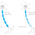
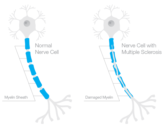
What is Myelin and How Does it Impact Neuropathy?

Myelin is the insulation that protects the axons of the neuron (in the brain) or nerve (in the body) to allow rapid conduction or messaging across the brain or through the body. Recall the axon is the part of the neuron/nerve that sends messages to other neurons/nerves, to other tissues, like muscle, or to organs like the heart and the liver. The dendrites receive messages and the nucleus processes messages. Myelin is like the plastic coating around the electrical wiring in your house. If the coating is too thick, conduction is blocked. If the coating is frayed or too thin, electrical sparks fly everywhere. Frayed myelin around axons is one of the mechanisms of neuropathic pain. Myelinated axons in the brain look white and therefore are called white matter. Whereas the grey matter, is where the nuclei of the brain reside. White matter in the brain consists of the oligodendrocytes – the type of cell that forms the myelin sheathing around axons. Myelin in the body, around the peripheral nerves, is made from cells called Schwann cells.

Progesterone, Myelination and the Nervous System

In the 1990s, Etienne-Emil Baulieu and colleagues recognized a role for progesterone (and other hormones) in central nervous system myelination. Over the next two decades, researchers uncovered the possible mechanisms and delineated more clearly for whom and in what types of injury progesterone seems most helpful. From studies of neurons (CNS) nerve cells (PNS), we now know that progesterone is key for myelination and neuron/nerve regrowth, at least in the acute stages. Progesterone stimulates myelination both directly by acting on oligodendrocytes and indirectly via actions on the neurons and the astrocytes that then message the oligodendrocytes to produce more myelin. Similarly in the PNS, progesterone aids in the remyelination and re-growth of nerve fibers, via the Schwann cells and via progesterone receptors located in what are called the dorsal root ganglia (DRG), the sensory neurons that carry information from the periphery to the brain. Whether in the CNS or the PNS, timing and length of progesterone administration are critical.

Animal Research – Progesterone, Nerve Injury and Neuropathy

The animal research has been mixed, but taken together, the results seem dependent upon the type of injury, the timing of the treatment and the methods of assessment. When treatment is begun early enough and extended long enough (this varies) and when the measure is neuropathic pain versus other potential outcomes (such as morphological changes to the nerve), there seems to be a favorable response. In rodents, single dose treatment does not seem to work, neither does treatment that is initiated too late after the injury or ended prematurely, though these criteria vary from study to study.

For example, using an induced model of diabetic neuropathy, researchers from Italy found that diabetes markedly reduced progesterone concentrations in male rodents within three months (females were not tested). This was the only study I could find that measured progesterone concentrations relative to treatment and outcomes. Chronic treatment (one month) with progesterone or one of its derivatives restored nerve function, increased key components of myelin production and reduced pain. Similarly, an induced model of trigeminal pain in male rodents found when progesterone was initiated early and at a high enough dosage, it tempered the experience of pain while increasing myelin producing proteins. Lower dosages did not work.

From Animals to Humans: Traumatic Brain Injury and Neuropathy

The research with animals, male rodents specifically, shows that progesterone treatment works best if given early enough, for long enough, and at high enough dosages. With acute or induced injuries under experimental conditions, early treatment is much easier than in real life where neuropathic pain develops much more gradually and often goes undiagnosed and untreated for some time. Would progesterone work in humans and would it work for chronic, well established neuropathy? The answers to those questions are not clear because the human research on progesterone and myelin focuses on acute injury, like the traumatic brain injuries. The human research also suffers from short duration dosing, includes mostly males, and without exception fails to address endogenous progesterone concentrations either pre or post treatment. Nevertheless, there are some indications that progesterone therapy may work.

Progesterone and TBI – Human Studies

In a smaller, single center open trial and two larger, double-blind, placebo-controlled, human trials, progesterone therapy was administered to individuals with severe traumatic brain injuries (Glasgow coma scale <8). In each case, the progesterone group did better, showed reduced morbidity rates than the placebo groups.

In the first study, 26 cases were treated with progesterone and 20 controls with placebo. At both 10 days and three months post injury and treatment, the progesterone treated group improved significantly more than the control group (abstract only).

In a second study, 159 patients, arriving to the treatment facility just eight hours post traumatic brain injury were randomized to receive either intramuscular injections of progesterone (82) or placebo at 1.0 mg/kg via intramuscular injection and then once per 12 hours for 5 consecutive days. Both intake neurological functioning and post treatment functioning were assessed and compared using a number of measures. Follow up assessment was conducted at 3 and 6 months post injury/treatment. The results were positive, albeit small. The progesterone treated group improved significantly across all measures showing consistently larger improvements compared to the placebo group. It should be noted that only 44 of the total subject population was female, 24 in the placebo group and 20 in the progesterone group. No analysis by sex was conducted and so it is not clear whether progesterone therapy works equally well in males and females.

In the third study, called ProTECT, a similar double-blind, placebo controlled, randomized methodology was used. Here, however, the randomization was 4:1 and favored progesterone treatment, whereas in the study cited above, the progesterone and placebo randomization was 1:1. Progesterone was given via IV for three days. The ProTECT study researchers found that patients in the progesterone had a lower 30-day mortality rate than controls (rate ratio 0.43; 95% confidence interval 0.18 to 0.99). While those who suffered more severe injuries had relatively poor outcomes at the follow up tests 30 days post injury, despite the treatment, and those who suffered only moderate traumatic brain injury and received progesterone were more likely to have a moderate to good outcome than those randomized to placebo (abstract only).

Two additional trials are on-going, hoping to test progesterone on thousands of patients: the ProTECT-III and SynAPSe studies.

Translating the TBI Research for Use with Neuropathy

What does improvement post TBI tell us about treating neuropathic pain from demyelination disorders? It is not clear, because even though researchers know that progesterone promotes myelination, the human research has focused narrowly on injuries where demyelination occurs but also where other factors are also involved in the outcome. We know from animal and cell culture research that progesterone attenuates the cascade of events that occur post TBI or post nerve injury via multiple mechanisms, inducing myelin regrowth is only one of those mechanisms. Progesterone reduces swelling of both vasogenic and cytotoxic sorts. It has anti-oxidant properties, upregulating enzymes that increase free radical elimination. Progesterone inhibits inflammation, stabilizes mitochondria, reduces neural excitoxicity and can limit apoptosis. Finally, progesterone promotes myelination. All factors that should point to consistent improvement in TBI and neuropathic pain syndromes, but the research is limited and mixed. Why?

The primary reason for mixed results is study design, almost all are short duration. Hormones are long acting molecules and the shorter duration may not be sufficient to generate the response, particularly when the injuries are severe or longstanding. Longer treatment regimes are likely in order.

Another reason for mixed results is the one-size-fits-all approach. None of the human studies and few of the animal studies, investigates why progesterone works in some subjects and not others. Almost all of the studies are predominantly male, rodent and human alike. None have investigated whether being female has anything to do with efficacy. None of the human studies measured circulating concentrations of progesterone, either pre-, during, or post-treatment and so there is no way to tell if those who responded had higher circulating concentrations or if improvement was contingent upon reaching a certain concentration.

Perhaps even more importantly, is the fact that progesterone, like any hormone, works within a vast and compensatory network of other hormones. The reductionist approach that utilizes a single hormone treatment protocol, while ignoring the potential cross-talk with other hormones and other variables is a consistent flaw these and other research protocols. Again, hormone measurement, progesterone and its metabolites, in addition to other key hormones, is imperative if one is to determine therapeutic efficacy.

I Have Peripheral Neuropathy, Should I Try Progesterone?

Progesterone therapy is generally safe, but as with everything there are risks. Women have been using it for generations in its bio-identical form to mitigate menstrual and menopausal symptoms. Since it is fat soluble, transdermal (skin) absorption is possible and progesterone creams have become popular. Some physicians prefer micronized progesterone, a pill form that reduces the molecule so it more easily passes through the liver without degradation. The pill form, and to a much lesser degree, transdermal progesterone, cause sedation and should be taken at night. Micronized progesterone has been shown to increase free thyroxine (T4) as well. For some women, and presumably men too, a gain of function mutation on the mineralocorticoid receptor can evoke very high blood pressure with any increase in progesterone concentrations (luteal phase of the menstrual cycle and during pregnancy especially). Although there are dosing references for progesterone relative to menstrual or menopausal therapy, the dosing is individualized and often includes the replacement of other hormones along with progesterone. Salivary hormone testing is used to monitor and hormone doses are adjusted regularly. Progesterone is also used predominantly for women. No such dosing considerations exist for men that I am aware of. Likewise, for peripheral neuropathy there are no references from which to design a treatment protocol and so it would be prudent to work with a functional medicine specialist, familiar with hormone management, to develop and monitor the course of treatment.

My Two Cents

I suspect, if progesterone therapy works for peripheral neuropathy, it will require a much longer term treatment period than is currently tested in the human trials. I suspect also, it will be difficult to ascertain whether it is the sole contributor to improvements in neuropathy symptoms, as neuropathy is a multi-factorial process that ought to be treated as such. Nevertheless, if you suffer from neuropathy and can find a physician to work with that is familiar with hormones and the research, progesterone therapy might provide a viable option, among other options like stabilizing thyroid hormones and supporting mitochondrial function.

Postscript

This article was first published February 19, 2014. Since then, a few more studies and review articles have been published and continue to support the role of progesterone in myelin regeneration, although the data are mixed. From a 2020 article.

Indeed, PROG and its metabolites modulate the expression of myelin proteins of the PNS, such as myelin basic protein (MBP), myelin proteolipid protein, glycoprotein zero (P0) and peripheral myelin protein (PMP22) as well as myelin formation [6,8,9,20,63,64]. In particular, the expression of P0 in the sciatic nerve of adult male rats, as well as that in Schwann cell culture, is increased by treatment with PROG, DHP or THP.

…Data here reported support the concept that neuroactive steroids, synthetic ligands acting on their receptors or inducing their synthesis, may improve PN symptoms, including neuropathic pain and consequently may represent an interesting possible therapeutic strategy. In addition, based on the sexual dimorphism of neuroactive steroids as well as of PN here discussed, a gender specific treatment based on these compounds may be also proposed.

We Need Your Help

More people than ever are reading Hormones Matter, a testament to the need for independent voices in health and medicine. We are not funded and accept limited advertising. Unlike many health sites, we don’t force you to purchase a subscription. We believe health information should be open to all. If you read Hormones Matter, like it, please help support it. Contribute now.

Yes, I would like to support Hormones Matter. 

This article was published originally on February 19, 2014. 

Vitamin D’s Role in Preventing and Treating Multiple Sclerosis

6971 views

Modern lifestyles are countering nature’s intentions to keep us healthy. Since the advent of the Industrial Revolution in the late 18th century, we have migrated from farms to factories and office buildings. Nature intended for us to live and work outdoors in the sun—without sunscreen. Today most of us live and work indoors—often wearing sunscreen, or cosmetics containing sunscreen. By doing so, we have denied our bodies one of the most fundamental sources of health: the ultraviolet B (UVB) rays of the sun that initiate vitamin D protection in our skin.

Compelling scientific evidence over the past century indicates the significant role vitamin D plays in protecting us from developing a wide variety of medical conditions including autism, autoimmune disorders, cancer, cardiovascular disease, diabetes, and thyroid disorders. It is not a coincidence that the prevalence of these diseases has emerged during “modern” times. These medical conditions, many of which are serious, chronic, and life-threatening, frequently result in health, financial, and social burdens to the patients and their families.

What is Multiple Sclerosis?

Multiple sclerosis (MS) befits a disease of modern civilization. First identified by French neurologist Jean-Martin Charcot in 1868, MS is a chronic, neurological autoimmune disorder that damages the myelin sheath, the multiple layers of fatty tissue that surround and protect the nerves in the brain, spinal cord, and optic nerves. When the myelin sheath is intact, electrical impulses are carried through the nerves with accuracy and speed. When the myelin sheath is damaged (sclerosis is the scar tissue formed by damaged myelin), the nerves do not conduct electrical impulses normally. The impulses are distorted or interrupted, resulting in a range of symptoms including numbness, blindness, paralysis, and brain damage. MS also can result in death.

Who is at Risk of Developing MS?

Despite the identification of MS almost 150 years ago, MS has no cure. Over 2.5 million people around the world have been diagnosed with MS including about 400,000 Americans. Women are to two to three times more likely to develop MS than men. Although MS is usually diagnosed between the ages of 20 and 50, the disease can strike at any age. In addition, Caucasian women of Northern European descent are more frequently diagnosed with MS than African Americans, Hispanics, and Asians.

As part of the Environmental Risk Factors in MS Study (EnvIMS), researchers at the University of Bergen in Norway sought to understand better the association between MS and sun exposure measures by studying a total of 1,660 MS patients and 3,050 controls from Norway and Italy. The researchers’ findings included significant connections between infrequent summer outdoor activity and sunscreen use and an increased risk of MS. Published in the January 10, 2014 issue of Multiple Sclerosis, the study’s conclusion stated, “Converging evidence from different measures underlines the beneficial effect of sun exposure on MS risk.”

It is not surprising that incidences of MS in the equatorial region occur much less frequently than at the higher latitudes. Epidemiological studies over the past several decades however indicate that women who live at higher latitudes have an increased risk of developing MS. For example, University of Oxford researchers studied MS patterns in Scotland by examining hospital admissions throughout the country between 1997 and 2009. The research team discovered a “highly significant relationship between MS-patient-linked admissions and latitude” across Scotland. This study was published in a 2011 issue of the Public Library of Science (PLoS) One journal.

In addition, a seasonal risk factor also exists for MS. Researchers at Queen Mary University of London conducted a systematic review of data for 151,978 MS patients to ascertain the link between month and location of birth, and the risk of developing MS. They found that babies born in April had the highest risk of development of MS, and infants born in October enjoyed the lowest risk of MS. The researchers also noted a direct correlation between the latitudinal location of expectant mothers and MS risk. The study, published in a 2012 issue of the Journal of Neurology, Neurosurgery, and Psychiatry, suggests the importance of maternal vitamin D supplementation in particular during the winter season.

What Causes MS?

The definitive cause of MS remains unknown but medical research suggests genetic and environmental factors influence one’s risk of developing MS. Interestingly enough, science has demonstrated that vitamin D plays a role in influencing environmental and genetic factors that may affect how likely one is to develop MS.

A landmark study at the University of Oxford, published in a 2009 issue of Public Library of Science (PLoS) Genetics, examined how genes and the environment interact in MS. A gene variant called HLA-DRB*1501 is associated with an increased risk of developing MS. The research team discovered how vitamin D influences the HLA-DRB*1501 gene variant. As we know, the amount of vitamin D synthesized by UVB sunlight exposure fluctuates from season to season. Therefore, women who give birth during the spring, carry the HLA-DRB*1501 gene variant, and have low vitamin D levels are more likely to produce children with a higher risk of developing MS.

The study’s author Dr. Sreeram Ramagopalan suggested that adequate vitamin D3 supplementation during pregnancies may decrease the risk of children developing MS in later life. The combination of carrying the HLA-DRB*1501 gene variant and lacking adequate vitamin D levels may impair the ability of the thymus, an immune system organ, to delete rogue T cells, a type of white blood cells, that play an important role in maximizing the immune cells. The rogue cells would attack the body, causing demyelination of the central nervous system.

How Can Vitamin D Protect Against MS?

MS is a neurological autoimmune disorder. Scientific research over the past few decades solidifies the connection between vitamin D and autoimmunity. Vitamin D plays an integral role in the regulation of the adaptive immune system.

Adequate vitamin D in our bodies can protect us from autoimmunity because adaptive immune cells contain vitamin D receptors (VDRs). These receptors are attached to the surface of the adaptive immune system’s antibodies and sensitized lymphocytes. When the VDRs receive adequate amounts of vitamin D, they enable the adaptive immune system to function properly by attacking new and previous invaders.

When the VDRs attached to the adaptive immune system’s cells do not contain sufficient vitamin D to attack invaders, autoimmunity may kick in, causing the death of healthy immune cells. Thus, vitamin D deficiency can contribute to the development of autoimmune disorders such as MS.

How Can Vitamin D Treat MS?

The scientific community is delivering hope to MS patients by investigating vitamin D intake as a treatment for the disease. Research suggests that higher vitamin D levels are associated with reduced disease activity in MS sufferers.

Dr. Alberto Ascherio of Harvard University’s School of Public Health and colleagues recently concluded that vitamin D appears to be connected with MS disease activity and progression in patients who experienced an initial episode suggestive of MS and were treated with interferon β-1b. The researchers found that 20 ng/mL-increases of vitamin D levels within the first 12 months of experiencing an initial episode predicted a 57 percent lower rate of new active lesions as well as a lower risk of relapse. In addition, the results included a 25 percent decrease in annual T2 brain lesion volume and a 0.41 percent lower yearly loss in brain volume over four years. The Harvard study was electronically published on January 20, 2014 in JAMA Neurology.

According to a study published in a 2012 issue of the Annals of Neurology, a University of California, San Francisco research team examined 469 male and female MS patients over five years to ascertain how vitamin D affected disease progression. The researchers discovered that for each increase of 10 ng/mL in vitamin D levels, the MS patients benefited from a corresponding 15 percent decrease in new brain lesions as well as a 32 percent lower risk in inflammation of the myelin sheath.

A Finnish study, published in a 2012 issue of the Journal of Neurology, Neurosurgery, and Psychiatry, concluded that vitamin D3 supplementation significantly reduced the number of brain lesions in MS patients undergoing interferon β-1b treatment.

Paving a Way to Better Health and Quality of Life

Adequate vitamin D levels in our body may indeed protect us from developing MS. If you have experienced a possible initial episode or have been diagnosed with MS, please consider how vitamin D3 supplementation may decrease the severity of your symptoms.

We must take ownership of our health by understanding the importance of vitamin D as well as other micronutrients. Why wait years, or decades, to garner the results of further studies and clinical trials to define the exact relationship between vitamin D status and MS. We can be proactive by taking daily vitamin D3 supplements and enjoying moderate sunlight exposure to increase our vitamin D levels.

It is imperative to take enough vitamin D so this essential nutrient will be stored in your cells to help regulate your immune system. The greater your vitamin D level (easily obtained from a simple blood test called 25(OH) vitamin D), the more likely you will benefit from a stronger immune system that will protect your body’s cells from attacking one another.

No one wants to endure the health, financial, and social burdens of a chronic debilitating disease. By empowering yourself with adequate vitamin D, you may not only reap lots of health benefits but enjoy a better quality of life.

We Need Your Help

More people than ever are reading Hormones Matter, a testament to the need for independent voices in health and medicine. We are not funded and accept limited advertising. Unlike many health sites, we don’t force you to purchase a subscription. We believe health information should be open to all. If you read Hormones Matter, like it, please help support it. Contribute now.

Yes, I would like to support Hormones Matter. 

To Learn More


Image credit: Stephanie021299, CC BY-SA 4.0, via Wikimedia Commons
This post was published here originally on March 4, 2014. 

Copyright © 2014 by Susan Rex Ryan. All rights reserved.

Vitamin D Plays an Integral Role in Adaptive Immunity

6315 views

Severe Adverse Reactions Include Vitamin D Deficiency and Autoimmunity

Hormones Matter researchers discovered that, inter alia, severe adverse reactions to any of the surveyed drugs trigger significant but varying autoimmune responses. Moreover, the research revealed an underlying consistency involving all reviewed drugs: vitamin D deficiency.

Vitamin D Helps Regulate the Adaptive Immune System

The adaptive immune system comprises the body’s intricate network of antibodies and special types of white blood cells (called sensitized lymphocytes ) to thwart new and previous invaders including viruses, bacteria, and drugs. When the adaptive immune system is not strong enough to endure external disruptions such as severe side effects of drugs, it can go awry by signaling antibodies and sensitized lymphocytes to attack healthy cells. This response is called autoimmunity—when the adaptive immune system’s cells do not recognize previous invaders and designate healthy cells as those invaders. In other words, the body’s immune cells attack its own healthy cells.

Scientific research over the past three decades solidifies the connection between vitamin D and autoimmunity. Vitamin D plays an integral role in the regulation of the adaptive immune system. Adequate vitamin D in our bodies can protect us from autoimmunity because adaptive immune cells contain vitamin D receptors (VDRs). These receptors are attached to the surface of the adaptive immune system’s antibodies and sensitized lymphocytes. The VDRs act as “gate keepers” by signaling what external substances, e.g., components of medications, can enter a cell. The VDRs must be replete with vitamin D to effectively regulate adaptive immunity. When the VDRs receive adequate amounts of vitamin D, they enable the adaptive immune system to function properly by attacking new and previous invaders.

When the VDRs attached to the adaptive immune system’s cells do not contain sufficient vitamin D to attack invaders, autoimmunity may kick in, causing the death of healthy immune cells. Thus, low vitamin D levels can lead to autoimmune diseases including thyroid disorders such as Hashimoto’s and demyelinating diseases including multiple sclerosis (MS).

Vitamin D and Hashimoto’s Autoimmune Thyroid Disease

The Real Women, Real Data research also uncovered another consistency among severe adverse reactions to the reviewed drugs: Hashimoto’s thyroiditis, an autoimmune disease caused by abnormal cells constantly assaulting the thyroid gland
.
Vitamin D receptors are present in the thyroid as well as the pituitary, the pea-shaped gland that controls the thyroid. Not surprisingly, low levels of serum vitamin D have been linked to Hashimoto’s thyroiditis, according to recent Turkish medical research:

Published in a 2013 issue of the journal Endocrine Practice, a study conducted at a training and research hospital in Ankara demonstrated that serum vitamin D levels of female chronic Hashimoto’s patients were significantly lower than healthy subjects. Furthermore, the researchers discovered a direct correlation between serum vitamin D levels and thyroid volume as well as an inverse correlation to the antibodies involved in the thyroid.

Researchers at Medeniyet University’s Goztepe Education and Research Hospital in Istanbul learned that 92 per cent of their 161 Hashimoto’s thyroiditis cases had serum vitamin D levels lower than 30 ng/mL (12 nmol/L), a value characterized as “insufficient.” Published in a 2011 issue of the journal Thyroid, the study reports an association between vitamin D insufficiency and Hashimoto’s thyroiditis.

Vitamin D and Demyelinating Disorders

Another disturbing outcome of the Real Woman, Real Data research is the reporting of neurological and neuromuscular symptoms, many which of are consistent with demyelinating disorders such as MS, an autoimmune disease. The development of MS occurs when a poorly functioning, adaptive immune system gradually attacks the protective covering of the nerve cells (called the myelin sheath) of the brain and spinal cord. This potentially debilitating process is called demyelination.

Scientific—primarily epidemiological—research indicates an association between vitamin D levels and the risk of developing a demyelinating disorder such as MS. VDRs exist on nerve cells and the myelin sheath. When the VDRs receive adequate amounts of vitamin D, they help protect the integrity of the myelin sheath. However, when the VDRs do not contain sufficient vitamin D, autoimmunity may occur, resulting in the death of healthy nerve cells. Numerous clinical trials are underway to assess the connection between vitamin D status and the likelihood of developing demyelinating disorders.

Low Vitamin D: The Chicken or the Egg?

The connection between low vitamin D status and the development of autoimmune disease is genuine. However, medical research has not yet determined if vitamin D deficiency plays a role in the development of autoimmune disease, if low vitamin D levels are a consequence of the disease itself, or if vitamin D deficiency acts as both a cause and effect. The authors of the aforementioned 2013 Hashimoto’s study concluded,

“Finally, our results suggested that there may be a causal relation between vitamin D deficiency and development of Hashimoto’s thyroiditis. On the other hand, there might be a possible relation between severity of vitamin D deficiency and progression of thyroid damage. However, further studies are needed especially about the effects of vitamin D supplementation on prevention and/or progression of autoimmune thyroid disease.”

Proactive Protection against Severe Adverse Reactions

We could wait years (or decades) to garner the results of further scientific studies and clinical trials to define the exact relationship between vitamin status and severe adverse reactions to vaccines and medications that culminate in autoimmune disorders. Or we could be proactive by taking daily vitamin supplements and enjoying moderate sunlight exposure to increase our vitamin D levels.

It is imperative to take enough vitamin D3 so this essential nutrient will be stored in your cells to help regulate your immune system. The greater your serum vitamin D level (easily obtained from a simple blood test called 25(OH) D, the more likely you will benefit from a stronger immune system that protects your body’s cells from attacking one another.

No one wants to endure severe adverse reactions to drugs such as Gardasil and Lupron, let alone an autoimmune disease. Attaining and maintaining adequate supplementation provides a safe, easy, and inexpensive approach to improved preventive health. By empowering yourself with adequate vitamin D, you may reap the benefits of avoiding disease and enjoying better quality of life.

Copyright © 2013 by Susan Rex Ryan. All rights reserved.

This article was published previously on Hormone Matter in September 2013.

We need your help.

Hormones Matter needs funding now. Our research funding was cut recently and because of our commitment to independent health research and journalism unbiased by commercial interests, we allow minimal advertising on the site. That means all funding must come from you, our readers. Don’t let Hormones Matter die.

Yes, I’d like to support Hormones Matter.

Anti-NMDAR Encephalitis and Ovarian Teratomas

4914 views

In 2005 researchers began documenting the existence of a new form of encephalitis, a brain disease that afflicts predominantly young women (80%) and children and attacks a critical set of brain receptors, the N-methyl-D-aspatate receptors (NMDAR). The disease, called Anti-NMDAR Encephalitis, produces a syndrome that over the course of several weeks to months progresses from flu-like symptoms, to psychosis, to catatonia, to the ICU and the need for mechanical ventilation. It is treatable, when identified in a timely manner, but because of the physiological importance of the receptor it attacks, if not treated in time or treated sufficiently, anti-NMDAR encephalitis can be fatal. Interestingly, there is an important connection to ovarian health that makes this disease process particularly relevant to women – 60% of the cases present with ovarian teratomas.

NMDA Receptors and Brain Function

NMDA receptors are the brain’s and the indeed the body’s primary mechanism through which activity is initiated. NMDARs are excitatory receptors that bind with glutamate, the excitatory neurotransmitter. NMDARs, along with the AMPA receptor (α-amino-3-hydroxy-5-methyl-4-isoxazolepropionic acid), a secondary excitatory receptor regulate all brain excitation. These receptors are located all over the brain, but are especially dense in the hippocampus, where learning and memory occur, and the frontal and prefrontal regions where planning, motivation, impulse control and emotional regulation take place.

NMDARs are also dense in subcortical regions, where all sorts of functions involving movement control, motivation and emotionality are controlled and in the brainstem, the region at the base of your brain where a set of nuclei called the medulla oblongata reside. The medulla oblongata control heart rate, respiration and vomiting reflex. Impair the functioning of the medulla oblongata by injury, by reducing NMDARs or even by alcohol poisoning, sedative or pain killer overdose, and heart rate and respiration will slow to a stop, until death becomes imminent.

Too much glutamate-NMDAR activity and seizures ensue. This is because brain’s major inhibitory neurotransmitter called GABA becomes ineffective at reducing brain excitation. Reduce glutamate/NMDAR activity and the perception of pain is also be reduced, but with far too many side-effects to make NMDAR antagonist likely therapeutics. Conversely, too little glutamate and NMDAR activity also will lead to seizures, psychosis, and even, cell death. It’s a complex balance between brain excitation and brain inhibition that must be maintained. When that balance is disrupted, serious illness occurs.

What is Anti-NMDAR Encephalitis?

As far as scientists can tell, anti-NMDAR encelphalitis begins with an illness, sometimes a virus or a vaccine, and in 60% of cases, an ovarian teratoma, that causes the body to have an immune reaction against the NMDA receptors. The immune reaction elicits the production of an antibody that tells certain types of NMDARs to involute into the cell so that they are no longer active. From the cases thus far, the disease process follows the path of the receptors attacked. It appears to begin in the frontal and temporal cortices and progress to the deeper brain regions and subcortical structures until it reaches the brain stem and mechanical ventilation is required. Flu like symptoms emerge first, hence the belief that the disease is triggered by illness, medication or vaccine. The flu-like symptoms are then followed by a memory deficits and rapid disintegration into psychosis, paranoia, delusions, hallucinations. Sometimes seizures occur, sometimes they do not. If untreated, within a period of weeks, the afflicted individual lands in ICU requiring mechanical ventilation. The mortality rate is approximately 4% and the median time from disease onset to death is 3-5 months. When treatment is initiated, the recovery process mirrors the disease onset stages, though in reverse. Recovery can take years.

The Connection between Anti-NMDAR Encephalitis and the Ovaries

One of the many striking components of this disease is the co-morbid presentation of ovarian teratomas, in 60% of the cases. Teratomas, sometimes referred to as dermoid cysts, are a unique type of tumor that contain germ cells that can grow into brain or nervous tissue, glands, fat, and even skin, teeth and hair. It is not uncommon for teratomas to have teeth or hair. Treatment and indeed survival of anti-NMDAR encephalitis is predicated upon tumor removal, in most cases.

Ovarian teratomas represent an error in germ cell division; germ cells being those cells handed down at birth from our parents that contain the genetic materials needed to form ovarian follicles (eggs) for women, sperm cells for men. The germ cells are pluripotent and contain all the ingredients to make skin, gland and other tissue, hence the nervous tissue, hair, nails and other components found in these tumors. Typically germs cells divide in a logical sequence that eventually results in oocyte, an egg, that will then become fertilized or not. In some women (and men), the cell division progresses unconventionally, producing the teratoma. In part, the teratoma develops as a result of epigenetic factors including the health and environmental exposures of our parents, even our grandparents. In utero exposures to medications, vaccines and other toxins can cause errors in germ cells, and as a result, many individuals are born with these errors, but not all are triggered. Germ cell division is very highly environmentally influenced suggesting that exposures later in life can trigger errors in germ cell development, as in a teratoma.

The Connection between Teratomas and Anti-NMDAR Encephalitis

What does an ovarian teratoma have to do encephalitis?  Researchers don’t know for sure, but think that because the teratomas express nerve cells with NMDA receptors, when the immune system recognizes the teratoma as foreign and begins to attack, it also attacks brain NMDARs, mistakenly so. What they have observed is that if the teratoma is not removed, survival is difficult. They have also observed that in cases where no teratoma is found, recovery is more complicated and arduous than in cases where the teratoma is found and excised.

Symptoms of Anti-NMDAR Encephalitis

Approximately, 70% of cases begin with flu-like symptoms that include: headache, fever, nausea, vomiting, diarrhea and upper-respiratory symptoms. Within a few days to two weeks, this progresses to psychiatric and cognitive symptoms that include everything from anxiety and insomnia to hallucinations, delusions, mania, memory deficits, delirium, language difficulties to frank mutism. This is followed by autonomic instability (heart rate, blood pressure and temperature instability, incontinence), alternating periods of agitation and catatonia, oral/facial tics, limb jerking, posturing. Motor and complex seizures may develop, including status epilepticus (continuous seizures), coma can occur and mechanical ventilation is required to maintain breathing. In all cases, hospitalization is required during the acute phase, which can last 3-4 months. During the recovery phase, which can last many more months, hospitalization and/or direct supervision may also be required because of an on-going need for nocturnal ventilation assistance and also because of a unique dis-inihibition of frontal cortex functioning with high degrees of uncontrolled, impulsive behavior.

Diagnosing Anti-NMDAR Encephalitis

Diagnosing anti-NMDAR encephalitis is difficult because many of the traditional first line tests come back negative. Brain MRIs are normal in 50% of patients and mostly normal or only transiently abnormal in the remaining patients. This is in direct contrast to the severity of the patient’s illness. Brain biopsies are also unremarkable. The electrical activity of the brain is often abnormal with electroencephalograms (EEG) showing slow, non-specific and disorganized activity in general, with electrographic seizure and/or rhythmic delta-theta activity during catatonia, but this pattern not necessarily solely indicative of anti-NMDAR encephalitis. Blood tests for the anti-NMDAR antibodies also are often not indicative of the illness. From the research thus far, it appears that the most accurate test involved measuring the antibodies involved in anti-NMDAR encephalitis via cerebral spinal fluid (CSF). Antibody titres appear to follow the course of the disease and recovery, even relapse and remission.

If anti-NMDAR encephalitis is suspected in women, imaging for ovarian teratomas should be conducted, and if found, the teratomas should be removed.

How is Anti-NMDAR Treated?

Because anti-NMDAR encephalitis is an immune response, the goal of treatment is to reduce the concentration of anti-NMDAR antibodies. This is done with corticosteroids to reduce inflammation, plasmapheresis or plasma exchange to clear out the antibodies and intraveneous immunoglobulin (IVIG) treatment to boost the immune response. If an ovarian teratoma is present, it must be removed. If the teratoma is not removed, prognosis is poor, recovery is possible, but takes significantly longer.  In general, treatment of the acute phase, where mechanical ventilation is required and recovery require months of hospitalization. Full recovery can take years. The disease also appears wax and wane with periods of remission and relapse.

Final Thoughts

The connection between anti-NMDAR encephalitis and ovarian teratomas is fascinating and though not fully delineated, presents one more bit of evidence that ovarian health is connected to total health. I suspect as the research progresses, our understanding of ovarian teratomas will expand exponentially and offer clues to a myriad of brain and autoimmune diseases currently unrecognized and often inappropriately treated. Who knows, perhaps the environmental factors, medication and vaccines influencing germ cell and teratoma development will garner more respect too.