fatty acid metabolism

Hyperemesis Gravidarum – Severe Morning Sickness: Are Mitochondria Involved?

14955 views

Hyperemesis gravidarum, more commonly known as severe morning sickness, is the type of intractable vomiting that lasts well beyond morning and well after the first trimester. It affects up to 2% percent of all pregnant women and often leads to serious maternal and fetal health complications, including mortality. Although many theories abound, hormone changes and psychosocial stressors among them, the research is extremely limited and, more often than not, steeped in tried and not-so-true aphorisms of the blatantly obvious. Of course hormones play a role and of course stress is involved but neither are requisite to evoke the continuous vomiting experienced by some women.

As a result of our fealty to the obvious, we have no idea what causes the vomiting or how to treat it; leaving women to suffer and their physicians and midwives few tools to alleviate the vomiting. Nevertheless, there are clues from other disciplines and other diseases processes that if we piece them together correctly might point us towards a cause, and more importantly, new treatment options.

If you have followed my work here on Hormones Matter, you’ll know that I spend a lot of time understanding pharmaceutically and environmentally induced damage to the mitochondria. Over the years, I have come to realize that every illness involves the mitochondria in some manner or another. In some instances mitochondrial impairment precipitates illness. In others, it is a consequence of the illness and, in yet other cases, the disease processes involved are a gobbled mess with mitochondrial cascades initially meant to be protective promoting a sort of self-perpetuating damage that is difficult to unwind much less assign fundamental causation. No matter the origins of mitochondrial distress, however, it is my belief that if we look to the mitochondria first we can solve a great number of previously unsolvable disease processes, including hyperemesis.

Outside the Box with Hyperemesis Gravidarum

Not known for coloring within the lines, I often look for clues about disease processes outside the given discipline. So disregarding most of the hyperemesis research, I looked for other ways into this condition. Specifically, I wondered if mitochondrial disorders that cause vomiting independent of pregnancy, like Cyclic Vomiting Syndrome (covered here) or pregnancy complications that fell outside of the hyperemesis classification, but caused severe vomiting nonetheless, such as Acute Fatty Liver of Pregnancy (AFLP), would provide clues and treatment opportunities for severe morning sickness. Indeed, they did.

In both of these disease processes (and a few others), severe, ‘unexplained’ nausea and vomiting are present and, more importantly, share mitochondrial components in the form of deficient fatty acid oxidation. It appears that with Cyclic Vomiting Syndrome (CVS) and AFLP, a critical component of mitochondrial energy production is impaired within the liver (and likely elsewhere) that hinders the liver’s capacity to metabolize fatty acids and detoxify metabolic waste products effectively. When hepatic mitochondria are defunct, liver function is compromised leading to the nausea and vomiting. We get deficits in mitochondrial bioenergetics (made worse by the increased energy demands of pregnancy), but also, a buildup of toxins (energy starved mitochondria cannot clear waste products effectively), and an accumulation of unprocessed fatty acids, all leading to the body’s only mode of clearance, vomiting.

Mitochondrial Fatty Acid Metabolism

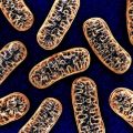
The mitochondria take nutrients from food, consume oxygen, and convert those nutrients into a fuel source (adenosine triphosphate ATP) that the cells use to function (learn more). There are three primary mitochondrial fuel pathways (and a whole bunch of secondary and tertiary pathways), one for carbohydrates, one for proteins, and the other for fats, disrupt one or more and all sorts of problems arise. Disrupt these pathways in the liver, the organ responsible not only for toxic waste removal but also for glycogen and fatty acid processing and storage, and the problems become exponentially worse. In the case of the severe morning sickness of pregnancy, I suspect that the mitochondrial beta oxidation pathway, the route for turning fatty acids into ATP, is disrupted.

How to Damage Mitochondria: Let Me Count the Ways

Mitochondrial function can be disturbed by a number mechanisms. Sometimes there are heritable genetic mutations, but not always. Heritable genetic mutations are called primary mitochondrial disorders and occur in up to 1 in every 200 individuals. Fortunately, not all mutations result in illness, but when they do, the results are often devastating.

More frequently, researchers are seeing what are called secondary, acquired, or functional mitochondrial damage evoked lifestyle variables. Epigenetic injuries, sometimes from generations past, have become increasingly common routes to disease. Epigenetic injuries do not induce mutations per se, but rather, aberrantly turn on or turn off gene activity that then influences mitochondrial function. Epigenetic activation or deactivation occurs relative to environmental influence, exposure to toxicants, stressors and/or other variables.

Among the least well recognized secondary mitochondrial injuries are those that are purely environmental; cumulative dietary and lifestyle exposures that damage multiple aspects of mitochondrial functioning. Many environmental and pharmaceutical chemicals evoke mitochondrial damage by leaching critical nutrients needed for mitochondrial energy production and other mitochondrial and cellular functions, but they also damage the structural or functional integrity of these organelles. The cumulative damage of everyday exposures when combined with genetic, epigenetic and/or poor dietary choices, render many individuals susceptible to mitochondrial illnesses. I suspect many of the idiopathic pregnancy complications, like hyperemesis, have their roots in mitochondrial dysfunction.

Although most of this paper, and indeed, most of the popular press focuses on mitochondrial bioenergetics, we must keep in mind that the mitochondria regulate a number of other important and endlessly reciprocal cellular functions, namely: steroidogenesis, immune signaling and cell death. Disturbances in mitochondrial bioenergetics, thus, would be expected impair hormone regulation, induce uncontrolled inflammation (chronic inflammatory and autoinflammatory diseases) and initiate tissue and organ injury. Individuals with mitochondrial issues would be expected to have a broad range of subtle and not-so-subtle health issues; many of which are endemic and epidemic in Western cultures.

Clues for Hepatic Mitochondrial Dysfunction in Hyperemesis Gravidarum

Backing up a bit, let’s connect some dots from the AFLP research. From the research on AFLP, we know that a mutation in the mitochondrial enzyme responsible for processing an important mitochondrial transporter evokes some, but not all of the cases of this disease process. Notably, in some women with hyperemesis, the fetus carries the mutation and evokes the vomiting, while mom is simply a heterozygous carrier.

The mutation (L-3-hydroxyacyl-CoA dehydrogenase deficiency – LCHAD) involves an enzyme (carnitine palmitoyltransferase I – CPT I) responsible for synthesizing the protein that acts as key transporter for fatty acids across the mitochondrial membrane. The protein involved is called carnitine.

When a fetus carries the CPT I mutation, the fetus’ inability to metabolize fatty acids and the associated bi-products are kicked back into maternal circulation effectively overriding the mom’s capacity to process these compounds. The increased load on the mom’s liver induces the vomiting, leading, in some cases, to the compensatory reaction of fat deposits within the liver cells – AFLP.  Since AFLP is relatively rare, developing in only 7-10 per every 100,000 pregnancies, is not present in all hyperemesis cases (50% of women with severe vomiting show some liver damage), and the fetal mutation is even rarer, we can deduce that neither AFLP nor the mutations that impair fetal fatty acid metabolism account for the totality of hyperemesis cases or even the morning sickness of early pregnancy.

Nevertheless, this research provides several important clues about hyperemesis. First, given the right set of circumstances, e.g. pregnancy or another high intensity stressor, carriers of a particular mutation may become symptomatic. We often view heterozygous carriers as being asymptomatic or less symptomatic than their homozygous counterparts. This may not be true. We may be simply viewing the symptom status incorrectly. Secondly, mitochondrial fatty acid metabolism is likely impaired and in some manner related to carnitine. Thirdly, maternal hyperemesis may not be a primary mitochondrial disorder in the classical sense (those definitions are changing, however). Even though there are a number of possible genetic mutations involved with the carnitine pathway, most are either severe enough to be identified during infancy (save except CPT II, which may remain latent until adolescence or early adulthood) and/or present differently (with muscular weakness and cardiomyopathy), and therefore preclude them from our differential. For all intents and purposes, hyperemesis presents during pregnancy, mostly in women with no known fatty acid oxidation or carnitine-related mutations, suggesting non-genetic mechanisms at play. In other words, I think we’re looking for functional mitochondrial disturbances in fatty acid metabolism related to carnitine.

What is Carnitine?

Carnitine is an essential micronutrient derived from the amino acid lysine with the help of methionine (an essential amino acid derived from diet). It is highly expressed in liver, testes and kidney. Dietary carnitine from meats, dairy and other sources yield carnitine. (L-carnitine is biologically active isomer. The research nomenclature varies considerably. For consistency, the word carnitine will be used throughout except when speaking of supplementation, where L-carnitine is more appropriate.) Carnitine is then shuttled off to skeletal and cardiac muscle where fatty acids are used as a primary fuel source. Although it is believed that endogenous carnitine homeostasis is maintained to some extent despite dietary contributions, there are number of conditions that override the internal synthesis of carnitine. These include genetic mutations that limit carnitine synthesis, difficulties with nutrient absorption (leaky gut or bacterial imbalances), kidney dysfunction which limits carnitine re-absorption, pharmacological inhibition of carnitine transporters, and nutrient deficiencies that disrupt any of the many enzymes involved in carnitine biosynthesis or metabolism.

In addition to its direct role in fatty acid metabolism, carnitine is also involved in glucose metabolism (the other major source of mitochondrial ATP) via its potentiating role in the pyruvate dehydrogenase complex, its modulation of  acyl-coenzyme A (CoA) and the storage of acylcarnitine. So when we disrupt carnitine availability, by whatever mechanism, not only is fatty acid metabolism derailed, but the other primary pathways for mitochondrial energy production are negatively impacted, as are the storage and clearance pathways.

Carnitine, Fertility and Pregnancy

We know very little about carnitine during pregnancy except that it generally declines. Below is a review the literature.

In women undergoing in vitro fertilization, higher maternal carnitine concentrations are associated markedly improved fertilization rates and overall better outcomes. Competent fatty acid oxidation is required for oocyte and embryonic development.

During pregnancy maternal carnitine concentrations diminish significantly. Indeed, at delivery, plasma carnitine concentrations have been reported 50% lower than in non-pregnant women. Researchers don’t know why carnitine decreases so much during pregnancy. There is some indication that carnitine concentrations are inversely related to iron status. Iron is needed for carnitine biosynthesis and so the increased demands for iron during pregnancy, if not met, may negatively impact carnitine synthesis. Since carnitine crosses the placental barrier, maternal carnitine deficiency would lead to fetal carnitine deficiency. The research, however, is all but nonexistent.

From animal research, we know that supplementing with L-carnitine, maintains carnitine concentrations across the pregnancy and improves a number of variables associated with reproductie function. Supplementation with L-carnitine also appears to offset liver damage and improve liver function in a mouse model of acetaminophen induced liver toxicity. Similar to the human IVF research mentioned above, L-carnitine supplementation improves oocyte development while increasing overall fatty acid oxidation capabilities.

Carnitine Deficiency with Nutrient Depletion

Population data for carnitine deficiency are unknown but nutrient deficiencies in general are postulated to be non-existent in the developed world, except with poverty. This assumption is erroneous and dangerous in the land of nutrient stripped processed foods. What little data exist for different nutrients, show that a significant portion of the Western population is deficient in one or more nutrients. Nutrient deficiencies impact enzyme function and the mitochondria’s ability to produce ATP and perform other critical functions. Carnitine synthesis alone requires five different enzymes, each with their own nutrient demands. This is in addition carnitine’s requirement for lysine and methionine. Given such demands, it is entirely conceivable, and in fact likely, that Western women come to pregnancy deficient, either marginally or grossly, in any one of the many nutrients involved in the carnitine pathway. Here are just a few.

Possible Nutritional Culprits in Functional Carnitine Defiency

Endogenous carnitine synthesis requires methionine. Methione concentrations in foods have steadily decreased (by as much as 60%) in parallel with the increase in glysophate (Roundup) used in conventional agricultural practices. Methionine synthesis also requires vitamin B12a nutrient deficiency common with the Western diet and exacerbated by many medications.

One of the only accepted treatments said to reduce the nausea in hyper-emetic women is vitamin B6 supplementation. Vitamin B6 is involved in carnitine synthesis. It is also an important anti-inflammatory, especially in the central nervous system.

The other nutrients required to maintain active enzymes for carnitine synthesis include: iron, niacin (B3) and vitamin C.

Finally, with pregnancy in general, but especially, with pregnancies involving severe nausea and vomiting, the risk of nutritional deficits is exacerbated as the intake of nutrients diminishes. Not only would we expect carnitine depletion but deficits in many of the other vitamins and minerals required by the mitochondria to produce ATP either via fatty acid metabolism or via glucose metabolism. The vomiting itself depletes nutrient stores, and thus, becomes self-propagating; fewer nutrients > more vomiting, more vomiting  > fewer nutrients.

Connecting the Dots: Potential Treatment Options for Hyperemesis Gravidarum

Thus far, the clues point to some sort of functional, epigenetic, or even an unrecognized, but latent, genetic derailment of fatty acid metabolism involving carnitine. The deficit in carnitine then precipites the severe morning sickness of pregnancy known as hyperemesis gravidarum. The nausea and vomiting worsen nutrient deficiencies and continue the cascade. If this is true, and I think it is, then the question becomes, can we support the carnitine system and mitochondrial function in general, to alleviate or completely eliminate the vomiting. I think we can.

I mentioned cyclic vomiting syndrome in the early sections of this post but haven’t spent any time on the topic. It is from the cyclic vomiting research that we find our treatment options. Specifically, Dr. Richard Boles has successfully treated pediatric patients who have cyclic vomiting syndrome with L-carnitine and Co-Enzyme Q10 (CoQ10), as have others. Indeed, we have personal experience with Dr. Boles’ work, as the daughter of one our writers had treatment refractory cyclic vomiting syndrome; that is, until the L-carnitine and coQ10 eliminated the constant vomiting. Cyclic vomiting syndrome is believed to be a mitochondrial disorder falling under a category of disorders called dysautonomias. And though a specific mitochondrial genotype has not been linked to CVS, Dr. Boles’ clinical data shows a clear association with mitochondrial fatty acid oxidation (L-carnitine supplementation) and the electron transport function (coQ10 supplementation).

Other Bits and Pieces

Fatty acid and carbohydrate metabolism within the mitochondria are closely tied to each other, with multiple interleaving levels of reciprocity. Both pathways demand nutrients to power their enzymes. A nutrient that is particularly high on food chain for mitochondrial function, is vitamin B1 or thiamine. We’ve written about thiamine deficiency repeatedly, as it seems to be leached from the mitochondria by a number of medications and vaccines and is implicated in a wide variety of adverse medication reactions. As a core nutrient in the pyruvate dehydrogenase enzymes, thiamine is critical for ATP production. Thiamine is also critical for fatty acid metabolism. A borderline thiamine deficiency would impair fatty acid metabolism and is linked to hyperemesis related liver damage and Wernicke’s Encephalopathy. Thiamine deficiency also impairs brainstem control of vomiting, thereby exacerbating the already difficult-to-control pregnancy hyperemesis. Thiamine supplementation should also be considered for hyperemesis gravidarum. Our own Dr. Lonsdale tells us that he has used thiamine in clinical practice to reduce cyclic vomiting in pediatric patients. The research on hyperemesis gravidarum, however, is extremely limited, focusing solely on the use of thiamine to curb the effects of hyperemesis-induced Werknicke’s syndrome.

Final Thoughts

Although there is little direct evidence linking a functional carnitine deficiency in pregnancy to hyperemesis gravidarum, there are a enough indirect data to suggest this may be a mechanism worth investigating. If this work bears fruit, L-carnitine, CoQ10, thiamine, vitamin B6 and likely other nutrients may be all that are needed to alleviate the nausea and vomiting across pregnancy.

Please note, I am not a medical doctor and this should not be construed as medical advice. Please speak to your healthcare practitioner before beginning any treatment protocol.

If there are any physicians or midwives who have used L-carnitine in patients with hyperemesis, please comment below.

We Need Your Help

More people than ever are reading Hormones Matter, a testament to the need for independent voices in health and medicine. We are not funded and accept limited advertising. Unlike many health sites, we don’t force you to purchase a subscription. We believe health information should be open to all. If you read Hormones Matter, like it, please help support it. Contribute now.

Yes, I would like to support Hormones Matter.

This post was published originally on Hormones Matter on July 22, 2015.

Solving the Medically Unsolvable: Gene, Nutrient, and Diet Interactions with Dysautonomia

11273 views

Years of Pain, Fatigue, and Weird Symptoms

I have had chronic fatigue syndrome, excessive Non-REM (NREM) dreaming, mood issues and muscle pain 24/7 for as long as I can remember. I have been to more than 100 health practitioners of various flavors, from the conventional doctors and specialists, to herbalists, energy medicine doctors, hypnotists and acupuncturists as well as taking pretty much every test available. Most tests were frustratingly normal. I had deficiencies in iron and B12 at various times but that came right after being diagnosed with Celiac Disease and starting a strictly gluten free diet.

Still, the fatigue and 24/7 pain persisted, and with the start of menopause, new symptoms emerged. I experienced periods of vertigo, brain fog, unexplained cold sensations in my chest, and after taking bioidentical progesterone for a few days I experienced daily dizziness and eventually got diagnosed with postural hypotension when the cardiologist measured a drop in systolic blood pressure of more than 30 upon standing. This daily dizziness continued for more than three years.

I tried all of the usual dietary interventions as well as the low oxalate diet, the anti-candida diet, the Failsafe diet and other elimination diets. All failed to make a dent in symptoms. The only time I noticed an improvement was when, after several bouts of gastroenteritis, I was forced to subsist on dry gluten free bread, a whey protein based meal substitute drink and skinless chicken. People commented that my skin looked good and I had improved energy, however, over time I returned to my normal diet and the benefits gradually disappeared.

Resolving the Dizziness, Brain Fog, Dysautonomia / POTS with B Vitamins and Diet

For more than three years, I have had daily dizziness pretty much all day as a symptom of dysautonomia/postural hypotension. An OAT test showed low thiamine and I had high hopes that thiamine would be the magic ticket that would get me out of dizzy brain fog hell. Thiamine and a reduced sugar diet did help a lot with energy, mood and general well-being, but unfortunately the reductions in postural hypotension were minor.

I experimented with supplements and lifestyle changes. I increased my meditation time, and did gratitude journaling and worked on taking in the good and rewiring my brain. This helped me better manage the stress of chronic illness and reduced some of my symptoms of depression and agitation.

I also found improvements in taking thiamine and riboflavin (B2) 3-4x a day along with high doses of the other B vitamins once a day. But, again the daily dizziness and brain fog persisted.

My big breakthrough came when I discovered that I had been taking the wrong form of niacin. I had been taking niacin and inositol hexanicotinate for the last three years, but it wasn’t until I returned to taking niacinamide that the symptoms dysautonomia dialed down. I started with a 50mg dose and by the end of the day I noticed that I had been less dizzy. I gradually increased the dose to the full 500mg and the symptoms kept reducing. I also got my brain back! No longer was I feeling constantly brain fogged, sluggish and mentally confused. Now that the niacinamide had my blood circulating properly and fueled my biochemistry things started working. My thyroid numbers had always been on the cusp of hyperthyroidism, yet I had a sluggish metabolism. Within a week, I noticed that with no other dietary changes my post-menopausal muffin top had reduced, my energy increased, and my skin was looking better.

The Missing Pieces: HACL1 and Phytanic Acid

I had high hopes that I would be able to completely eliminate the symptoms of dysautonomia, however, there is still some lingering dizziness. Over the last few weeks, I have been experimenting and have noticed two interesting associations.

The first, is sugar intake. Additional fruit or anything high in sugar increases my symptoms of postural hypotension. This could be linked to thiamine or niacin.

The second, is a reaction to foods high in phytanic acid. I first learned about the HACL1 gene from the Hormones Matter blog and I quickly realized that this made sense of the fact that I reacted to both A1 and A2 cheeses and yogurts as well as butter, but am fine on whey protein. I also react to oily fish and red meats but I am fine with pork and chicken. I live in New Zealand where all of our lamb and beef are grass fed, so all of our dairy products and red meat are higher in phytanic acid than the same products from grain fed animals.

In the past, I had noticed that any of these foods that are high in phytanic acid trigger feelings of rage and anger. There seems to be a threshold, so I can do an elimination diet and reintroduce butter and be fine, but over time, I believe that they phytanic acid accumulates and then the symptoms appear. Once I reach the threshold, I “hulk out” within minutes of eating beef, lamb, fat containing dairy products and oily fish. I have also had similar reactions in the past when I ate sugar or drank alcohol. I had in the past noticed that my dysautonomia was worse with all of these things. It would appear that thiamine is required to process all of these things, either directly in the case of sugar and alcohol or through the HACL1 gene for the other foods. This suggests that my body struggles to maintain thiamine levels and get the thiamine to where it is needed.


HACL1 rs17485390 (C) TT
HACL1 rs6784844 (T) CT
HACL1 rs6797119 (T) CT
HACL1 rs7648958 (A) AG

Feeling confident after increasing my niacinamide to 1,000mg spaced throughout the day, I reintroduced foods high in phytanic acid and the dizziness increased fairly quickly. I am now sticking to a low phytanic acid with only occasional red meat, fish or butter. (Yogurt and cheese are gone for good and maybe the other foods will need to be completely eliminated too.)

I found an old test that showed that my urinary l-lysine was low. After more research, I discovered that lysine helps maintain tryptophan activity and reduces the draw on niacin in the body. My tryptophan levels were normal on both urinary and blood tests but perhaps a lysine deficiency was indirectly affecting my niacin levels. After an initial dose of lysine I felt almost euphoric. This effect quickly leveled off. I am wondering if, after decades of fatigue, my body likes homeostasis and is counteracting the effects of nutrients that I clearly need. This has happened in the past with medications. After a few doses, they are basically rendered useless. This applies to antihistamines, psychotropics, painkillers, and so on.

Possible Secondary Pellagra

Is it possible that I have secondary pellagra? I initially dismissed the idea of pellagra as the symptoms seemed more severe than mine. My dermatitis was minor compared to the pictures online, I had an explanation for the dizziness (diagnosis of dysautonomia), the diarrhea has been an issue on and off, so it didn’t seem significant, and my mental confusion didn’t seem enough to qualify as dementia and yet I can now see that I did have the 3 Ds of pellagra despite adequate niacin intake. I don’t eat corn and rarely eat grains and have a diet high in niacin but I had many of the symptoms of pellagra including sensitivity to light, dermatitis, diarrhea, dizziness, feeling cold all the time, brain fog and mental confusion, difficulty falling asleep and weakness.

Interestingly, some of these symptoms overlap with thiamine deficiency symptoms and I feel very sure that I have had severe thiamine deficiency because I have also had tingling sensations and muscle pain, as well as a history of high intake of sugar, carbs and alcohol and a very positive response to thiamine and benfotiamine.

Going Forward: More Questions

My plan is to continue with my supplements and a low sugar diet and low phytanic acid foods. I am hopeful that this will completely eliminate the dysautonomia and leave me free to work on my other symptoms. My brain function is good when the dizziness is kept at bay and I feel more optimistic and happy and have a small but noticeable uptick in energy and strength.

Although I have made huge strides in my health, I am left with some lingering questions:

  1. I have been on high doses of many B vitamins for years and yet it seems that my body still craves them. Could years of undiagnosed Celiac Disease have affected the enzymes that take vitamins and converts them to the active form and transports them into organs and tissues? Is it realistic for this to still be happening after eight years of being gluten free?
  2. The literature glosses over the conversion from niacin to niacinamide as something that the body can easily do, however, I have taken high doses of niacin and inositol hexanicotinate without benefit and eat a diet rich in niacin foods without getting the benefits that I got from small doses of niacinamide. Is it possible that some people have challenges converting niacin to niacinamide? I have yet to find any research to support this other than a study suggesting that niacinamide is twice as effective as niacin. However, I was taking triple the dose of niacin with no benefits. I believe that my body is inefficient at converting niacin to niacinamide. If anyone knows of a specific illness that may cause this I would be interested in learning more.
  3. Is my reaction to phytanic acid foods due to a deficiency in thiamine (despite taking very large doses for years) or is there another reason that my body appears not to tolerate phytanic acid foods?
  4. Are there still more vitamin or amino deficiencies that I am yet to discover? In the future I will probably do another OAT or Nutreval to see whether my levels have improved but for now I want to let my body get used to the lower phytanic acid levels and see if things settle.
  5. My body seems to like homeostasis. For the first couple of days that I took niacinamide I noticed that I felt very warm, but I have returned to feeling cold all of the time. The dizziness has improved and it had almost disappeared but then crept back in. Could this be due to more vitamin or amino deficiencies that I am yet to discover, problems with my enzymes or is there some sort of ANS wiring issue that is better addressed by neural retraining?
  6. Sleep is another big issue for me and until I consistently sleep well without excessive NREM dreaming it is possible that these other issues will not fully resolve, but progress is exciting and I am hopeful that the last puzzle pieces will fall into place.

We Need Your Help

More people than ever are reading Hormones Matter, a testament to the need for independent voices in health and medicine. We are not funded and accept limited advertising. Unlike many health sites, we don’t force you to purchase a subscription. We believe health information should be open to all. If you read Hormones Matter, like it, please help support it. Contribute now.

Yes, I would like to support Hormones Matter. 

Thiamine Deficiency and Aberrant Fat Metabolism: Clues to Adverse Reactions

18195 views

Over the last several months, the writers and researchers at Hormones Matter have posted a number of articles about mitochondrial dysfunction and thiamine deficiency.  Thiamin, or thiamine as the internet search engines prefer, is critical to mitochondrial function. We’ve learned that thiamine deficiency can emerge gradually due to dietary inadequacies or more suddenly as a result of a medication, environmental or surgical insult. Regardless of the cause, deficits in thiamine evoke devastating health issues that can be treated easily if identified. More often than not, however, thiamine deficiency is not assessed and symptoms are left to escalate, mitochondrial damage increases, and patient suffering continues. Because thiamine deficiency is rarely considered in the modern scientific era, mild symptoms are ascribed to other causes such as “an allergy” or “it’s all in the patient’s head”. If, however, the cause is not revealed, the same old dietary habits will continue and can be guaranteed to produce much more severe and difficult to treat chronic disease.

Although there are a myriad reasons why mitochondria are damaged, medication or vaccine reactions paired with latent nutritional deficiencies seem to be common. Predicting who and how the mitochondrial dysfunction might appear, however, is more complicated. Quite often, athletes and individuals considered healthy are hit harder by a stress factor such as a vaccine than those whom we might not regard as particularly healthy. There are several potential reasons for this, some of which have been outlined previously. In this post, I would like to add one more reason why highly active, high performing individuals might be hit harder and more quickly than their less active counterparts with vaccine or medication reactions that induce thiamine deficiency.

Mitochondria are the Engines of the Cell

To use an analogy, the usefulness of a car obviously depends upon its engine. Mitochondria are the “engines” of each cell within our bodies, all 70 to 100 trillion cells that make up an adult body. They are known as organelles and are so small that their structure can only be seen with an electron microscope. But we can take this analogy further by comparing each cell to a different car model. A high powered car uses more gasoline than a low powered one and there are many models of each type of car. So some cells in the body require more energy than others, depending on the special function of the cell.  The most energy consuming cells are in the brain, the nervous system and the heart, followed by the gastrointestinal system and muscles. That is why those organs and tissues are most affected in the disease known as beriberi, the thiamine deficiency disease that we have discussed previously in other posts. The function of other organs is affected by the deficiency because of the changes in the control mechanisms originating in the brain through the autonomic (automatic) nervous system.

It has been pointed out that this disease in its early stages affects the autonomic nervous system by causing POTS. Beriberi and POTS, both being examples of dysautonomia (abnormal activity of the autonomic nervous system), can only be distinguished by finding evidence of thiamine or other nutrient deficiency as a cause. Thiamine is but one factor whose deficiency causes loss of cellular energy, resulting in defective brain metabolism and dysautonomia.  Although the relationship with vaccination is conjectural, some individuals with post Gardasil POTS were found to be thiamine deficient and had some relief of symptoms by taking supplementary fat soluble thiamine, an important derivative that occurs in garlic and has been synthesized. Not all of these thiamine deficient individuals have benefited to the same degree, suggesting that other deficiencies might also be involved. This post is to provide some information about more recent knowledge concerning the action of thiamine and the incredible, far-reaching effects of its deficiency, particularly in the brain. Experimental work in animals has shown that thiamine deficiency will damage mitochondria, a devastating effect for an acquired rather than a genetic cause. Far too much research has been devoted to genetic cause without sufficient attention to the way genes are influenced by diet and lifestyle.

The Importance of Enzymes to Mitochondrial Function

Before I provide this new information, let me remind the reader that enzymes, like cogwheels in a man-made machine, enable bodily function to occur. The importance of thiamine is that it is a cofactor to many of the enzymes that preside over energy metabolism. Without its cofactor an enzyme becomes inefficient. Perhaps it might be compared with missing teeth in a cog wheel. With missing teeth the cog wheel may still function but not nearly as well as it would with all of its component parts.

In previous posts we have discussed how thiamine deficiency can be caused by an excess of sugar in the diet. I have likened this to a “choked engine” in a car where an excess of gasoline, relative to insufficient oxygen concentration in the mixture, makes ignition of the gasoline extremely inefficient. Bad diet, one that is rich in sugary, carbohydrate laden foods may be one of the more common contributors to latent thiamine deficiencies. Excessive intake of processed fats and the concomitant changes to mitochondrial function and energy metabolism may be another important contributor.

Thiamine and Fat Metabolism

All the enzymes affected by thiamine deficiency have a vital part to play in obtaining cellular energy from food by the process of oxidation. Most of them have been known for many years but in the nineties a new enzyme was discovered. It has a very fancy name that has been simplified by calling it HACL1.  Only in recent years has it been found that HACL1 is dependent on thiamine as its cofactor. Although not reported, it may mean that it is also dependent on magnesium. This is exceedingly important because it introduces the fact that thiamine is involved in fat as well as carbohydrate metabolism, something brand new, even to biochemists.

Here I must digress again to describe another type of organelle called a peroxisome that occurs in our cells.  Like mitochondria, they are infinitesimally small. Their job is to break down fatty acids and they have a double purpose. One purpose is to synthesize very important substances that construct and maintain cells and their function: they are particularly important in the brain. The other purpose is what might be called fuel preparation. As the fatty acids, consisting of long carbon chains, are broken down, the resulting smaller fragments can be used by mitochondria as fuel to produce energy.  Failure to break down these fatty acids can result in the accumulation of natural components that may be toxic in the brain and nervous system or simply result in lack of one type of fuel. That is why feeding medium chain triglycerides by administration of coconut oil has been reported useful to treat early Alzheimer disease. They can be oxidized in mitochondria.

The Important Use of Fatty Acids in Mitochondrial Health

Here, I want to use another analogy. Imagine a lake that admits water to a river through a sluice gate that has to be opened and closed by a farmer who regulates the supply of water. If the gate is open the river will supply water to the surrounding fields. If however the gate is closed, the river will begin to dry up and the crops in the fields will suffer. Perhaps the farmer half closed the gate during a rainy period and has forgotten to open it when a dry period follows. High temperatures in the dry period results in insufficient water to meet the growth needs of the crops.

In this analogy, the lake represents food, the sluice gate is the HACL1 enzyme and the farmer who controls the gate represents thiamine. The water in the river represents the flow of fatty acids to the tissues for the double purpose of cellular construction and fuel for oxidation. The half open gate represents a minor thiamine deficiency, more or less sufficient for everyday life but not enough when there is greater demand. A high temperature that increases the water needs for crops represents Gardasil and many other medications as a stress factor, placing a greater demand on essential metabolic action.  The analogy also implicates the nature of the crops, some of which require more water than others. The crops, of course, represent body tissues and organs.

If we consider high performing individuals, whether academically or athletically, like high performance cars or crops that demand more nutrients, we can see how a previously unrecognized minor deficiency might trigger clinical disease by the stressful demands of a vaccine or medication. Some pharmaceuticals can attack thiamine directly, like Gardasil and the fluoroquinolones, while others attack different pathways within the mitochondria.

No matter the pathway, high performing individuals, with high energy needs not covered by diet, may be hit harder when a medication attacks mitochondrial energy.

The Outcome of Defective Fatty Acid Metabolism

Returning back to the HACL1 enzyme, we now know that HACL1 is the first thiamine dependent enzyme to be discovered in peroxisomes. It is research news of the highest importance, affecting us all. Its action is to oxidize a diet related fatty acid called phytanic acid and fatty acids with long carbon chains that cannot be used for fuel until they are broken down. Phytanic acid is obtained through consumption of dairy products, ruminant animal fats and some fish. People who consume meat have higher plasma phytanic acid concentrations than vegans. If the action of HACL1 is impaired because of thiamine deficiency the concentration of phytanic acid will be increased. The river in the analogy actually represents a series of enzymatic reactions that may be thought of as down-stream effects, whereas thiamine deficiency, being up-stream, affects all down-stream phenomena. One of the reasons thiamine deficiency is such an important contributor to illness is because its effects are broad.

These enzymatic reactions, known technically as alpha oxidation, involve four separate stages. It has been known for some time that if another enzyme at stage two is missing because of a gene defect, the result will be damage to the neurological system known as Refsum’s disease. Symptoms include cerebellar ataxia (also reported after Gardasil vaccination), scaly skin eruptions, difficulty in hearing, cataracts and night blindness. Other genetic mutations in alpha oxidation, resulting in various biochemical effects, result in a whole variety of different diseases. This places thiamine deficiency as a potential cause for all the down-stream effects resulting from defective alpha oxidation, for it has been shown in mice that this vitally important chemistry is totally dependent on presence of thiamine. Since its complete absence would be lethal, we have to assume that it is mild to moderate deficiency, equivalent to a partial closure of the sluice gate in the analogy.

Sources of Phytanic Acid: How Diet Affects Thiamine

In ruminant animals, our source of beef, the gut fermentation of consumed plant materials liberates phytol, a constituent of chlorophyll, which is then converted to phytanic acid and stored in fat. The major source of phytol in our diet is, however, milk and dairy products.  It raises several important questions. If thiamine deficiency is capable of causing an increase in phytanic acid in blood and urine, it might be a means of depicting such a deficiency in a patient with confusing symptoms. It might also explain why some individuals who have been shown to have thiamine deficiency by means of an abnormal transketolase test have symptoms that are not traditionally accepted as those of such a deficiency, perhaps because of loss of efficiency in HACL1.

If an excess of sugar in the diet gives rise to a secondary (relative) thiamine deficiency, we are provided with an excellent view of the extraordinary danger of empty simple carbohydrate and fat calories, perhaps explaining much widespread illness in Western civilization. Interestingly, it would also suggest that something as benign as milk could give rise to abnormal brain action in the presence of thiamine deficiency, because of phytanic acid accumulation. Our problems with dairy products may go well beyond lactose intolerance and immune dysregulation.

In sum, the discovery of HCAL1 enzyme and its dependence upon thiamine suggests one more mechanism by which thiamine deficiency affects mitochondrial functioning. As emerging evidence indicates a myriad of environmental and pharmaceutical insults impair mitochondrial functioning, thiamine deficiency ought to be considered of prime importance. Deficits in thiamine evoke devastating health issues that can be treated easily if identified.  If, however, thiamine deficiency is not identified and the same old dietary habits continue, the latent thiamine deficiency can be guaranteed to produce a much more severe and difficult to treat chronic disease. Moreover, individuals with thiamine deficiency who do not respond sufficiently to thiamine replacement might also have aberrant fatty acid metabolism. This too should be investigated and dietary changes adopted.