hypertension

Diet Induced Pseudo-Hypoxia and Hypertension

7829 views

Although hypertension has become almost exclusively used to indicate high blood pressure, it is worth examining the true underlying meaning. The prefix “hyper” is from the Greek, meaning over or above. Tension is defined as “the state of being stretched tight”. Perhaps then it is worth looking at how this applies to blood pressure.

When a blood pressure is measured, there are always two figures represented. The higher number is known as “systolic” and the lower one as “diastolic”. The systolic is when the heart is contracting and indicates the ability of the arterial system to expand enough to accommodate the pressure from an increased volume of blood. The diastolic indicates the pressure in the arterial system when the heart is resting between beats. We are therefore looking at the highest and lowest pressures in a closed tube system that must be capable of expanding and contracting.

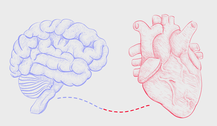
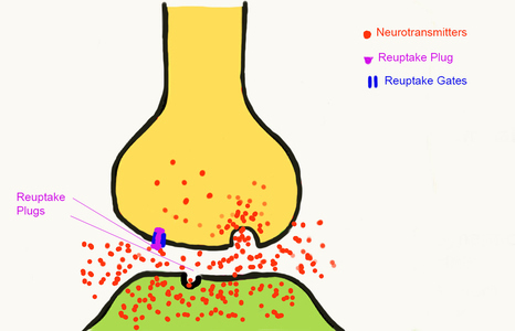
Since the system is made from live cells, it does not behave like a rubber tube with elastic recoil. The arteries where blood pressure is measured are lined with muscles. It is the contraction and relaxation of these muscles that control the capacity of the artery to accommodate the amount of blood arriving from the heart. The muscles are controlled by nerves carrying messages from the brain. These muscles are completely different from those that are activated willingly, such as those in the limbs. They are contracted and relaxed automatically by a part of the brain that acts more like a computer. The body muscles are activated by a nervous system known as “voluntary”. The arterial muscles are activated by a completely separate an involuntary nervous system known as autonomic (ANS). We therefore have to examine the control mechanisms.

Understanding the Autonomic Nervous System

I have discussed this nervous system many times in Hormones Matter because, when it goes wrong, it is a potent source of disease. The nerves of this system go to every organ within the body. The control system is in the lower part of the brain. It consists of two channels. One is known as sympathetic: the other is known as parasympathetic. Although they work together, their actions oppose each other and I will try briefly to outline this dichotomy.

Sympathetic. The sympathetic nervous system is designed for both physical and mental action through a reflex mechanism known as the fight-or-flight. It prepares us to meet an enemy or escape from danger. One of its actions is to raise the blood pressure. It does this by contracting the arterial muscles already described.

Parasympathetic. When the action is completed, the brain controls automatically withdraw the activity of the sympathetic and initiate those of the parasympathetic nervous system. When this happens, the body is prepared for resting.

Chronic Activation of the Sympathetic Nervous System

There is a large amount of evidence in the medical literature that this is the primary cause of chronically high blood pressure. If the system is healthy, the blood pressure will go down on completion of the action. If not, the blood pressure remains elevated. From here, I am going to hypothesize why this happens. Please remember as you read it that it is a hypothesis, not a proven fact.

Hypoxia. This word simply means lack of oxygen. Obviously, this is a dangerous state for the brain and it is not surprising that it will activate the sympathetic component described above, including raising the blood pressure.

Pseudo-hypoxia. The prefix “pseudo”, meaning false, or sham (from the Greek, lying, false) has been used in the medical literature to describe a state that is exactly like that of hypoxia when the presence of oxygen is normal. In order to understand this, focus on the fact that oxygen must be introduced to the body but is completely useless unless it is consumed. Therefore we must try to indicate how oxygen consumption occurs.

Oxidation and burning. All forms of burning are derived from oxygen combining with a fuel, liberating heat energy. That is why we are warm blooded, but other forms of energy are produced to drive physical and mental function. Because the burning is incomplete, ash is formed. Our cells derive their energy by the oxygen, delivered in the blood from the lung, combining with glucose. The “ash” is carbon dioxide and water, discarded in the breath. The oxidation of glucose is governed by a set of enzymes that require the vitamin B complex for their action. The leader of this orchestration appears to be vitamin B1 (thiamine). That is why many papers have appeared in the medical literature that describes thiamine deficiency as a cause of pseudo-hypoxia. Its function is to catalyze the enzymes essential for oxidation. Its deficiency results in lack of sufficient energy. It is therefore not surprising that one of the symptoms of thiamine deficiency is fatigue.

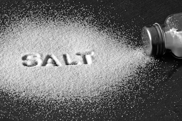
Calorie/thiamine ratio. A healthy diet provides us with calorie producing elements that are broken down to glucose and used as fuel. The amount of thiamine provides a normal calorie/thiamine ratio that enables efficient oxidation. If we load the diet with empty calories (calories without essential non-calorie nutrients that include thiamine) the calorie/thiamine ratio becomes abnormal. Measuring the concentration of thiamine in the blood would be normal for a healthy diet but inadequate to meet the demand of the empty calorie load. The laboratory method for identifying thiamine deficiency is by measuring it in the blood. If the result is reported by the laboratory as normal, the relevant symptoms produced by inadequate oxidation may well be ascribed to causes other than thiamine deficiency.

Hypothesis: High Calorie Malnutrition Induces Chronic Sympathetic Overdrive

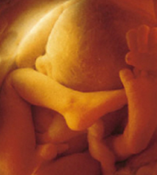
I suspect that a common cause of hypertension is high calorie malnutrition, inducing a state of chronic sympathetic overdrive. It may be why obesity in children often foretells their rise in blood pressure. Perhaps another cause is the gradual diminution of oxidation associated with aging. There are genetic mechanisms that are turned on by hypoxia and these also may be activated by pseudo-hypoxia, e.g. thiamine deficiency.

Spontaneously Hypertensive Rats

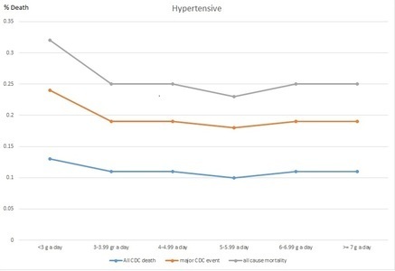
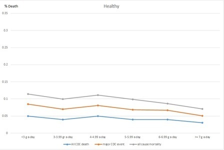
Lipothiamin is a synthetic derivative of thiamine. Its biologic properties enable it to be used as a drug. A rat known as SHR (spontaneously hypertensive rat) is used as the animal model for studying the effect of antihypertensive drugs. Many years ago I took a group of these rats and treated them with Lipothiamin to see if it would prevent the rise in blood pressure that always occurs in these animals. There was a statistically significant difference between the experimental rats and the controls, indicating that this thiamine derivative did indeed prevent the spontaneous rise in blood pressure. This experiment is published in our book (Lonsdale D, Marrs C. Thiamine Deficiency Disease, Dysautonomia and High Calorie Malnutrition). It obviously requires human subjects to research the use of this completely non-toxic, nutrient/drug derivative but nevertheless provides us with solid clues about hypertension.

Conclusion: Diet Matters

It has been said that simplicity must be distilled out of complexity in order to make complex issues usable. The brain/body, whether we like to recognize it or not, is an “electrochemical machine” that must obey all the physical laws designed by Mother Nature. Health is governed by only three factors:

  1. Genetics: the enormous complexity is dictated by a code written in DNA. Passage from generation to generation makes mistakes and represents our inheritance.
  2. Stress: defined as anything that requires physical/mental defensive response. The response, designed for relatively short term action, demands a huge consumption of energy.
  3. Nutrition: this is the only one of the three issues that we can control. It must supply both fuel and the multiple factors that enable the fuel to be turned into energy.

We Need Your Help

More people than ever are reading Hormones Matter, a testament to the need for independent voices in health and medicine. We are not funded and accept limited advertising. Unlike many health sites, we don’t force you to purchase a subscription. We believe health information should be open to all. If you read Hormones Matter, like it, please help support it. Contribute now.

Yes, I would like to support Hormones Matter. 

Why Are We So Scared of Salt?

9927 views

Over the past several decades, the general consensus of health professionals has been to recommend that all people lower their salt intake. Without the recognition of the effects of lifestyle and dietary choice differences, this avalanche of low salt advice hit the general public and as a direct result many became ill. Differences in individual genetic, lifestyle, and dietary factors have completely been ignored in the broad-brush campaign for lowering salt intake. Today, it is unmistakably obvious that a large segment of the population followed the low salt regimen with disastrous consequences.

The professionals who first introduced and propagated the low salt diets had good intentions. They did not know any better. Now we do know better and there is no excuse for not revising a failed treatment regimen in the face of new countervailing evidence. The process of correction needs to begin on a large scale. My work is part of this very much needed correction.

Why Are We Scared of Salt?

In the 1960’s, scientific studies linked salt consumption to hypertension and obesity. I am not quite sure why it was salt they picked on as “enemy number one.” I suspect the reason was the proliferation of precooked and canned food, all of which were salt preserved. To me, it was not logical that only salt was picked on. There were many other dangerous food items that could have been singled out: sugar, margarine, preservatives, pesticides, etc. The American Heart Association still has some of these salt reduction articles on their website. Even today, when waiting for an appointment at my medical institution, the forever-on TV was showing how to cut salt out of kids’ daily lunch to be “healthy.” Indeed, once something is ingrained in our brains, it is habit forming. Habits are very hard to break, particularly when the medical research relied upon showed that salt is something dangerous that may kill you.

Is Salt or Sugar the Enemy?

The problem is that hypertension and obesity are not and have never ever been caused by salt! They are caused by sugar—I am saving the sugar discussion for my next article.

Why not salt? Consider: human fetuses are floating in salt water and are typically not born with heart attack or hypertension. Our bodies are made of over 7% salt, our brains, heart, and all of our cells use salt to function. Humans have always consumed salt. Do they all have hypertension and heart attacks? No, they don’t. In fact, for some time now, studies have been surfacing suggesting that reduced salt does not eliminate the chances for hypertension and heart attack but may even contribute to the problem.

It is scientifically irresponsible to analyze biological processes in the human body involving salt without accounting for the effects of sugar and sugar substitutes and the amount of water consumed.

Probably not many of you have the handbook “Harrison’s Manual of Medicine” (18th edition McGraw Hill Medical by Longo et al.,) but I do. Page 4

…serum Na+ [sodium] falls by 1.4 mM for every 100-mg.dL increase in glucose, due to glucose-induced H2O efflux from cells.

Let me explain this sentence for you: Sodium is part of salt. Salt is Sodium (Na+) and Chloride (Cl-) where the + and – represent the ionic state in which there is either one extra or one fewer electron (electrons have negative charge) and so the atom is looking for another atom it can attach to and form a bond creating a molecule. According to the medical handbook, Na+ drops if glucose, which is blood sugar, increases. If you eat glucose, it causes “H2O efflux from cells” which means that sugar attracts water to the point that it pulls it out of the cells, thereby emptying the cells of sodium, and thus, the cells are dehydrated.

Sugar causes a very serious problem that can result in hypertension and heart attack. The volume of blood inside the cells reduces by dehydration of the sugar and higher pressure is required to pass the dehydrated blood to traverse the same route and be able to oxygenate organs at the same rate as hydrated blood cells. Think of a water hose when suddenly the pressure drops (unfortunately we cannot replicate reduced water molecule size the same way dehydrated cells become smaller). You instinctively squeeze the hose end to increase pressure so the water can continue to reach to the same distance. You have just given a hypertension to your water hose!

Note that if sodium (page 3 in same book) falls below 135 mmol/L, it is an electrolyte abnormality whose symptoms include “nausea, vomiting, confusion, lethargy, and disorientation”; if Na+ falls below 120 mmol/L it is a life threatening emergency that may cause “seizures, central herniation, coma, or death.” Not having enough salt (sodium) in the body is called hyponatremia and is “primarily a disorder of H2O homeostasis” meaning too much water and not enough salt. In common parlance, this is called water toxicity. Water toxicity can be caused by drinking too much water—e.g. drinking only water.

Interestingly, in the same book under the section of hypertension (page 834-835), the causes of hypertension are listed. Increased salt (or sodium) is not mentioned at all, but glucose intolerance is. However, under treatment, on page 836, it recommends lifestyle modifications that include lowering salt intake. So increased salt did not cause hypertension but lowering will cure it? I do not understand. Do you? Seems the authors of even this highly respected medical reference book could not escape the fallacy of the low salt campaign. Hypertension is clearly listed to be caused by sugar under the causes. So for heaven’s sake, if something is caused by sugar, treat it with removing sugar from our diet and not salt.

Confusion in the Ranks

In recent years a major fight started between the academic groups, not-for-profit organizations, and the government. Test after test shows that earlier hypotheses were all wrong about salt. Not only is added salt not hurting us, reduced salt does. Even the American Heart Association (AHA) and other heart organizations are in complete confusion. Next to the article of “lower your salt for health” are articles saying “that is all wrong and increase your salt.” I find this kind of funny. Here is an article from the AHA suggesting to increase salt. Here is another from the HealthAffairs organization; one from the American Journal of Hypertension, one from the Journal of the Association of American Medical Colleges, and there are now dozens more proving that indeed, reduced salt is actually bad for you.

How Bad is Reduced Salt on Health?

This particular article is my favorite because it shows how bad reduced salt diets really are on the heart. In detail, for a healthy individual reduced salt diet reduces BP by 1% (that means your systolic BP of 120 just dropped to oh my 118.5!!! gasp) and in patients with hypertension it reduced their BP by 3.5% (that is if it is say 160 systolic, which is high, it is reduced by a whopping 5.6 to 154.4! gasp again) but at the same time triglycerides, which contains the accurate measure of the sticky type of bad cholesterol in the LDL increased by 7% in people with hypertension (triglyceride should be less than 149). So if an individual with hypertension and triglyceride levels at 150 went on a low salt diet, that low salt diet would increase their triglycerides by 10.5 to 160.5, which is a significant jump for bad cholesterol. In a healthy individuals, the triglycerides jumped by 2.5%. Armed with such details, do you still believe that salt is bad for you?

Which Would You Rather Eat?

If I handed you 2 teaspoons: one was full of table sugar and the other full of table salt, which would you chose? For taste, we all would choose the sugar. What happens to our salt levels when we eat sugar? Refer back to the Harrison’s Medical Manual I mentioned earlier: eating glucose drops salt in our body because it sucks up all water and dehydrates. Eating a teaspoon of sugar will effectively dehydrate you and put you at risk of hyponatremia. By contrast, what will happen if you chose the teaspoon of salt? You will be thirsty, drink a couple of glasses of water and will feel like you are on top of the world.

My Recommendation

Stop being scared of salt and start being scared of sugar!

Sources

Longo et al., Harrison’s Manual of Medicine; 18th Edition, 2013; McGraw Hill Medicine

We Need Your Help

More people than ever are reading Hormones Matter, a testament to the need for independent voices in health and medicine. We are not funded and accept limited advertising. Unlike many health sites, we don’t force you to purchase a subscription. We believe health information should be open to all. If you read Hormones Matter, like it, please help support it. Contribute now.

Yes, I would like to support Hormones Matter.

Image by 28366294 from Pixabay.

This article was first published on June 13, 2015.

The Lyme Spiral

2873 views

Two of the most commonly asked questions I face as someone with Lyme disease is “when were you bitten by a tick?” and “when did you get Lyme?” To be honest, while I have my suspicions, I really have no idea. Many people are surprised to hear that the majority of people with Lyme disease never recall being bitten by a tick. The bullseye type rash that people assume is the hallmark of Lyme symptoms also doesn’t occur in many of us with Lyme, hence why we have no idea when we first contracted the disease.

Early History

I was an active child and a competitive swimmer. By high school, I was swimming at the state and national level. I was in great shape with great health, or so I thought, and had no obvious reason to be experiencing any medical abnormalities. However, over my freshman year in high school, I started experiencing widespread joint pain. Because I was in such good health and swimming and training up to four hours per day, it came as a surprise to my parents, coaches, and doctors when I was experiencing such pain. During this time, I did get tested for Lyme by my pediatrician. While he was fine testing for rheumatoid arthritis and lupus, we had to beg him to test for a Lyme test, even though it was common in our area. It was years later when we learned that traditional doctors typically do not test for or treat Lyme disease.

My test showed that I had one positive band of Lyme. He said that this was not enough for a diagnosis, and I was sent home without any answers. My sophomore year of high school, the joint pain continued and even began to get a bit worse. I went to a rheumatologist at the Children’s Hospital of Philadelphia and was diagnosed with Amplified Musculoskeletal Pain Syndrome, also known as AMPS. Their main treatment plan was exercise, which, with swimming, I was already doing. There wasn’t much else that could be done, but by junior year it seemed like I had grown out of AMPS. Whether it was really AMPS or had it been Lyme disease all along, I have no idea.

College Dorm Flu Masked My Infections – Until My Vocal Cords Went Haywire

My freshman year of college living in a dorm I was sick constantly. I missed several of my college swim meets from contracting the adenovirus, developing severe swimmer’s ear, and coming down with strep throat and bronchitis. Another strange thing I began to notice was that I was having low grade fevers nearly every day. I got tested for the mono virus, as many college students are, at one point or another, but the test came back negative. With the constant infections, along with many headaches and slight fatigue, a blood test showed that I had an underlying Epstein Barr virus from prior contraction. I do think they missed mono, and I can’t help but wonder if Lyme played a role.

During my sophomore year of college, I began the swimming season feeling strong until I developed a nasty virus along with a bad cough within the first few weeks of the semester. Luckily, the virus died down, however the cough never went away. The cough became worse as I swam and I was led to assume I had developed sports-induced asthma. I just couldn’t get in a good full breath. During fall break, I went to an allergy and asthma specialist. When I told her I couldn’t fully breathe in, she pointed out that with asthma, you typically have trouble breathing out. She performed a rhinoscopy, where they stick a camera in your nose down into your throat, and examined my vocal cords on a very fuzzy screen before diagnosing me with vocal cord dysfunction. This was a difficult diagnosis because it was hard to explain to friends and family. It is not commonly known, and somewhat misunderstood. People genuinely had no idea that when I was breathing in, my vocal cords were spasming closed. The only thing I could link this to was the virus I had a month prior.

It took months of my vocal cord dysfunction being taken lightly or just blatantly disbelieved until I went to an otolaryngology specialist in New York City. The specialist performed a rhinoscopy as well, but recorded everything on a crystal clear screen. He was able to visibly show my parents that my vocal cords were, in fact, spasming closed when I breathed in. My parents have since admitted they didn’t fully believe me until that appointment. The recommended treatment was Botox. This meant that I had to go into the city every few months to get Botox injected into my vocal cords. Eventually, the doctor recommended surgery because folds of tissue were leaning into my airway over my vocal cords, a condition known as laryngomalacia, making my vocal cord dysfunction worse. I got surgery and continued with my Botox appointments and my vocal cord dysfunction finally calmed down a bit. Again, here I had suspicions that this was related to Lyme disease as well. My first Lyme specialist commented that vocal cord dysfunction could have been a result of the disease. After hearing Shania Twain’s story about how Lyme disease affected her vocal cords, my suspicions were stronger, but, again, I will never know for sure.

Looking For a Zebra: Fatigue, Tachycardia, and Hypertension

I took a year off of school for a few different reasons, some of them being medical. I was still experiencing low grade fevers constantly, fatigue, tachycardia, and hypertension. I was still very active, going to OrangeTheory Fitness classes a few times a week, along with lifting weights and swimming. Currently, I can barely walk my dog and my exercise consists of physical therapy. A few times, I went to OrangeTheory Fitness with my mom, and when I looked up at my name on the screen, I saw the number 212, which I thought was my score (I thought I was winning), but my mom realized it was actually my heart rate. At another point during this year, my blood pressure was in the 200s over the 200s. I went to an endocrinologist for my fevers, fatigue, and the general feeling that something was very off. She told me I was on so many psychiatric medications that they were making me “sound slow,” and that I just had fever of unknown origin. She also said that she was going to visit her family in India and could bring me back herbal supplements for the fever of unknown origin.

That being said, I did have severe anxiety at the time and was dealing with post-traumatic stress disorder. I had no idea what this meant at the time, but my psychiatrist told me to go to my primary care doctor and tell him “we’re looking for a zebra,” and even a pheochromocytoma. I saw several doctors that year, each who ordered different types of blood work. One doctor was so unsure what was happening that she ordered twenty two vials of blood. Yet, I was still left without any answers.

And Then Came Covid

After my mental health and anxiety had not improved with medication and therapy, my mom felt there still had to be an underlying issue. This is when she found and took me to a Lyme specialist in the fall of 2019, who immediately diagnosed me with Lyme disease and bartonella, and later, mast cell activation syndrome (MCAS). Physically, my symptoms were manageable. However, I contracted the first strain of COVID-19 in May of 2020, and my life has never been the same. Within the next few months, I started experiencing joint pain, severe migraines, vertigo, severe fatigue, and continued mental health issues. I even had to drop all of my classes in the fall semester of 2020 because I missed nearly two weeks of school from having a fourteen day long migraine. I began developing neurological and cognitive issues, such as trouble focusing and thinking and I had with memory. I switched to a different Lyme specialist, and this time I even tested CDC positive, which is more uncommon than not in those with Lyme. I started yet again another regimen of antibiotics and herbs. I spent most of that year couch ridden, sleeping much of the day, and constantly in hot Epsom salt baths to ease the joint and muscle pain. It felt like my mind and body were completely outside of my control, almost like my autonomy was taken from me. I was on several combinations of antibiotics until I eventually switched doctors again. This new physician was recommended by my Lyme literate dietitian, and was extremely knowledgeable of complex cases. I still see her to this day.

The next year I went back to school part time, only taking two classes each semester. Before the fall semester, I was diagnosed with babesia, and a few months into the semester I was diagnosed with hypothyroidism. These diagnoses often come together in the case of Lyme disease, but a new diagnosis is always a lot to take in. In class, I would forget what I was saying mid-sentence, which made me self-conscious and hesitant to participate. I struggled to balance the fatigue, physical pain, and brain fog with school. Yet, I successfully completed those two semesters.

Medication-Induced Pancreatitis

Finally, my senior year began: Fall of 2022. I was still attending part-time, but was now taking three classes as well as a lab each semester. In late September, early October I started having these acute episodes of severe pain in my upper abdomen that spread to my back. Because I have been medically gaslit so many times and I also have a high pain tolerance, I was nervous to go to the doctor. These episodes felt like I should have been in the emergency room, but because they only lasted an hour I was afraid to go to the hospital in fear that they would brush me off as a young female having stomach aches. I dealt with the pain for almost four weeks before I finally went to a doctor.

As it turns out, I had pancreatitis. I had to go on a mostly liquid diet for two weeks, stop taking my antibiotics and other supplements for Lyme, as doctors believed it was drug induced, with the culprit suspected to be rifampin, or the combination of too many other medications and supplements. I had been on various psych meds for years. Initially they were prescribed for severe anxiety and panic attacks and when I began to get sick, it only worsened. Among the medications I have been prescribed and used are several SSRIs/SNRIs (Prozac, Zoloft, Lexapro, Luvox, and Effexor). I have used sleeping medications such as Prazosin, Seroquel, and trazodone, as well as a tricyclic antidepressant, some benzodiazepines, Wellbutrin, Buspar, hydroxyzine, and different ADHD medications (Concerta, Ritalin, Vyvanse, Adderall, Straterra).

While pancreatitis mostly resolved, my Lyme started to continue to progress. I was having many more neurological and cognitive symptoms, my whole body was tremoring nearly every day, and I knew that we had to take action before the Lyme and coinfections progressed any further. I went back on antibiotics at the beginning of the spring semester, but had many Herxheimer reactions. I was out of it and sweating with fevers in class, all while also managing an internship. Within a few months my tremors became much better, indicating that the antibiotics were working. It was a long and stressful year, but I graduated Magna Cum Laude after having periods of extreme sickness where I questioned whether I would ever graduate.

Right now I’m on Anafranil, Buspar, Trazodone, Hydroxyzine, and Valium. As for Lyme and coinfections, I’m on clarithromycin, Tinidaloze, Valtrex, Mepron, methylene blue, (recently started), low dose Naltrexone, liposomal oil of cinnamon clove and oregano, cryptolepis, Researched Nutritionals MycP, phosphatidylcholine, vit D and C, omegas, ATP360 and CoQ10 for mitochondrial dysfunction (my doctor has suspected mitochondrial death/dysfunction after having COVID the first time, followed by long covid). I’m on a whole bunch of other supplements too to support my immune system, help with mast cell activation/histamines, digestion, brain function, etc. I’m taking between 50-70 pills per day with a few liquids mixed in there. I’m also on thyroid Armour for hypothyroidism as well as migraine medications and Zofran.

Chronic Illness Limbo: Neither Disabled Nor Able Bodied

Something that healthy people may not think about is having to choose your battles in terms of symptoms and treatments. For example, I started methylene blue shortly after graduation. Methylene blue, which is a medical dye, is a relatively newer, yet promising treatment for Lyme disease, but it is a monoamine oxidase inhibitor (MAOI). This means it can interact with psychiatric medications, and, therefore, I had to go off of all of my ADHD medications. The combination of an MAOI and a serotonergic ADHD medication can lead to both serotonin syndrome and a hypertensive crisis. The idea is that over time and with building up the dosage of the methylene blue it will minimize brain fog, a huge Lyme symptom, and help to treat ADHD symptoms. However, it takes quite a while to build up to an adequate dosage of methylene blue, and it also takes some time for it to actually show effects and improvements. Methylene blue treatment has been a bit brutal here and there with Herxheimer reactions.

One of the most daunting and frustrating things about Lyme disease, especially when it becomes chronic, is that there is rarely a trajectory or expected timeline to start feeling better or to reach remission. I’m currently still trying to find my way to remission, but cannot say I am close. Lyme Disease has turned my life upside down. At this point in my life, I thought I would be living up my twenties with either having already started graduate school or a big girl job. Something that people with Lyme disease, and those with other chronic illnesses as well, deal with on a regular basis is not being able to keep up with productivity culture leading to feelings of failure, laziness, or character flaws. Chronic illness is isolating, and, for many, it may feel like there is no place in society for us when we’re neither fully able bodied nor fully disabled. Instead of living up my twenties, I spend most days with my parents and my dog, I rarely drink alcohol or go out with friends, as it is difficult to maintain friendships when chronically ill. I am still trying to find a place to fit into society post bachelor’s degree, without a master’s or doctoral degree. I feel resentful of all of the doctors who misdiagnosed me or gaslit me, letting me go longer and longer undiagnosed, and therefore, untreated, allowing the Lyme to progress. However, I have found friends in the chronic illness community that, even though we have a variety of illnesses and different symptoms, understand the common struggles of chronic illness.

If you suspect you have Lyme, the best thing you can do is go to a Lyme literate doctor in your area, whose names and locations are usually spread through word of mouth due to the politicization of Lyme Disease, which can lead to possible repercussions for doctors who treat Lyme without CDC positive tests, which, like I said earlier, are inaccurate and are not often used as a basis for diagnosis.

We Need Your Help

More people than ever are reading Hormones Matter, a testament to the need for independent voices in health and medicine. We are not funded and accept limited advertising. Unlike many health sites, we don’t force you to purchase a subscription. We believe health information should be open to all. If you read Hormones Matter, and like it, please help support it. Contribute now.

Yes, I would like to support Hormones Matter.   

Hypertension, the Autonomic System, and Thiamine

14529 views

Physiological studies have long demonstrated the vital role of the autonomic nervous system in controlling blood pressure values. Beriberi, the classical disease due to thiamine deficiency, is the prototype example of dysautonomia in its early stages. The limbic system and brainstem are peculiarly sensitive to thiamine deficiency. Hypoxia is known to initiate sympathetic nervous system activity. Because thiamine deficiency has effects similar to those of hypoxia, these effects have been referred to in the literature as arising from pseudo-hypoxia.

There is much evidence that the common indulgence of empty calories, particularly those derived from simple carbohydrates and processed fats, is producing pathophysiologic brain effects due to what has been termed high calorie malnutrition. This form of malnutrition is totally different from that produced by starvation, the traditional form of malnutrition. Although the concentration of thiamine in the blood may be considered to be normal, it is only normal in the presence of a healthy diet. The excess of empty calories results in a high calorie/thiamine ratio that overwhelms the normal oxidative capacity associated with the presence of thiamine.

Patients suffering from high calorie malnutrition represent the “walking sick” of America. In its early stages it results in a polysymptomatic disease that defies our present concept of diagnosis. Traditional laboratory results may be quite normal or nonspecifically abnormal and the symptoms “written off” as psychosomatic. It may well be that some cases of hypertension, affecting millions, is part of the clinical effect produced by the dysautonomia of thiamine deficiency. The text that follows provides support for this hypothesis. It may also be true that failure to recognize the true etiology may lead over time to chronic brain disease in the untreated patient.

Autonomic Control of Blood Pressure

It has been well documented that the autonomic nervous system plays a key role in controlling blood pressure values. The hypothesis has been put forward that the origin, progression, and outcome of human hypertension are related to dysfunctional autonomic cardiovascular control, especially to abnormal activation of the sympathetic division. There is a consistent association between hypertension and pro-inflammatory cytokines of the innate and adaptive immune system. The sympathetic nervous system, a major determinant of hypertension, is pro-inflammatory, whereas the parasympathetic nerve activity dampens the inflammatory response through α7-nicotinic acetylcholine receptors. In the Goldblatt model of renal hypertension and the use of renal denervation in the treatment of drug-resistant hypertensive patients, autonomic mechanisms underpin the maintenance of this hypertension. Autonomic mechanisms initiate the development and maintenance of renal vascular hypertension. Obesity-associated overnutrition leads to neural inflammatory molecular changes, particularly in the hypothalamus, leading to elements of the metabolic syndrome. Increased sympathetic activation is a critical mediator.

Children, Hypertension, and Obesity

There are an increasing number of children diagnosed with primary hypertension, mainly in association with obesity. One of the factors considered is dysregulation of the autonomic nervous system. Emerging evidence suggests that the sympathetic branch plays a much broader role in the regulation of blood pressure, including the development and maintenance of sustained hypertension by a chronically elevated central sympathetic tone. A recent study examined the relationship between the autonomic nervous system dysfunction, anxiety and depression in hypertension. The sympathetic nervous system and arterial baroreceptor reflex control of renal sympathetic nerve activity has been proposed to play a role in long-term control of arterial pressure.

Abundant evidence supports the role of the sympathetic nervous system in the pathogenesis of obesity -related hypertension. The mechanisms are incompletely understood. A study supports the concept that increased renal sympathetic activity is the critical mechanism by which increased central sympathetic outflow initiates and maintains reductions in renal excretion function, resulting in obesity hypertension. Hypertension and obesity are risk factors for coronary heart disease in adults. Childhood weight problems and high blood pressure increase the risk of subsequent obesity and hypertension as an adult.

The Role of Hypoxia in Blood Pressure Regulation

Obstructive sleep apnea is one of the most common causes of hypertension in Western societies, causing chronic intermittent hypoxia. The normal homeostatic balance between HIF-1α-dependent pro-oxidant and HIF-2α dependent antioxidant enzymes is disrupted. Hypertension affects one in three United States adults and involves the integration of the actions of multiple organ systems, including the central nervous system, driving enhanced sympathetic outflow and increased blood pressure. There is increased understanding of hypothalamic activity in hypertensionChanges in the baroreflex and chemoreflex may contribute to the development of chronic hypertension observed in obstructive sleep apnea patients. Animal studies have identified induced hypoxic hypertension, but this is still conjectural in humans.

Thiamine Deficiency and Pseudo-hypoxia

Because mild to moderate thiamine deficiency results in pseudo-hypoxia in the limbic system and brainstem, emotional and stress reflexes of the autonomic nervous system are stimulated and exaggerated. Like chronic intermittent hypoxia that activates HIF-1 and suppresses HIF-2-mediated transcription, thiamine deficiency induces HIF-1α mediated gene expression similar to that observed in hypoxic stress. Thiamine deficiency induces an early, functionally significant central muscarinic cholinergic lesion that may be at least part of the reason for sympathetic overdrive. Details of the genetic determination in hypertension are poorly understood. A novel blood pressure locus that encodes a previously uncharacterized thiamine transporter has been reported, again perhaps emphasizing the importance of thiamine deficiency in hypertension. Evidence supports the likelihood that an increased cytosolic ratio of NADH/NAD+, caused by hyperglycemia, results in playing an important role in the pathogenesis of diabetic complications. Thiamine deficiency occurs in individuals with diabetes, leading to hyperglycemic-induced damage and oxidative stress. The potential benefit of long-term replacement is not yet known but may well reduce the cardiovascular risk and angioplasty complicationsThiamine deficiency should be considered in all patients with pulmonary hypertension of unknown origin.

Maternal hypertension is the most common medical disorder of pregnancy, varying from 4 to 6% of all pregnancies. The daily dose of 100 mg of thiamine has been given to over 1000 non-selected prenatals starting in the second and third trimesters. The expected number of toxemia patients in this group was well over 150, but the actual occurrence was zero.

Hypertension and High Calorie Malnutrition

Optimum nutrition is the level of intake that should promote the highest level of health. A deficit in nutrition may result in tissue depletion of essential nutrients that can lead to biochemical changes and eventually to clinical signs and symptoms. Adverse conditions prenatally increase the risk of cardiovascular disease, including hypertension. Obstructive sleep apnea increases over the course of pregnancy and is common during the third trimester. These adverse conditions have reportedly been completely prevented by the use of 100 mg of thiamine beginning even before pregnancy is confirmed. Thiamine deficiency, resulting in impaired oxidative metabolism, leads to a multifactorial cascade of events in the brain that include focal decreases in energy status, oxidative stress, blood-brain barrier disruption, glutamate-mediated excitotoxicity, amyloid deposition, immediate-early gene induction and inflammation.

Thiamine Tetrahydrofurfuryl Disulfide and Hypertension

This disulfide derivative of thiamine is the synthetic counterpart of allithiamine that occurs naturally in garlic. Allithiamine was discovered in Japan in 1951 and extensively researched by Japanese scientists. Experimental work in animals and human subjects revealed that its metabolic effect was much more powerful than the thiamine from which it was derived. Almost unknown in America, its therapeutic actions have been reviewed.

SHR rats are widely studied as a model of hypertension, exhibiting metabolic abnormalities that share features with the human metabolic syndrome. The SHR rat becomes hypertensive early in life and blood pressure continues to increase with age, often resulting in a cardiovascular event. Over the course of four months, blood pressure and body weight of two groups of SHR rats were monitored. The control group was fed normally and the TTFD group was given increasing doses of TTFD from 5 to 15 mg. There was a statistically significant reducing effect on the treated rats as compared with the controls. Of the 13 TTFD-supplemented rats, five showed a definite response with no rise in the expected blood pressure measurements, while the control group showed a 20 to 25 mm Hg increase in blood pressure across the experiment. The results showed that TTFD has a preventive effect on blood pressure in these genetically abnormal rats.

Thiamine Deficiency > Hypoxia > Hypertension

It has been shown in the above text that hypoxia and pseudo-hypoxia have virtually identical effects in the area of the brain that is peculiarly sensitive to thiamine deficiency. Since any phenomenon that causes inefficient oxygen consumption is dangerous to the organism, activation of the fight-or-flight reflex seems to be entirely logical. Repeated hypoxia in sleep apnea syndrome induces persistent chronic sympathetic dominance and is obviously an abnormal state of metabolism. It is possible that sleep apnea represents a combination of brainstem thiamine deficiency and recurrent obstructive hypoxia. Lonsdale and Marrs have published evidence that thiamine deficiency is widespread in the American population and that its effects in the brain are diverse and the resulting morbidity prolonged. Evidence could be accumulated by proving thiamine deficiency and attempting clinical trials with TTFD.

We Need Your Help

More people than ever are reading Hormones Matter, a testament to the need for independent voices in health and medicine. We are not funded and accept limited advertising. Unlike many health sites, we don’t force you to purchase a subscription. We believe health information should be open to all. If you read Hormones Matter, like it, please help support it. Contribute now.

Yes, I would like to support Hormones Matter. 

This article was published originally on November 17, 2020. 

Silent Death – Serotonin Syndrome

317030 views

It started very slowly; at an almost non-existent rate. My mother, then about 84 years old, broke her ankle. She had been extremely active, playing table tennis regularly in a senior club; she was also a bridge champion almost all her life. She even joined online bridge groups and beat everyone on the internet too. When they asked how old she was, her partners and competitors just flipped that she was in her 80s and a bridge champion. She was sharp as a tack!

The ankle that broke needed surgery with plates and screws. She was restricted to bed for 6 months and then to wheelchair for life. While her ankle was healing she was in bed and could not play bridge, she lost her skills and partner. She was also dependent on others and became depressed. I would rather say she was angry with life for what happened to her rather than depressed but she insisted that she was depressed. She paid a visit to a neurologist begging for an antidepressant.

The neurologist prescribed half of the smallest possible dose of Mirtazapine, a simple serotonin that on its own is capable causing major damage but she received a very small dose. As she started taking the medicine, very tiny changes developed in her personality but they were so mild as to almost unnoticeable. In retrospect, we see what happened – hindsight is always 20/20.

First Signs of the Impending Doom

The first sign that she had too much serotonin in her brain was that rather than feeling calmer and happier she became more agitated; she was unhappy with people around her, criticized everything, nothing was good enough. Then bowel incontinence started and she had trouble holding her stool until she reached the bathroom; her bowel incontinence further limited where she dared going so she felt angrier. She became very easy to irritate and was pissed at the whole world.

What I have just described took four years to evolve so we did not see the connection of all these changes to the serotonin medicine. Then one day as I was refilling her medicine, the drugstore ran out of Mirtazapine and they placed her on an SSRI called Zoloft instead—the doctor changed her prescription.

An SSRI (Selective Serotonin Reuptake Inhibitor) is a very different medicine from the old small dose serotonin my mother received. While Mirtazapine merely provided a small extra dose of serotonin to the brain, Zoloft forced her brain to make serotonin 24/7.

How SSRIs Work in the Brain

To understand what SSRIs do, envision a sink with an overflow hole on the top, in case you left the water running. This will allow the extra water to flow back into the drain and if you have an automated sink that is connected to this backflow, the sink would know it is full and would turn the faucet off. This little overflow hole in the brain cell is called reuptake. It does exactly what the overflow does. If it senses that enough serotonin was made, it shuts down serotonin manufacturing of the cell until it senses that more is needed. However, SSRIs inhibit the reuptake receptor, i.e. plug it up. Just as your sink will flood your house with water if the overflow is plugged up, so does the brain fill up with serotonin as long as the reuptake is inhibited. This makes the brain cell manufacture serotonin forever, regardless how much is needed and how much it already has made. Reuptake inhibitors serotonin syndrome

Only a small percentage of serotonin is made in the brain, less than 10%, and 90% is in other parts of the body. The intestinal tract uses most of the serotonin to pass the food through the intestines with proper speed—this explains why having too much serotonin in one’s body causes bowel incontinence. Serotonin also functions as part of memory and cognition, and it is also a vasoconstrictor. Serotonin is a dangerous substance that predisposes the patient to diabetes 2. Thus it is no surprise, in retrospect, that we saw changes slowly from Mirtazapine but very fast changes as my mother was moved to take an SSRI. Suddenly changes took place at a drastic pace:

  • Day one of the change to SSRI was a confusion day. She was clearly agitated, confused, and bowel incontinence became a permanent feature
  • Day two she was angry staring up at the ceiling all day in bed, refused to eat or do anything. The commode had to be moved into the bedroom though she barely made it that far without accident.
  • Day 3 she fought the whole world, nothing was right. She set in a corner totally agitated
  • Day 4 she called me on her cell phone at 5 am (we lived in the same house, with me right above her) asking when breakfast was served in this house. I rushed down and found her sitting at the edge of her bed in total confusion. I put her back in bed and told her breakfast will be served at 9 am so she should go back to sleep.
  • Day 5 is when the moment of recognition hit me. She called me again on the cell phone at 5 am. I ran downstairs. She was seated at the edge of her bed, totally naked with her bathrobe barely on. Her entire closet was on the floor; she pulled everything off every single hanger and shelf. I ran up to get the blood pressure meter. Her blood pressure was so high the cuff gave me error twice before I was finally able to read her blood pressure. The systolic was over 180 (120 is ideal), I don’t remember the diastolic but it was over 100. I called the ambulance and off she went to the hospital.

In the hospital, I tried to tell every doctor what her history was with the SSRI. I am a medically trained professional in neuroscience and though not a medical doctor but a researcher, I can identify a serotonin syndrome when I see one as long as I know the history that led up to it.

For my biggest surprise, and why I am writing this article, is that physicians rarely recognize serotonin syndrome. No one believed me when I told them that I suspected that my mother was suffering from serotonin syndrome. No one listened to me when I asked that they test for serotonin syndrome. I received comments like this from a psychiatrist: “Your mother cannot have serotonin syndrome, it is too rare.” Serotonin syndrome is not rare but the doctors who identify it are, and he was one of the many who did not recognize serotonin syndrome when he saw it. Another doctor told me that “she may have serotonin syndrome but we cannot test for that and cannot treat for it.” In fact, testing and treatment are both available for serotonin syndrome. The problem is with the doctors who do not ask any questions and only make assumptions based on the patient’s age (she was 88 at this time) using profiling assume that anyone over the age of 80 must have dementia. They diagnosed my mother with Alzheimer’s type dementia (something we were able to see via autopsy to have been the wrong diagnosis). She was misdiagnosed and mistreated with the wrong medicines until she died. There was nothing I could do. I suspect that for those of you who are not scientists like I am, the task is even more daunting. So prepare for the fight of a lifetime.

Unfortunately, the symptoms of many illnesses or conditions resemble that of the symptoms of serotonin syndrome. The surest way of knowing if you or your loved one has serotonin syndrome, is if serotonin medicines have been taken for a long time and symptoms slowly worsened over time or if new serotonin medicine was just introduced. If three of the following symptoms appear, take the patient to the nearest hospital via ambulance immediately, stand guard and get ready for a fight to save a life!

  • Agitation or restlessness
  • Confusion
  • Rapid heart rate and high blood pressure
  • Dilated pupils
  • Loss of muscle coordination or twitching muscles
  • Muscle rigidity
  • Heavy sweating
  • Diarrhea
  • Headache
  • Shivering
  • Goose bumps
  • High fever
  • Seizures
  • Irregular heartbeat
  • Unconsciousness

The importance of this long introduction is that today more people take SSRIs than ever before hence the increased odds of ending up with serotonin syndrome, and that serotonin syndrome is misdiagnosed. More people take multiple types of SSRIs or mix SSRIs and other medicines with serotonin, such as triptans that are so often prescribed for migraineurs. Serotonin syndrome is fatal if it is not attended to very quickly. Unfortunately, it was indeed fatal for my mother.  I run a large migraine group and one of the first things each member has to do is answer a few questions via private messaging. One of the questions is about the list of medications they take. I go through every single medicine and provide a full analysis and if I find they are at risk of serotonin syndrome they are given all information to talk to their doctors. A very large percent of the new migraineurs joining take two or more serotonin medicines at once. Checking for possible serotonin syndrome is essential.

Additional information to help you to select a good hospital for your care: Medicare has created a program aiming to reduce mismanagement of patient care. They provide a score to each hospital based on the number of mismanaged cases, which includes hospital induced delirium as well as other cases. Hospital induced delirium is the new name for serotonin syndrome in many hospitals and you may find it listed as the official cause of death. Medicare assigns a score to each type of condition and sums up the incidences of misdiagnosis and mismanagement per hospital. Those hospitals that rank over the 75 percentile receive a reduction of payment from Medicare until they improve the care.

I wish that doctors were just as well trained in recognizing serotonin syndrome as they are trained to write prescriptions for serotonin. Since doctors are so unaware on how to recognize serotonin syndrome and because the consequence of that oversight is fatal, it is best to consider your options carefully before accepting serotonin prescriptions. Serotonin medicines are prescribed for everything, but when we look at what they actually help is very minimal.

To get serotonin without medicines, eat those foods that put you to sleep after lunch: turkey has lots of serotonin. Head out to the sun. Sun releases serotonin. If you live in a cold region where sun is rare in the winter, invest in a home sun-lamp. The light it releases initiates serotonin release in your body. Enjoy a pleasant walk; go shopping; watch children play in a park; go to social gatherings. Anywhere full of happy friends or people in general will supply you with feel-good hormones that will help ease any depression. There are many treatments on their way for depression and one of them is the same treatment as for migraine and anxiety. Join my migraine group to learn more.

This article was published originally on Hormones Matter on November 30, 2015. 

We Need Your Help

More people than ever are reading Hormones Matter, a testament to the need for independent voices in health and medicine. We are not funded and accept limited advertising. Unlike many health sites, we don’t force you to purchase a subscription. We believe health information should be open to all. If you read Hormones Matter, like it, please help support it. Contribute now.

Yes, I would like to support Hormones Matter.

Why Academic Research is Dead

3202 views

The Perils of Academic Publishing

I am an avid student of cardiology since migraine, my field of interest, is connected to electrolyte imbalance, renin-angiotensin-aldosterone-system, blood pressure problems, and metabolic disorders, all of which collectively fall into the bucket of SyndromeX. SyndromeX is the disease researchers of the world have been trying to prevent and/or cure—or at least treat successfully—from the middle of the 20th Century through today and likely to continue for the next several decades. Why so long? There seems to be three trends in academic research:

  1. Poor study design and analyses often compromised by financial interests. 
  2. Only incremental findings are published.
  3. Paradigm shifting research is rarely, if ever published.  

Researchers who cannot publish often say the most important things.

These researchers are shifting the paradigm. When research goes against the current dogma, academic journal editors and reviewers have little interest in publishing. In these fields, particularly in the field of nutrition, researchers can lose their careers, face lawsuits and worse, when they dare to attempt to publish their work (see this page where I quickly saved a publication before it was removed–it is in PDF form) or here where a doctor (not a nutritionist) advised his patients on how to reverse metabolic syndrome by a new diet or here where one of the most famous researchers in the field of nutrition is sued by the same organization (Australian Health Practitioner Regulation Agency (AHPRA)). By contrast, those researchers who stand on false dogmatic premise always get published because their findings support the findings of the editors, reviewers, and the dominant industry (see here for or here for two sample articles that stand on completely erroneous research but support the dogma; and there are thousands more like these). An example of an historical review of the nutrition industry and how they misled us for over 60 years is published by the British Medical Journal here but it was not without an attack by (I counted) 199 academicians from the school of “Dogma” that demanded the article to be retracted by the journal. You can find their retraction demand here. Some of the signatories–if you are familiar with the nutrition war–will be familiar to you. Luckily the article was not retracted only one sentence was corrected. Another exception that published just two days ago is here. I have yet to see the comments and if it will stay published.

Historically, this has been the case too. Einstein, for example, could not publish any of his papers today–all his papers (except one) were published without the peer review process. Without his findings where would we be today? The one paper that went through peer review was not accepted in that journal and he published without peer review elsewhere. Today, a paper published without peer review is considered to be junk and equal to not having published anything at all. On the flip side, a published peer reviewed article in a top journal does not mean the paper is not junk. Published scientific papers are often junk even if published in the top journals.

In my research field of migraines and how increased dietary salt, for example, reduces migraines, I see this all the time. Migraines are preventable by increased sodium and thus research on sodium and health matters. Hundreds of studies show that it is sugar, and not salt, that increases blood pressure–implying safety in increased salt intake. I found no research that showed the effect of salt on blood pressure that discusses the magnitude of the shift by salt, only that there is a minor shift. The magnitude is tiny; only 2-8 systolic point changes are observed, while a normal daily blood pressure variation considers 39 such point changes as normal. Thus, the change of 2-8 points is statistically not significant at all. I submitted a paper stating this to The Lancet and received the message that “it is not a priority.” I published the paper elsewhere. Less than a year from the rejection, a paper was published in The Lancet along on the same subject, except that it contained an unexpected twist. The article tried to cross the dogmatic line of the “less dietary salt is healthier” while presenting findings supporting the notion that more dietary salt is actually better. Let me show you what I mean. Here is a portion of the abstract.

“Increased sodium intake was associated with greater increases in systolic blood pressure in individuals with hypertension (2·08 mm Hg change per g sodium increase) compared with individuals without hypertension (1·22 mm Hg change per g; pinteraction<0·0001). In those individuals with hypertension (6835 events), sodium excretion of 7 g/day or more (7060 [11%] of population with hypertension: hazard ratio [HR] 1·23 [95% CI 1·11–1·37]; p<0·0001) and less than 3 g/day (7006 [11%] of population with hypertension: 1·34 [1·23–1·47]; p<0·0001) were both associated with increased risk compared with sodium excretion of 4–5 g/day (reference 25% of the population with hypertension). In those individuals without hypertension (3021 events), compared with 4–5 g/day (18 508 [27%] of the population without hypertension), higher sodium excretion was not associated with risk of the primary composite outcome (≥7 g/day in 6271 [9%] of the population without hypertension; HR 0·90 [95% CI 0·76–1·08]; p=0·2547), whereas an excretion of less than 3 g/day was associated with a significantly increased risk (7547 [11%] of the population without hypertension; HR 1·26 [95% CI 1·10–1·45]; p=0·0009).” [Note they state they measured sodium all through the abstract but in the article they used these same numbers for salt. Sodium is 40% of salt. Salt is made of sodium chloride, so “7 gr sodium” would thus be equal to 17.5 gr salt, which is not something they measured or mentioned. Thus the abstract is misleading and confused]

Aside from the almost total incomprehensibility of the text, the abstract appears to suggest that less dietary salt intake is better, when in fact, if we translate and read the rest of the article, it indicates the exact opposite, that more salt is better. Here is my translation–replacing sodium with salt where it is due:

“This study found that increased sodium intake was associated with an increases in systolic BP in individuals with hypertension (2.08 mm Hg change per each gram of sodium increase) compared with healthy individuals. In hypertensive individuals, 7-gram salt [2.8 gr sodium] per day or higher amount, or less than 3-gram salt [1.2 gr sodium] a day were both associated with increased heart risk—meaning some heart event, such as a heart attack. Thus, the ideal daily sodium intake for a hypertensive individual has a definite lower end of 1.2 gr sodium and an upper end of 2.8 gr sodium, with the best outcome reached by hypertensive subjects at 4–5 gr salt [1.6 – 2.0 gr sodium] a day, which also did not harm the healthy population. Healthy individuals suffered when they excreted 3 grams salt [1.2 gr sodium] a day. Healthy individuals had a significantly increased risk of a negative heart outcome (heart attack or similar) from 1.2 gr sodium a day and also greater than 7 grams salt [2.8 gr sodium] a day.”

This was a sly move to suggest two things at once. The strategy worked. The article was published. Unfortunately, the evaluation of the data was flawed and since the presentation of said data obfuscated their real findings, the publication became a confused mess. Moreover, subsequent to the article’s publication, hundreds of other articles have referred to it, as if the findings were correct, deepening the damage this badly analyzed study created.

What the Data Actually Demonstrated

In order to fully illustrate the findings in this study (Associations of urinary sodium excretion with cardiovascular events in individuals with and without hypertension: a pooled analysis of data from four studies), I created two charts using their data.

Figure 1. Death rates per sodium change in hypertensive patients

Hypertensive death per sodium change

Figure 2. Death rates per sodium change in healthy individuals

Healthy Subjects Death per Change in Sodium

While Figure 1, shows the hypertensive and diabetic patients with a mixed curve-like relation to urinary salt, Figure 2 shows that healthy individuals tend to remain healthier the more salt they have in their urine. The two graphs show that both groups (healthy and hypertensive) tended to die or have cardiac events when less urinary salt was found. Thus, we may conclude that too little urinary salt is unhealthy and that for the healthy, the more salt in the urine the better. Do we know why the unhealthy had low or high urinary salt? No clue. Do we know why the healthy had low or high urinary salt? Nope. Can we conclude anything at all? Yes: in both groups, less urinary salt found in the urine meant earlier death.

The authors of the article did not publish these simple graphs. Why not? Perhaps because these graphs show that the data of the sick and the healthy cannot be combined. Instead, they ran a regression that cancels these differences to some degree, with which the whole article passes on a very confused message. Unfortunately, since The Lancet is the top academic journal, every single medical practitioner, cardiologist, and nutritionist who may gain some insight from an article like this will put it down and head for a walk instead.

These findings could have been important if properly analyzed, since they would represent a paradigm shift in medical understanding of the role of salt in our diet, why the sick are getting sicker and those on low salt diet die faster!

My Take on the Research

The study was flawed from the onset.

  1. The database they used, PURE, has data from thousands of individuals for general epidemiological analysis and so the control mechanism to conclude any causal relationship is impossible. In particular, reading salt amount in morning fasting urine samples tells nothing about how much salt the individual consumed the day before let alone in general. One cannot conclude a meaningful association between salt amount in urine and salt amount consumed because there are so many factors interfering with the clearance of salt from the body (1-9). Even correlation is dubious; deriving causation is impossible, yet that is precisely what this paper did.
  2. The authors combined findings of the sick and the healthy, regardless of what heart or diabetic medications the sick were taking (both alter urinary salt content), and created a combined graph representing the recommended daily dietary intake for all people (sick, healthy, children, elderly).

Back to the Perils of Academic Publishing

I wrote a commentary about the flaws and sent it to The Lancet. It was rejected after the editors sat on it for over two months. Why was it rejected? Interestingly, while most academic articles are followed by comments and debates, The Lancet did not allow any comments for or against this particular article. This is odd since without a healthy debate, science heads nowhere. When researchers discover errors or flaws and cannot publish these finding, less discerning readers will continue to misunderstand the research. In this case, most will think that less salt is better for cardiovascular health, when it is absolutely not.

References

  1. Unger T & Jun Li (2004) The role of the renin-angiotensin-aldosterone system in heart failure. Journal of Renin-Angiotensin-Aldosterone System 5(1 suppl):S7-S10.
  2. McQuarrie EP, et al. (2014) Association Between Urinary Sodium, Creatinine, Albumin, and Long-Term Survival in Chronic Kidney Disease. Hypertension 64(1):111-117.
  3. Christensen BM, et al. (2010) Sodium and Potassium Balance Depends on αENaC Expression in Connecting Tubule. Journal of the American Society of Nephrology : JASN 21(11):1942-1951.
  4. Ragot S, et al. (2016) Dynamic Changes in Renal Function Are Associated With Major Cardiovascular Events in Patients With Type 2 Diabetes. Diabetes Care 39(7):1259-1266.
  5. Mannucci E, et al. (Cardiac safety profile of rosiglitazone. International Journal of Cardiology 143(2):135-140.
  6. Longo DL, et al. (2013) Harrison’s Manual of Medicine 18th Edition (McGraw Hill Medical, New York).
  7. DiNicolantonio JJ & Lucan SC (2014) The wrong white crystals: not salt but sugar as aetiological in hypertension and cardiometabolic disease. Open Heart 1(1):e000167.
  8. Verbalis JG (Disorders of body water homeostasis. Best Practice & Research Clinical Endocrinology & Metabolism 17(4):471-503.
  9. Catterall WA (2000) From ionic currents to molecular mechanisms: the structure and function of voltage-gated sodium channels. Neuron 26.


High Blood Pressure in Women: Could Progesterone be to Blame?

78036 views

High blood pressure develops for a number of reasons, with poor diet, lack of exercise and medication key contributors to chronic hypertension. Explaining high blood pressure in young, otherwise active, athletic and healthy individuals is difficult at best, but when the blood pressure seems to wax and wane for no obvious reason, diagnosing and treating this form of hypertension can be downright impossible.

This is where an good percentage of the female population find themselves; with treatment refractory high blood pressure, that worsens across the menstrual cycle, during pregnancy and in response to certain formulations of oral contraceptives. Unbeknownst to many of these women and their doctors, they are carriers of a genetic mutation that raises their blood pressure relative to circulating progesterone concentrations. For these women, when progesterone concentrations are high, as during the luteal phase of menstrual cycle, and especially during pregnancy, blood pressure skyrockets uncontrollably. When progesterone concentrations are low and provided lifestyle variables are in check, blood pressure is more manageable.

Mineralocorticoids and Blood Pressure

Blood pressure is controlled in large part by what is called the mineralocorticoid system. This is a set of both steroid and peptide hormones that interact with the vasculature and the kidneys to increase or decrease blood flow. The mineralocorticoid hormone receptors are at the center of this system. Binding to these receptors is the mineralocorticoid hormone called aldosterone. Aldosterone regulates salt homeostasis and whether the kidneys store salt and water or release it.

Sensors within the kidneys monitor sodium concentrations. When there is not enough salt, they release the peptide hormones renin and then angiotensin I and II. The renin-angiotensin system then activates the release of the steroid hormone aldosterone. Aldosterone binds to the mineralocorticoid receptors and signals the kidney to reabsorb more salt. More water is also reabsorbed, and with those changes, plasma volume increases. Increased plasma volume, increases vascular volume which in turn requires a higher cardiac output. Increased cardiac output then leads increased blood pressure. The reverse is also true, decrease salt reabsorption and we get hypotension or low blood pressure.

Regulating Blood Pressure

At every junction there can be errors or mutations in this system and compensatory reactions to environmental or dietary changes. Add those possibilities to the variety of other factors that control blood pressure (baroreceptors, natriutetic peptides, kinin-kallikrein, adrenergic receptors, nitric oxide and endothelin) and it is easy to see why hypertension is sometimes difficult to manage. Women, however, have the added bonus of constantly changing hormone concentrations, across the menstrual cycle, pharmaceutically, across pregnancy and across menopause. Since the mineralocorticoid receptors, like all steroid hormone receptors, bind promiscuously to a bunch of different hormones, it is easy to see how drastically changing hormone concentrations can impact salt and water homeostasis (premenstrual swelling, pregnancy swelling, oral contraceptive swelling), and ultimately, blood pressure. Add a little stress to this mix and we have a recipe for hypertension. Indeed, it should be noted that both cortisol, the stress hormone, and progesterone, bind more strongly to the mineralocorticoid receptor than aldosterone.

Progesterone Binds to Mineralocorticoid Receptors

Progesterone, the reproductive hormone that prepares the endometrium for implantation during the luteal phase of the menstrual cycle and supports the pregnancy once underway, binds to mineralocorticoid receptor. The relationship between progesterone and blood pressure is complex, but mostly progesterone blocks the mineralocorticoid receptors and lowers blood pressure. In some women, however, the shape of the mineralocorticoid receptor is altered, allowing for increased progesterone binding in such a way that it activates the signals sent from the receptor. So rather than block the mineralocorticoid-aldosterone pathway, it launches it. Progesterone then becomes the conduit for increasing salt and water retention and increasing blood pressure.

Progesterone and High Blood Pressure

For women who carry this gain-of-function mineralocorticoid receptor mutation, high blood pressure emerges early, before the age of 20 and, for all intents and purposes, is refractory to the normal lifestyle changes and many medications that reduce blood pressure, except perhaps diuretics. The high blood pressure often becomes severe during pregnancy. It may also become severe with any drug that increases progesterone or decreases aldosterone including with oral contraceptives, synthetic progestins (medroxyprogesterone, micronized progesterone, prempro and related), and blood pressure medications that block aldosterone (spironolactone) and are derived from 17a- spirolactone. Fourth generation progestin, contraceptives or hormone replacement therapies (HRT) containing drospirenone are particularly dangerous, as they both bind to progesterone receptors strongly and block mineralocortiocoid activity simultaneously.

It should be noted that boys and men also carry this mutation and are susceptible to early onset hypertension.

The Problem with Progesterone Mediated Blood Pressure

The problem with progesterone mediated high blood pressure is one of recognition. High blood pressure in general is not recognized in young women. Blood pressure mediated by progesterone concentrations, whether via endogenous and menstrually-related changes or the use of synthetic progestins, is all but completely unrecognized. Genetic testing in this population is unheard of. Consequently, many young women are at risk for cardiac and thromboembolic events and do not know it. Could the sudden deaths reported in association with drosperinone oral contraceptives be related to unrecognized gain of function mineralocorticoid mutations? Possibly.

During pregnancy, the risk is magnified exponentially as progesterone concentrations increase several hundred fold. Hypertension accounts for 6% of all pregnancy complications, one wonders what percentage are related to progesterone? No one knows, because it is not studied.

For older women, though blood pressure is routinely monitored, connections between synthetic progestins in HRT and blood pressure elevations may only be cursorily understood, leaving many women open to inadvertently increasing their risk for heart attack and stroke coincident with attempts to manage menopausal symptoms. Although, hormones naturally decline during menopause, the re-supplementation of progesterone and progestins can be problematic for these women.

Fourth Generation Progestins: The Dangers of Drospirenone

For women with the gain of function mutation, certain forms of oral contraceptives and HRT can be deadly. The 4th generation progestins contained the Yas, Yasmin, Ocella series of birth control pills and in the Angeliq brand of HRT, are derived from a synthetic progestin that blocks the mineralocorticoid receptor directly.

Drospirenone is a progestin and mineralocorticoid antagonist derived from 17a- spirolactone. 17a- spirolactone blocks aldosterone. Under normal circumstances, blocking aldosterone’s ability to bind with mineralocorticoid receptors might be a good thing and reduce high blood pressure, particularly in folks who have aldosterone based hypertension or who are in heart failure. In other populations, however, blocking aldosterone might not be such a great idea.

Drospirenone based oral contraceptives and HRT formulations contain progestins that bind with equal affinity to the progesterone receptor compared to endogenous progesterone, but bind 500X more strongly to the mineralocorticoid receptors  than aldosterone. This is problematic for most women, even if they don’t carry the gain-of-function mutation. Drospirenone based contraceptives increase the rate of serious cardiovascular events by as much as 6X compared to the older contraceptives. In women who carry the gain of function mineralocorticoid receptor mutation, taking a drosperinone based oral contraceptive or menopausal replacement therapy, the results can be deadly.

Drospirenone – Mineralocorticoid Receptor Connection

If one has the gain of function mutation, where the mineralocorticoid receptors preferentially bind with progesterone and its metabolites, blocking what remaining aldosterone controlled feedback loops of salt/water balance, skews the receptor activity in such a way that it is always on, and always telling the kidneys to reabsorb more salt and water. Similarly, increasing progesterone concentrations unnaturally, with drosperinone, or any other progestin, quantitatively increases binding with the mineralocorticoid receptors, displacing aldosterone, removing important feedback controls and creating a state of perpetual progestin-mineralocorticoid activation.

Epigenetic Factors in Blood Pressure

Environmental toxicants and pharmaceuticals induce what are called epigenetic changes in protein expression. Epigenetic means beyond genetics. These types of changes don’t alter DNA per se, just alter whether a certain protein codes are activated or deactivated. There are number technical mechanisms by which epigenetic variables can alter receptor expression and function. For other receptors, especially the glucocorticoid and estrogen receptors, many of these epigenetic mechanisms have been elucidated. Not so for the mineralocorticoid receptors. Certainly, with environmental exposures, which are all endocrine disrupting in some manner or another, it is possible to create constitutively open mineralocorticoid receptors, much like the gain of function mutations. This would mean that millions of women could be a great risk when using drospirenone based oral contraceptives or HRT, progesterone increasing or mimicing drugs (most oral contraceptives and HRT), aldosterone blocking agents from spirolactone and during pregnancy. Even across the menstrual cycle, blood pressure would be expected to wax and wane relative to circulating progesterone concentrations.

Final Thoughts

Maybe it is time to move beyond the one hormone, one receptor, one function view of endocrinology. It’s certainly time to address the role of cycling hormones on human physiology. Cycling progesterone concentrations impact blood pressure; recognizing this simple fact would help women and their physicians develop more reasonable and effective approaches to managing high blood pressure.