microbiota

Hormones, Gut Bacteria, and Autoimmune Disease

6949 views

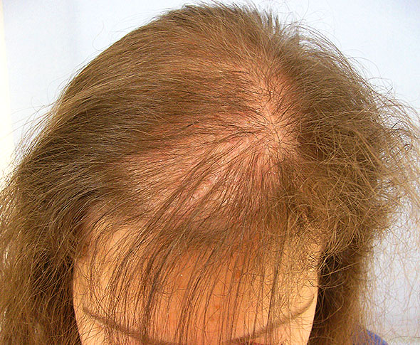
Women disproportionately suffer from autoimmune diseases at a rate of 3 to 1 compared to men. Most believe hormones influence the risk of autoimmune disease but determining the mechanisms has eluded researchers for some time. In animal research, when male rats are castrated, the incidence of autoimmune disease goes up, but when female rats are castrated – their ovaries are removed, the results are mixed. New research finds that the bacteria in the gut interact with hormones and provide the key to understanding the sex differences in the risks for and onset of autoimmune disease.

In a study published in the journal Immunity, Gender Bias in Autoimmunity is Influenced by Microbiota, researchers tested the potential signals between hormones and immune function, gut microbes and immune function, and hormones plus gut microbes and immune function in male and female mice. They wanted to delineate the relationship between hormones, specifically testosterone, gut bacteria and autoimmune factors. Is it solely the hormone signal that influences the types and distribution of gut bacteria that then initiates or regulates the immune response or do the gut microbes regulate the hormones which then influence the immune response, or is it some combination of the two? It appears to be the latter; hormones and gut microbiota interact synergistically to regulate the expression of various immune factors and each other.

In a series of experiments, researchers were able to show that the gut bacteria in post pubescent male mice, both quantitatively and qualitatively differed from those of the female mice and testosterone was the key. In male mice, there appeared to be a feedback loop in which testosterone increased gut bacteria and the gut bacteria in turn increased testosterone, to some degree. Once the minimum threshold was reached, higher concentrations of testosterone did not influence gut bacteria any longer. Because these bacteria in turn control the balance between inflammatory and anti-inflammatory factors researchers postulate that the relationship between gut microbiota and testosterone may be key in understanding the gender bias in autoimmune disease.

Questions remain. Testosterone is not the only androgen within the hormone pathway. There are other androgens that have noted and direct influence on immune factors. Moreover, while testosterone may be implicated that does not necessarily rule out a role for the estrogens, as testosterone is the precursor hormone for estrone, estradiol and the other estrogens. The downstream conversion from testosterone to estrone or estradiol may be an important consideration for future research. Nevertheless, this study highlights the importance of hormones in disease, and more specifically, the critical cross-talk between hormones, gut bacteria and inflammatory/anti-inflammatory factors.

We Need Your Help

More people than ever are reading Hormones Matter, a testament to the need for independent voices in health and medicine. We are not funded and accept limited advertising. Unlike many health sites, we don’t force you to purchase a subscription. We believe health information should be open to all. If you read Hormones Matter, like it, please help support it. Contribute now.

Yes, I would like to support Hormones Matter. 

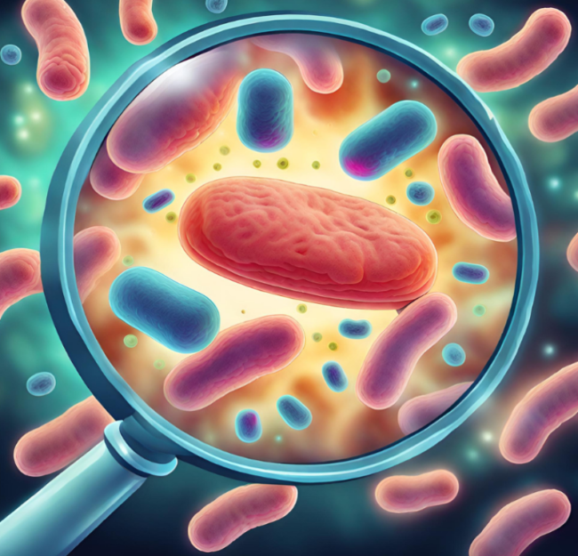
Image created using Canva AI.

This article was originally published on September 12, 2013. 
Glyphosate Induced Obesity?

12118 views

Are you struggling with your weight? Are you eating well and exercising but still not losing weight? Well then, it might be time to consider what’s on or in what you are eating or what you are eating eats. Sound complicated? It’s not. An emerging body of evidence shows a strong link between eating foods sprayed with commercial herbicides and eating meats raised on commercial feedlots (that are born and bred on a cocktail of chemicals) and obesity.

After years of eating highly processed and chemically laden fruits, vegetables and meats, the bacteria in our guts shift radically towards a species that emit what are called endotoxins. These endotoxin releasing bacteria induce inflammation, which then shifts a series biochemical pathways that favor fat storage as a protective and compensatory reaction to the steady state of chemicals coming from our diet and the lack of nutrients contained within these foods. Indeed, what we now call autoimmune reactions, the continued elevation in inflammation and antibodies, may be a result of the food we eat (and the other pharmacological and environmental chemical exposures). It turns out, that the constant state of inflammation many of us find ourselves in is the body’s way of trying to clear those toxins.

With obesity in particular, there have been several interesting studies published over the last couple years providing clear links between chemical exposures and fat storage. Whether the body stores fat or uses fat depends upon the balance of good and bad bacteria in the gut and that balance is predicated heavily upon nutrient availability and toxic exposures. High calorie, low nutrient, chemically dosed foods, shift bacterial communities that increase fat storage and inflammation. Not only that, but since gut bacteria metabolize dietary vitamins and even synthesize vitamins from scratch on their own, the high fat, low nutrient, chemically laden diet downregulates the vitamin producing bacteria, in favor of the more pathogenic and opportunistic bacteria. This further depletes nutrient stores while enhancing inflammation. The cycle becomes very difficult to end, as anyone struggling to lose weight knows all too well. There is hope, however. New research from disparate sources demonstrates how reducing the toxic load and increasing nutrient availability can re-calibrate fat usage and storage parameters.

Gut Bacteria and Obesity

Just a few years ago, researchers from Shanghai, China identified one of the gut bacterial over growths associated with obesity and published their results in a paper entitled: An opportunistic pathogen isolated from the gut of an obese human causes obesity in germfree mice. Called enterobacter clocae, the endotoxin producing bacteria was found overpopulated in the gut of a severely obese patient who was also insulin resistant, hypertensive and suffered from the array of obesity related health issues. The enterobacter clocae pathogens made up 35% of the total bacterial content in this patient’s gut; a huge bacterial load. Knowing that enterobacter emitted endotoxins and that endotoxins were associated with inflammation and insulin dysregulation, the researchers speculated that a reduction in the enterobacter population would correspond with a reduction in weight and the other health issues. They were correct. With a special diet and traditional Chinese herbs, weight loss and health parameters changed along with the reduction in toxic load. After 9 weeks, enterobacter represented only 1.7% of the total gut bacteria and at 23 weeks, .32%. The total weight loss during that period was 50kg or 110lbs.

Could something as simple as reducing the opportunistic enterobacter via diet be the solution to obesity? To answer this question, the researchers went back to lab and designed an experiment to test the hypothesis, only they did it in the reverse. They asked if enterobacter was a causative factor in obesity, could they induce obesity in mice bred specifically to resist excessive weight gain simply by increasing the bacterial load?

From the fecal matter of the obese patient, the researchers isolated the particular strain of enterobacter clocae called B29. They took the B29 and inoculated four groups of seven, germ-free mice; B29 inoculated plus normal diet or high fat diet and non-inoculated normal or high fat diet. Germ-free mice are a strain of mice that are microorganisms free and raised in isolates. They are resistant to obesity even when fed a high fat diet.

One mouse from each of the inoculated groups died immediately after the inoculation indicating the toxic nature of this bacteria. Remember, this strain of bacteria represented 35% of the original patient’s gut bacteria, likely acquired gradually over the course of lifetime. During the first week, all of the inoculated mice lost weight, again indicating the mounting immune response. Anorexia, is often a sign of illness as the body reallocates resources towards fighting an infection.

Subsequently, and after the immediate anorexic responses, both groups of inoculated mice gained excessive weight, whereas the non-inoculated mice did not. The inoculated plus high fat diet group not only gained significantly more weight but expressed higher levels of enterobacter inflammatory markers and insulin resistance showing an interaction between diet and bacterial growth. The researchers speculate that the high fat diet facilitates the transfer of this bacteria to the bloodstream and increases the systemic inflammatory reaction. The inflammation then shifts the body towards fat storage via a range biochemical cascades meant to fight the infection but that also induces other reactions along the way; reactions we consider hallmarks of metabolic disease including high cholesterol, insulin resistance, liver damage, decreased adiponectin (satiety hormone – low adiponection means one is always hungry) and even increased amyloid A proteins associated with Alzheimer’s. This study, albeit small and in need of replication, shows us that when the balance of good to bad bacteria shifts, obesity is induced. It doesn’t tell us, however, how environmental chemicals in and on food impact this bacterial shift. For that we have to go to a couple other reports.

Nutritional Perils of the Western Diet

The Western diet has become a synonymous with highly processed foods that barely resemble actual food in nutrient and DNA composition. Indeed, in our efforts to produce the largest and prettiest produce, we’ve cultivated out 95% of the genetic variation from food crops; reducing to almost nothing the ~200,000 plant metabolites that provide nutrition. To make matters worse, we have substituted nutritionally rich and diverse crops with ones that originate from plant seeds engineered with bacterial RNA and DNA and are laced with glyphosate, adjuvants and other chemicals. In addition, all commercial meat production relies heavily on genetically modified, glyphosate-doused feed to grow the cattle, combined with prophylactic antibiotics, growth hormones and a cocktail of other chemicals that compensate for the deplorable conditions under which Western foods are produced. The genetically modified, chemically laden food stuffs are then sold to the consumer as fruits, vegetables, meats and dairy or processed even further into other food-like products. From beginning to end of the food chain are exposures to chemicals and foreign bacterial DNA that our bodies cannot accommodate and that provide only limited nutrients.

So, in addition to the direct exposure to chemical toxicants, conventionally grown Western foodstuffs also impair health by reducing vital nutrient content required for even the most basic cell functioning. By disrupting the balance between good gut bacteria and bad or pathogenic bacteria conventionally grown further disrupts nutrient availability while increasing inflammation and the cascade of ill-health is set in motion.

Metabolic Starvation in the Face of Obesity

As we’ve covered previously, every cell in the body requires energy to exist and function. That energy comes in the form of mitochondrial adenosine triphosphate or (ATP). The production of ATP requires nutrients as co-factors and for enzyme functioning. Many of these nutrients come from diet and others are produced de novo or from scratch by the bacteria in our gut. Glyphosate grown foods attack both. Glyphosate reduces the nutrient availability of foodstuffs, even in the less processed, presumed healthy fruits and vegetables, while simultaneously killing the good bacteria in our guts. Glyphosate is a potent bactericide that in a perverse twist of design preferentially targets the beneficial bacteria while leaving untouched the opportunistic and pathogenic bacteria, like enterobacter clocae. So while eating a healthy diet might lead to weight loss and improved health outcomes under normal circumstances, when that diet consists of conventionally grown foods, with genetically engineered seeds capable of withstanding the toxic insults of glyphosate and its adjuvants, neither the diet nor the disrupted intestinal flora can produce the nutrients required to enable healthy cellular metabolism. The GM-glyphosate combo induces a state of metabolic starvation and through a number of survival pathways and shifts towards fat storage rather than fat loss as a secondary source of energy.

Critical to this entire equation is the fact that the bactericidal properties of glyphosate disrupt normal gut microflora.  Glyphosate directly shifts the balance of power away from the healthy, vitamin and mineral factories that feed the body’s enzymes and mitochondria, towards more pathogenic bacteria that are resistant to glyphosate and may even feed on it, further evoking metabolic starvation. As the bacterial balance continues to shift, disease appears and inflammation ensues. Those diseases are then treated pharmacologically with drugs that also disrupt gut bacteria, deplete nutrient stores and damage mitochondria. The cascade of ill-health becomes more and more difficult to end using traditional approaches. Moreover, where and how disease appears is as much based upon individual predispositions as it is on nutrition and other exposures, making the complexity of modern illness something modern medicine is not accustomed too. In other words, these diseases do not fit neatly into the one disease, one medication model, and thus, very rarely respond favorably to treatment.

To Lose Weight, Feed the Body What it Needs: Nutrients.

Despite the complexity of the interactions that come together and create the chronic health issues we face today, there is one variable that can be controlled that will mitigate obesity and ill-health directly: eating, or more specifically, what is eaten. The simple act of cleaning up one’s diet, of moving away from processed foods and away from conventionally grown foods towards organics, can have a tremendous effect on reducing the body’s toxic load and subsequent inflammation, weight gain, and disease. Similarly, replacing needed micronutrients so that bacterial and mitochondrial functioning can come back online and switch from fat storage to fat/energy burning will be critical. This will take time, however, and the transition towards health may be slow. Obesity and ill-health did not emerge overnight and they will not disappear overnight. Finally, we have to recognize that there is no one-size-fits-all, silver bullet, diet vitamin or diet pill. Each of us adapts to chemical exposures and the lack of nutrition individually and uniquely. So each of us requires a different cocktail of nutrients to move forward. Which nutrients and at what doses should be determined individually and may involve some degree of trial and error. As the Western diet is devoid of critical vitamins, minerals and amino acids, it is likely many individuals are suffering from broad based deficiencies. It is also likely, that restoring what has been absent chronically will go a long way towards health and healing, regardless of one’s particular health issues. So if you are struggling with obesity and other health issues, feed your body what it needs to function – nutrients.

We Need Your Help

More people than ever are reading Hormones Matter, a testament to the need for independent voices in health and medicine. We are not funded and accept limited advertising. Unlike many health sites, we don’t force you to purchase a subscription. We believe health information should be open to all. If you read Hormones Matter, like it, please help support it. Contribute now.

Yes, I would like to support Hormones Matter. 

Photo by i yunmai on Unsplash.

This post was published originally on Hormones Matter on July 28, 2014. 


Thoughts on Inflammation, Vaccines and Modern Medicine

6550 views

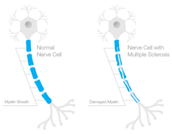
One of the core components of an HPV vaccine adverse reaction inevitably includes some degree of seemingly unexplainable but observable brain inflammation and white matter disintegration. The brain inflammation falls under a number of different names and diagnoses, some are regionally specific, cerebellar anomalies for example, while others demarcate a more diffuse injury including, acute disseminated encephalomyelitis (ADEM), myalgic encephalomyelitis (ME), sometimes known as chronic fatigue, multiple sclerotic (MS) type lesions and, the newest and perhaps more prescient among them, a set of conditions designated as Autoimmune/Inflammatory Syndrome Induced by Adjuvants or ASIA that denote chronic inflammation both centrally and peripherally relative to vaccine adjuvant exposure.

Is the Brain Immune Privileged?

Despite the observance of brain inflammation in many post HPV vaccine victims, many practitioners, and indeed, the FDA and CDC, seem loathe to recognize that an aluminum lipopolysaccharide adjuvanted virus vector might induce a neuro-inflammatory response, leaving patients with little recourse post injury. The difficulties attributing brain inflammation to a vaccine reaction stem from a long held belief that the blood brain barrier is stalwart in its protection against peripheral trespassers.  The brain has long been considered, ‘immune privileged’ having little to no communication with peripheral immune function. Indeed, the perceived impenetrableness of the blood brain barrier is so extensive that brain-body separation might as well be complete, with a brain in bottle and a decapitated body.

Logically, we know this cannot be true. There must be crosstalk between the immune systems of brain/central nervous system and that of the body. How else could we survive if the two modalities were segregated so completely? It turns out, that logic may be prevailing. A decade of research suggests that the long held notion brain immune – privilege is completely and utterly incorrect. Indeed, the immune system not only guides early neurodevelopment (and so mom’s immune function matters) but communicates and affects brain morphological changes chronically. Likewise, signals from the brain continuously influence peripheral immune function.

The immune system appears to influence the nervous system during typical functioning and in disease. Chronic infection or severe illness may disrupt the balance of normal neural–immune cross-talk resulting in permanent structural changes in the brain during development, and/or contributing to pathology later in life. The diversity, promiscuity, and redundancy of “immune” signaling molecules allow for a complex coordination of activities and precise signaling pathways, fundamental to both the immune and nervous systems. 

It should not be surprising then, that nutrient status and toxicant exposures in the periphery, in the body, affect central nervous system function and are capable of inducing brain inflammation and vice versa. And yet, it is; perhaps even more so than any of us realize.

Re -Thinking Brain Inflammation

When one reads through the definitions, research and case reports of ADEM, ME, MS or other instances of brain inflammation, the notion that biochemical lesions in the periphery are linked to observed neuro-inflammatory reactions is far from center stage. Nevertheless, if we can accept the premise what happens in and to the body does not stay in the body, then we can begin to re-frame our approach to brain inflammation. Specifically, we can look at inflammation more globally and ask not only what triggers inflammation, but allows inflammation to persist chronically, regardless of its location. If there is an on-going peripheral inflammatory response, is it not prudent to suspect that a similar response might be occurring within the central nervous system, even if our imaging tools are not yet capable of visualizing the inflammation; even if it is too premature to observe demyelination, neuronal, axonal swelling or other telltale signs of chronic brain inflammation?  I think it is.

Vaccine Adjuvants: A Pathway to Brain Inflammation

With the HPV vaccine, and indeed, any vaccine, the deactivated viral vectors come with a cocktail of additional chemical toxicants and a metal adjuvant to boost the recipient’s immune response, as measured by the increase in post vaccine inflammatory markers. It is believed that without these adjuvants (and data back this up), the recipient’s immune response is insufficiently activated to merit ‘protection’ against the virus. The strength or size of the immune response is then equated with success and protection.

By this equation, an excessive immune response that continues chronically and is eventually labeled ‘autoimmune’ as innate systems begin to fail, is in some way not a failure or side effect, but an example of extreme success; the larger the immune response, the stronger the vaccine. And so, skewed as this observation may seem, within the current vaccine-paradigm there can be no ‘side-effects’, not really. By design, there should be inflammation, even brain inflammation; the more the better. Also by design, metal, lipid soluble, adjuvants cross the blood brain barrier and directly induce brain inflammation. To say vaccines don’t or somehow couldn’t induce brain inflammation is ignorant, if not, utterly negligent, and quite simply, defies logic. Again, for prudence and safety, shouldn’t we assume that an inflammatory reaction in the body might also ignite some concordant reaction in the central nervous system?

Why Aren’t We All Vaccine Injured?

What becomes apparent though, is even with exposure to the most toxic brew of vaccines, not all who receive vaccines are injured, at least observably. (I would argue, however, even those who appear healthy post vaccine, had we the tools to observe brain inflammation more accurately, would show a central inflammatory response, at least acutely, and likely, progressively). So what distinguishes those individuals who seem fine post vaccine, particularly post HPV vaccine, from those who are injured severely and sometimes mortally?

More and more, I think that the fundamental differences between vaccine reactors and non-reactors rest in microbial and mitochondrial health. Indeed, all vaccines, medications, and environmental toxicants damage mitochondria, often via multiple mechanisms, while altering microbial balance. Whether an individual can withstand those mitochondrial insults depends largely upon a balance struck among three variables: 1) heritable mitochondrial dysfunction, genetic and epigenetic; 2) the frequency and severity of toxicant exposures across the lifetime; and 3) nutrient status. Those variables then, through the mitochondria, influence the degree and chronicity of inflammation post vaccine. With the HPV vaccine in particular, the timing of the vaccine, just as puberty approaches and hormone systems come online, may confer additional and unrecognized risks to future reproductive health.

Mitochondria and Microbiota

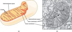
The mitochondria, as we’ve written about on numerous occasions, control not only cellular energy, but cell life and death. Every cell in the body, including neurons in the brain, require healthy mitochondria to function appropriately. Healthy mitochondria are inextricably tied to nutrient concentrations, which demand not only dietary considerations but balanced gut microbiota. Gut bacteria synthesize essential nutrients from scratch and absorb and metabolize dietary nutrients that feed the mitochondria. Indeed, from an evolutionary perspective, mitochondria evolved from microbiota and formed the symbiotic relationship that regulate organismal health. Disturb gut bacteria and not only do we get an increase in pathogenic infections and chronic inflammation, but also, a consequent decrease in nutrient availability. This too can, by itself, damage mitochondria.

When the mitochondria are damaged, either by lack nutrients and/or toxicant exposure, they trigger cascades of biochemical reactions aimed at conserving energy and saving the cell for as long as reasonably possible. When survival is no longer possible, mitochondrial sequestration, and eventually, death ensue, often via necrosis rather than the more tightly regulated apoptosis. Where the mitochondria die, cells die, tissue dies and organ function becomes impaired. I should note, as steroid hormone production is a key function of mitochondria, hormone dysregulation, ovarian damage and reduced reproductive capacity may be specific marker of mitochondrial damage in young women.

Mitochondria and Inflammation

Mitochondria regulate immune system activation and inflammation and so inflammation is a sign of mitochondrial damage, even brain inflammation. Per a leading researcher in mitochondrial signaling:

The cell danger response (CDR) is the evolutionarily conserved metabolic response that protects cells and hosts from harm. It is triggered by encounters with chemical, physical, or biological threats that exceed the cellular capacity for homeostasis. The resulting metabolic mismatch between available resources and functional capacity produces a cascade of changes in cellular electron flow, oxygen consumption, redox, membrane fluidity, lipid dynamics, bioenergetics, carbon and sulfur resource allocation, protein folding and aggregation, vitamin availability, metal homeostasis, indole, pterin, 1-carbon and polyamine metabolism, and polymer formation.

The first wave of danger signals consists of the release of metabolic intermediates like ATP and ADP, Krebs cycle intermediates, oxygen, and reactive oxygen species (ROS), and is sustained by purinergic signaling.

After the danger has been eliminated or neutralized, a choreographed sequence of anti-inflammatory and regenerative pathways is activated to reverse the CDR and to heal.

When the CDR persists abnormally, whole body metabolism and the gut microbiome are disturbed, the collective performance of multiple organ systems is impaired, behavior is changed, and chronic disease results.

Reducing Inflammation

Instinctively, we think reducing inflammation pharmacologically, by blocking one of the many inflammatory pathways, is the preferred route of treatment. However, this may only add to the mitochondrial damage, further alter the balance of gut microbiota and ensure increased immune activation, while doing nothing to restore mitochondrial and microbial health. In emergent and acute cases, this may be warranted, where an immediate, albeit temporary, reduction in inflammation is required. The risk, however, is that short term gains in reduced inflammation are overridden by additional mitochondrial damage and increased risk of chronic and/or progressive inflammation. The whole process risks becoming a medical game of whack-a-mole; a boon to pharmaceutical sales, but devastating to those who live with the pain of a long-standing inflammatory condition.

In light of the the fact that damaged mitochondria activate inflammatory pathways and that vaccines, medications and environmental toxicants induce mitochondrial damage, perhaps we ought to begin looking at restoring gut microbial health and overall mitochondrial functioning. And as an aside, perhaps we ought to look at persistent inflammation not as an autoinflammatory reaction, but for what is it, an indication of on-going mitochondrial dysfunction.

We Need Your Help

Hormones Matter needs funding now. Our research funding was cut recently and because of our commitment to independent health research and journalism unbiased by commercial interests we allow minimal advertising on the site. That means all funding must come from you, our readers. Don’t let Hormones Matter die.

Yes, I’d like to support Hormones Matter.

Image by Pavlo from Pixabay.

This post was published originally on Hormones Matter on September 22, 2014.

Traveling Super Bacteria in Commercial Livestock

2354 views

With the trend in commercial livestock to raise large numbers of animals in small and confined spaces, fed a regular diet of antibiotic doused feed, it should be no surprise when those animals develop infections, even antibiotic resistant infections. According to the third annual report released by the FDA’s Center for Veterinary Medicine in 2012, antimicrobials sold or distributed for food-producing animals are increasing, rapidly, far and above human use, and despite recommendations to the contrary. In 2011:

  • 29.9 million pounds of antibiotics were sold in the US for meat and poultry production
  • 7.7 millions pounds of antibiotics were sold in the US for humans

Along with that increased usage comes an increased incidence of antibiotic resistant strains of bacteria found in the animals, the retail meats sold for human consumption and in the humans who raise these animals and consume these meats with overuse of fluoroquinolone antibiotics being one the main culprits.

More recently, researchers investigating the persistence of livestock associated antibiotic resistant staphylococcus aureus among the industrial hog workers in North Carolina, found that fully 86% of the workers (n=22) carried the livestock staph bacteria nasally over the course of the two week testing period that sometimes included 96 hours away from the hog farms. One worker carried methicillin resistant staphylococcus aureus (MRSA) persistently and 46% of the workers carried the more virulent strain of mutli-drug resistant staphylococcus aureus (MDRSA) at some point during the test period. The researchers observed that carriage of the bacteria continued even when the workers were away from the farms. Although it should be noted, most workers were at the farms over 50 hours per week and rarely had more than 24 hours off unless ill (in itself, a cause for concern).

Traveling Bacteria

As we learn more about the microbial environment, it becomes clear that organisms, human and animal, are walking bacterial ecosystems, with trillions of microbes upon and inside us. Which populations of bacteria predominate have as much to do with the host organism’s health and habits as with his/her exposures. Bacteria and other microbial pathogens are emerging as a flexible interface of sorts, between us and the environment.

From birth onward which bacteria thrive is moderated by the environment. A child born by cesarean in a hospital adopts the bacteria of the hospital setting, even the pathogenic ones, whereas a child born vaginally develops much of the mom’s microbial influence. More interestingly, a child born vaginally and at home, adopts the microbial patterns of her surrounds, even from that of the family pet. Indeed, across the lifespan, our bacterial exposures influence our microbiome. Living with pets, eating habits and environmental exposures change our microbial ecosystems, regularly and continuously, as we adapt to our surroundings – and these changes happen quickly.

Change the diet and the gut microbiome changes within days. Spend time in the hospital, your microbiome changes within hours. Buy a dog, enter a relationship, ditto. The totality your environment influences your microbiome. And so, it should be no surprise that the animals raised in deplorable conditions, would carry dangerous and deadly bacteria more frequently than animals raised on organic feed and in a more healthy environment. Similarly, it should no surprise that those who work with those animals carry those same deadly bacteria and that their health and the health of their families could be impacted by microbial environment in which they work, but it is.

Even though the importance of the microbiome has only recently come to light, common sense should tell us that the way we raise and grow our food is not healthy for the animals or for us. I suspect, our hunter-gatherer ancestors would not consume diseased animals, understanding the risk of illness to themselves, but we do, every day. Perhaps it is because we do not see the animals and the connection between their health and ours is lost; perhaps it is because some of these animals look healthy bolstered by growth hormones, antibiotics and other chemical toxicants; perhaps it is because we don’t want to see or to think about what we put in our mouths. Whatever the reason, commercial livestock practices are becoming increasingly dangerous to human health. Whether we recognize those dangers or not, we will bear the costs in the chronic illnesses that raising and eating those animals initiate.

Endometriosis and the Gut – What Monkeys Can Tell Us About the Disease

3600 views

Female monkeys bred in captivity develop endometriosis at rates similar, if not a little higher (~20%) than those of the general population of women. Histologically, the endometriotic tissue in monkeys is identical to that in human women. Clinically, however, by the time symptoms present in monkeys or are recognized by their caregivers, the endometriosis has fully invaded the peritoneal cavity and the severity of the disease is often significant. With similarities in disease expression and because full medical necropsy (autopsy) reports are kept on all animals raised in captivity, investigating endometriosis in monkey may provide insight into this complicated disease process. Indeed, the few studies that have addressed endometriosis in monkey populations demonstrate clues overlooked in human research.

Risk Factors for Endometriosis in Monkeys

Like women, the risk factors in monkeys include, familial history, age, estrogens and environmental exposures. The link between estrogens, specifically estradiol implants, increases significantly with repeated exposures. Monkeys who have had more than three estradiol implants over the course of their lifetimes were nine times more likely to have the disease than their counterparts. Similarly, connections between environmental toxin exposures and risk of endometriosis have been noted.

An interesting finding in monkeys, perhaps not identified in humans, is a link between uterine surgery (hysterotomy), such as c-section or abortion and increased risk of ectopic endometriosis, by as much as five-fold. The theory being that the surgery ‘seeds’ the disease process by releasing the endometrial cells into the peritoneal cavity. Laparoscopic surgeries do not appear to increase risk, but the data are limited.

Gut Microflora

Like human women, female monkeys with endometriosis often have intestinal involvement with bowel movement irregularity, and as the disease progresses, visible endometrial implants along the large and small intestines. The presumption is that the damage to intestines arrives from the outside in; that is, from the endometrial tissue growth bearing down on the intestines and other organs. What if, there are factors within the gut that predispose or at least increase the risk or severity of intestinal involvement?  It turns out, there may intestinal factors that influence disease progression – the intestinal microflora.

In recent decades, our understanding of the importance of gut bacteria has increased significantly. We know that there are good bacteria and bad bacteria and that shifting the balance between those bacterial populations can lead to a host of health problems. In monkeys with endometriosis, the balance of bacterial power is significantly shifted in favor of yeast over-growth, gram-negative microbes and general intestinal inflammation. Compared to healthy controls, monkeys with endometriosis had significantly lower concentrations of lactobacillus, the good bacteria combined with significantly higher concentrations of gram negative bacteria such as E.Coli, Salmonella, the families of Enterobacteria, Klibsiella and Proteus. Additionally, intestinal inflammation was more prevalent in monkeys with endometriosis than in healthy monkeys. Is there a connection between gut bacteria and endometrial pain or gut bacteria and IBS? With the former, there is still too little research to tell, but relationship between gut bacteria and IBS is becoming more clear.

Gut Microflora, Irritable Bowel Syndrome and Probiotics

Irritable bowel syndrome (IBS) and endometriosis often co-occur. IBS can be misdiagnosed or pre-diagnosed ahead of endometriosis. Research suggests altered intestinal microflora in IBS similar to that identified in the monkey research described above, with reduced lactobicillus bacteria in combination with increased gram-negative bacteria. Endometriosis was not a variable. Could probiotic treatment improve IBS and by extension endometriosis related intestinal symptoms? Possibly.

A recent double-blind, placebo controlled study investigated whether a six month treatment protocol with probiotics relieved the common IBS symptoms of abdominal pain, distension, bowel movements (diarrhea and constipation), flatulence and stomach rumble. The researchers found a median reduction of symptoms 42% in the probiotic group versus the placebo group. The largest and statistically significant reductions were observed in abdominal pain and distension with few significant improvements in the quality or constitution the the bowel movements.

Though probiotics are common supplements in complementary care, no studies that I can identify have looked at either the gut bacteria in human endometriosis patients or evaluated the use of probiotics with these patients. With limited research from the monkey studies, however, perhaps we ought to look at the role of intestinal microflora and in women suffering from endometriosis. At the very least, restoring the balance of gut microflora could improve overall health. Moreover, I can’t help but think that as more and more studies link endometriosis and autoimmune diseases to alterations in micronutrients like vitamins A, B and D, damage to and/or changes in gut microflora might be involved.

We Need Your Help

More people than ever are reading Hormones Matter, a testament to the need for independent voices in health and medicine. We are not funded and accept limited advertising. Unlike many health sites, we don’t force you to purchase a subscription. We believe health information should be open to all. If you read Hormones Matter, like it, please help support it. Contribute now.

Yes, I would like to support Hormones Matter.