migraine ehlers danlos

Migraine Energy Metabolism: Connecting Some Dots

8898 views

I have been reading some of the fascinating posts by Angela Stanton PhD concerning her research in migraine headaches. I regard the substance of her discussions as somewhat like dots on a chart that need to be connected. I learned a great deal about the chemistry involved in migraine. One of her comments that involves ion homeostasis in brain metabolism is fascinating. She noted that “serotonin is created by a normally functioning brain. Why it decreases or increases in the brains of migraineurs has always puzzled me. Should we not try to find out why?” That simple three letter word is the heart and soul of research and I believe that I may be able to add some information that might provide an answer.

Ehlers Danlos and Migraine

In one of Angela’s posts she discusses a subject which has been of interest to me for many years, the overlap of symptoms in disease. She noted that 60% of migraineurs have one type of Ehlers Danlos syndrome (EDS) and 43% of EDS have minor changes in DNA (SNPs) found in migraineurs. She concludes that they must be related. Over 70% of migraineurs have Raynaud’s disease and there is an overlap with EDS and Raynaud’s. Therefore, she concludes that these three diseases are variants. In  fact, there is an association between EDS, Postural Othostatic Tachycardia Syndrome (POTS) and a group of conditions known as mast cell disorders. EDS-HT, (one of the manifestations of this disease), is considered to be a multisystemic disorder, involving cardiovascular, autonomic nervous system, gastrointestinal, hematologic, ocular, gynecologic, neurologic and psychiatric manifestations, including joint hypermobility. Many non-musculoskeletal complaints in EDS-HT appear to be related to dysautonomia, consisting of cardiovascular and sudomotor dysfunction. Many of the clinical features of patients with mitral valve prolapse can logically be attributed to abnormal autonomic function. Myxomatous degeneration of valve leaflets with varying degree of severity is reported in the common condition of mitral valve prolapse.

A woman, with what was described as a “new” type of EDS, died after rupture of a thoracic aortic aneurysm. Autopsy revealed myxomatous degeneration and elongation of the mitral and tricuspid valves. Patients with POTS, a relatively common  autonomic disorder, may have EDS, mitral valve prolapse, or chronic fatigue syndrome and are sensitive to various forms of stress, as depicted in the clinical treatment of a dental patient affected by the syndrome. Dysautonomia has been described in the pathogenesis of migraine, featured by nausea, vomiting, diarrhea, polyuria, eyelid edema, conjunctival injection, lacrimation, nasal congestion and ptosis. In general, there is an imbalance between sympathetic and parasympathetic tone.

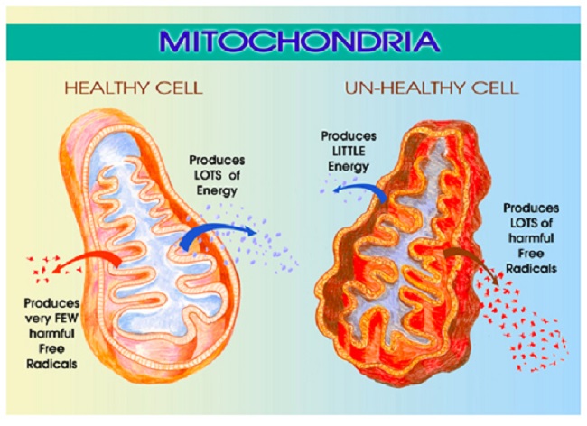
Energy Metabolism and Migraine

Technological studies have confirmed the presence of deficient energy production together with an increment of energy consumption in migraine patients. An energy demand over a certain threshold creates metabolic and biochemical preconditions for the onset of the migraine attack. Common migraine triggers are capable of generating oxidative stress  and its association with thiamine homeostasis suggests that thiamine may act as a site-directed antioxidant. It strongly suggests that migraine is a reflection of an inefficient use of brain oxygen.  An intermediate consumption of oxygen between deficiency and excess appears to be a necessity at all times. In fact,” moderation in all things” is an important proverb

Backing up energy deficiency, two cases of chronic migraine responded clinically to intravenous administration of thiamine. However, the authors are in error when they state in the abstract that “nausea, vomiting and anorexia of migraine may lead to mild to moderate thiamine deficiency”. An otherwise healthy 30-year-old male acquired gastrointestinal beriberi after one session of heavy drinking. Nausea, vomiting and anorexia relentlessly progressed. He had undergone 11 emergency room visits, 3 hospital admissions and laparoscopy within 2 months but the gastrointestinal symptoms  continued to progress, unrecognized for what these symptoms represented. When he eventually developed external ophthalmoplegia (eye divergence), he received an intravenous injection of thiamine which reversed both the neurologic and gastrointestinal symptoms within hours.

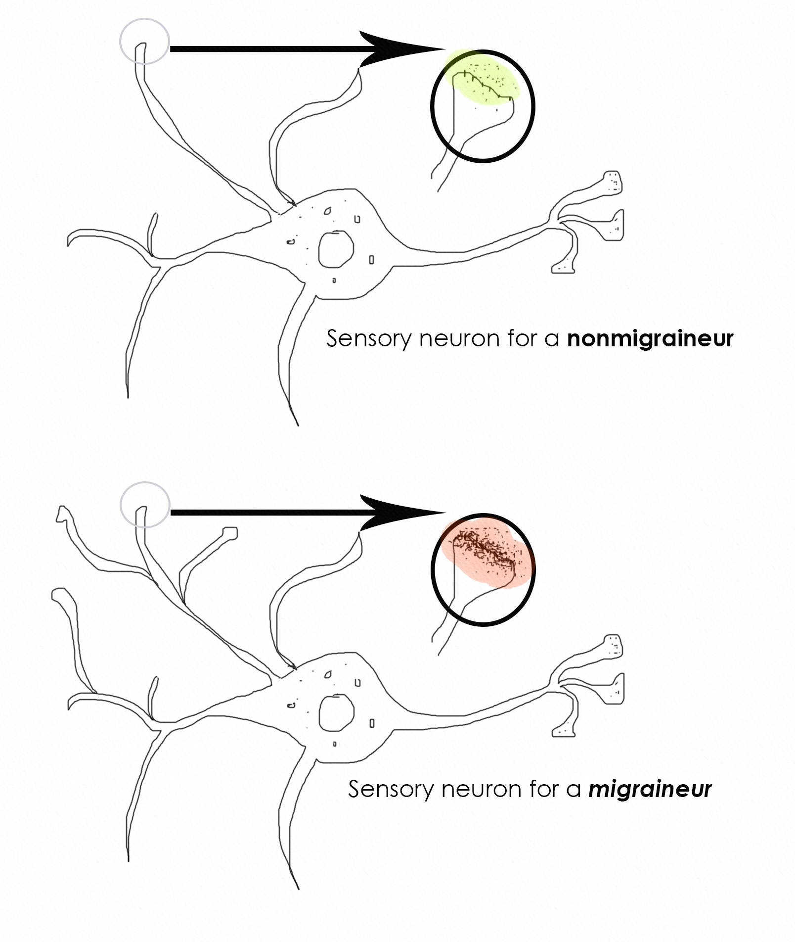
In other words it is important to be aware that nausea, vomiting and anorexia are primary symptoms of beriberi due to pseudohypoxia in the brainstem where the vomiting center is located. Chronic migraine has a well documented association with insulin resistance and metabolic syndrome. The hypothalamus may play a role. One of Angela’s comments concerns ion homeostasis in migraines. Thiamine triphosphate (TTP) can be found in most tissues at very low levels. However, organs and muscles that generate electrical impulses are particularly rich in this compound. Furthermore, TTP increases chloride (ion) uptake in membrane vesicles prepared from rat brain, suggesting that it could play an important role in the regulation of chloride permeability. Although this research was published in 1991, the exact role of TTP is still unknown. It has been hypothesized that thiamine and magnesium deficiency are keys to disease.

Angela wondered why serotonin might be increased or decreased in migraineurs. I strongly suspect that it is due to brain thiamine deficiency as the ultimate underlying cause of the migraine. In a review of thiamine metabolism, it was pointed out that metabolites could be high or low according to the degree of the deficiency. Victims of beriberi were found to have either a low or a high potassium according to the stage of the disease. If they were found to have a low acid content in the stomach, treatment with thiamine resulted in a high acid content before it became normal. If the stomach acid was high it would become low before it became normal. Since low and/or high potassium levels may be found in the blood of critically ill patients, thiamine deficiency should be a serious consideration in the emergency room or ICU Thiamine deficiency may be the answer for the fluctuations of serotonin observed in migraine.

Redefining Disease Models

According to the present medical model, each disease is described as a constellation of symptoms, physical signs and laboratory studies, each with a separate etiology. The overlap discussed by Angela suggests that the various conditions nominated have a common cause and that they are indeed nothing more than variations. If energy metabolism is the culprit, it would make sense of the infinite variations according to the degree and distribution of cellular energy deficiency. EDS-HT, described above is reported as a multi-system disease, exhibiting cardiovascular, autonomic gastrointestinal, hematologic, ocular, gynecological and psychiatric symptoms as well as the joint mobility. It seems to be impossible to explain this multiplicity without invoking energy deficiency as the cause. People with prolapsed mitral valve and a patient with a “new” form of EDS, reportedly have myxomatous degeneration as part of their pathology and it is tempting to suggest that such an important loss of structure might well be because of energy deficit.

The controls of the autonomic nervous system are located in the lower part of the brain that is particularly sensitive to thiamine deficiency and beriberi is a prototype for thiamine deficiency in its early stages. Dysautonomia is frequently reported as part of many different diseases, offering energy deficiency as the etiology in common. Yes, it is true that thiamine is not the only substance that enables the production of ATP. Nevertheless, it seems to dominate the overall picture of energy metabolism. It has long been considered the essential focus in the cause of beriberi, even though all the B complex vitamins are found in the rice polishings. Milling and the consumption of white rice was the prime etiology of the disease when it was common in rice consuming cultures.

We Need Your Help

More people than ever are reading Hormones Matter, a testament to the need for independent voices in health and medicine. We are not funded and accept limited advertising. Unlike many health sites, we don’t force you to purchase a subscription. We believe health information should be open to all. If you read Hormones Matter, like it, please help support it. Contribute now.

Yes, I would like to support Hormones Matter. 

Image by Chen from Pixabay.

This article was published originally on June 22, 2020.

Migraine: Do We Have It All Wrong?

22277 views

The combined APS (American Physiological Society) and EB (Experimental Biology) conference was full of excitement for me. This was my first migraine-associated presentation to scientists, MDs, and premed students. I have never presented a poster at any conference—I have usually given lectures. I was concerned. Will my posters catch any interest? Then I arrived at Chicago’s McCormick Conference Center and was shocked by its size and the size of this conference. There were 1500 poster boards, each with new posters every day for three days, in addition to booths and lectures. I took a deep breath.

Here I share with you what I presented and some of the questions attendees asked, answers I provided, and offer a more comprehensive explanation of what migraine is. I am also including one of the two posters I presented.

The first day, I presented a poster under the EB session so most visitors were curious about the biological aspects of migraine. The second was presented under the Electrolyte Homeostasis session, which brought very different visitors and very different questions. I find it important to define what migraine is and then continue with my hypothesis, which is drawn in the middle of the poster, and I highlight it here for easier viewing.

What is Migraine?

The answer is not pain. I found that the majority of those who have no migraines or never met anyone with migraines know migraine as a pain in the head. The truth is pain is just one – and not even mandatory – aspect of migraines. For me, presenting the real definition of migraines equals to explaining the conclusions of my many years of research.

Migraine is the manifestation of an energy (voltage) shortage in the brain, caused by insufficient electrolyte quality. This actually makes migraine into a symptom. The insufficient electrolyte mix is caused by certain genetic variances that result in some electrolyte mineral deficiency. Therefore, the electrolyte imbalance is also a symptom. The direct cause of migraine lies hidden in critical genetic variances that should operate the voltage gated channels of neurons but cannot. Why not? This is the heart of migraine: migraineurs are glucose sensitive with a brain that lacks capability to handle the huge electrolyte disruption when glucose enters the brain cells. As we know, adaptation is an important part of evolution. It seems that a very large percentage of the human population was able to adapt to the dietary circumstances of contemporary life but people with the migraine-brains could not. I call the migraine brain an ancient brain that is not able to utilize glucose. Modern carbohydrate-rich diets overload this ancient brain with glucose. Migraineurs are not the only ones with this problem. People with seizures, multiple sclerosis, Alzheimer’s disease, and many more conditions, have very similar—if not the same—problem. Not every human can eat birthday cakes, potatoes, and toast for breakfast. Migraineurs cannot.

The Migraine Brain Difference

sensory neuron comparison

Image copyright by Angela A. Stanton, Ph.D.©

Figure 1. Neuron: non-migraineur vs migraineur

The top image represents a single neuron in an average human brain, highlighting an enlarged section showing the receptors and the neurotransmitters in the circled region. The bottom image is the same for a migraineur. The two are obviously very different—look at the density of the receptors and neurotransmitters (the layer of black dots under red). The questions one must ask:

  1. Why do the migraineur’s neuron contain so many more receptors and neurotransmitters?
  2. Does that have anything to do with pain? And if so how and why?

A migraine brain’s sensory neurons are hyper sensitive (1) and hyper alert in comparison to the sensory neurons of a non-migraine brain. More alertness is explained by the more receptors (2) and neurotransmitters. But why do migraineurs have more receptors and hyper sensory brains? To answer this question I present a few hypotheses:

Hypothesis 1: If you look at many mammal species in the wild, you find they are always on alert for predators. If they are not on alert, they are likely to become food. For these species the hyper-sensory alert brain is the default. The migraine-brain is a hyper-sensory alert brain.

Hypothesis 2: If hyper-sensory brain can be the default then such a brain, the migraine-brain, is the default mammalian brain that all human ancestors must have had at one time. Since this brain is still very prevalent in the human population (15%), it must have represented a significant survival advantage in ancient times. Today, however, the majority of humans do not have this brain type.

Hypothesis 3: While the majority of humans were able to adapt to more modern lifestyles and food supply, some could not; their brain remained unchanged, still holding onto the ancient traits and retained hyper sensory organs. These then are today’s migraineurs.

Hypothesis 4: The tendency for electrolyte imbalance is a sign that the migraine-brain is greatly compromised under modern nutritional challenges. While the majority of humans adapted to the use of carbohydrates for fuel during our recent evolution, migraineurs did not. Up until about 15,000 years ago carbohydrates formed minimal part of the human diet.

Hypothesis 5: The ancient brain (in some of its traits) may go back as far as the Euarchontoglires (100 million years ago), the first common ancestor to all mammals and primates. The early mammals had a few special traits that appear to be connected to migraine-brain. Two of these are the Ehlers-Danlos Syndrome (EDS) and Raynaud’s Syndrome. Both of these are highly connected to migraine and to each other and are ancient traits that some small mammals still carry today—and apparently a very large percentage of migraineurs do as well, although in the general population the percentage is minuscule.

Hypothesis 6: Migraine is a neurovascular condition (not a disease) that is an evolutionary throwback. Neuro because it is neuron voltage energy generation problem and vascular because of the different vascular structure migraineurs have from non-migraineurs. Both EDS and Raynaud’s are vascular in nature and EDS is also associated with hyper mobility and being disjointed, which was a very important adaptation in early mammals and still are in many, such as cats, often referred to as floating shoulders. Having a different vascular system allows for more flexibility without vascular damage.

Hypothesis 7: Migraineurs have a very different vascular system from non-migraineurs. I propose the vasculature segments are shorter and more numerous. This can often be seen on the skin of people with EDS because they also have very thin and transparent skin. Shorter vascular regions allow for more flexibility along with the hyper flexibility of those afflicted with EDS.

Hypothesis 8: Migraine brain is of the era when being disjointed and having different vascular system was an evolutionary benefit. From evolutionary throwback perspective then it makes sense that migraineurs are glucose sensitive and carbohydrates intolerant, since at that time, carbohydrates and glucose were not consumed.

Support for the Hypotheses

Support for these hypotheses come from genetics, which shows us that the migraine brain has different voltage gated ionic channels for calcium, ATPase, the sodium/potassium pump, sodium channel, and potassium pump, implying that all electrolyte modulation for a migraineur is different from a non-migraineur, in addition to glucose and insulin regulatory variances. See below the top few genetic variances. I used GeneCards database to find all gene variance associated with migraines—sorted by importance called “score”—and removed some that are not relevant to my point:

GeneCards Migraine Genes

I only listed 16 gene variances from the 1293 currently associated with migraines (highlighted important words), though as more research is conducted, this number is certain to grow. Some variances that have low score—such as insulin resistance—will likely move to higher scores since migraineurs are associated with a very high incidence of metabolic disorders, specifically migraine is associated with insulin resistance (3) as a result of being glucose sensitive.

Migraines, Drugs, Nutrition

Pharmaceutical companies have spent much of their migraine-allocated time developing drugs that block migraineurs’ voltage gated channels that are different from that of the standard population, without any concern why they are different. As a result, these channels cannot work at all when medicated. Of course, just as there is no point in medicating blue eyes, a genetic variance, so is medicating a type of voltage gated channel is futile. It takes a bit of understanding why. At the conference I spent eight hours a day for two days explaining this to doctors, scientists, and even representatives of big pharma. It took me 10 years to figure it out.

Since “…serum Na+ falls by 1.4 mM for every 100-mg/dL increase in glucose, due to glucose-induced H2O efflux from cells” (4) (page 4), you can immediately see that as glucose enters the cells, sodium (Na+) falls and water (H2O) leaves the cell, causing major electrolyte disruption.

Regular human brains that have adapted and are able to reset electrolyte homeostasis with ease have no trouble. The migraine brain cannot because all associated voltage gated ionic pumps and channels are in their ancient forms, where carbohydrate consumption was too minimal to matter in electrolyte dysregulation. The long epoch in which the hyper sensory mammalian brain was still the dominant variant of humanity, sugar and grains were not part of the diet at all and very little if any carbohydrates were consumed.

The migraineurs’ exaggerated reactions to carbohydrate consumption shows that they have a problem with glucose metabolism. With this major negative, is there a way to prevent migraines without the use of any medicines? Absolutely. Before I detail the proper migraine nutrition, I would like to answer a question I received from some doubters during the conference. Facing a skeptic is great, it forces the scientist to come up with the appropriate answers.

The most common comment (particularly from those representing pharmaceuticals) was that “whatever is done without medicine must be placebo”. This was extremely easy to defend by the simple logic that goes as follows:

If A > B AND B > C then A > C 

This applies to pharmaceuticals since their clinical trials for medicines are placebo tested. Therefore, migraine medicines, M, are proven to be better than placebo, P. M > P. The medicine-free treatment available is MF. Given that the MF treatment has been successful for thousands of migraineurs who also quit taking all their migraine medicines and remained migraine free, we can state the following:

If MF > M AND M > P then MF > P – that is medicine free is better than placebo.

Medicine & Migraine Free

So what is medicine free and migraine free? It is quite simple: since migraineurs cannot use glucose as energy and if they try they end up with major electrolyte disruption as a result, it implies that by stopping the consumption of carbohydrates migraines can be prevented. Certain amount of protein also converts to glucose (gluconeogenesis) so controlling protein amount is also important but less critical.

Can migraineurs eat any carbohydrates? Yes, they can but minimal and highly fibrous. Carbohydrates of up to 5 net carbs grams (total carbs – fiber = net carbs grams) per meal seems to be tolerable by all migraineurs. For example, a migraineur can eat green leafy salads, cucumbers, zucchini, avocado, and many similar foods. However, she cannot eat a slice of bread, an apple, or drink smoothies, fruit juices, or soft drinks. See below the poster I showed in Chicago, summarizing what I just wrote and also showing what a migraineur should or should not be eating.

Image copyright by Angela A. Stanton, Ph.D.©

Conclusion

So we are dealing with a brain that is not used to eating carbs and this leads to glucose sensitivity, leading to major reaction to glucose with edema and sodium leaving cells. This leads to electrolyte dysregulation. Migraineurs on the Stanton Migraine Protocol®, LCHF, or the ketogenic diet, slowly taper off their medicines, their insulin reverts to normal and migraines typically vanish.

Sources

  1. Schwedt TJ (2013) Multisensory Integration in Migraine. Curr Opin Neurol:248-253.
  2. Wei Y, Ullah G, & Schiff SJ (2014) Unification of Neuronal Spikes, Seizures, and Spreading Depression. The Journal of Neuroscience:11733-11743.
  3. Fava A, et al. (2014) Chronic migraine in women is associated with insulin resistance: a cross-sectional study. European Journal of Neurology 21(2):267-272.
  4. Longo DL, et al. (2013) Harrison’s Manual of Medicine 18th Edition (McGraw Hill Medical, New York).

We Need Your Help

Hormones Matter needs funding now. Our research funding was cut recently and because of our commitment to independent health research and journalism unbiased by commercial interests we allow minimal advertising on the site. That means all funding must come from you, our readers. Don’t let Hormones Matter die.

Yes, I’d like to support Hormones Matter.

Image created using Canva AI.

This article was published originally on May 3, 2017.