mitochondria thiamine

Dementia and Thiamine Deficiency

8310 views

In this post I will try to indicate how you might detect cognitive changes in a loved one and how you might help defer the onset of severe neurological disease. Every person developing dementia goes through mild cognitive impairment first. A given symptom might be short-term memory loss, difficulty in finding a particular word or forgetting well-known procedures. We expect such changes in people in their nineties, but if they develop in the fifties, you can suspect that this is the beginning of dementia that is a disease, not normal aging.

What Do We Know About Dementia?

A study published in 2017 reviewed the observational studies examining associations between non-genetic factors and dementia. A total of 76 unique associations were examined, of which seven presented convincing evidence. They found an association with use of benzodiazepines (common drugs for mental disease), depression at any age, late-life depression and infrequency of social contacts for all types of dementia. Late-life depression and type  2 diabetes were associated with onset of Alzheimer’s disease (AD). Emotional stress causes brain function that consumes energy.

A review assessed the effect of the drug metformin (used in the treatment of type 2 diabetes) on the risk, progression and severity of AD and other forms of dementia, as well as any measures of cognitive performance or impairment. A case of encephalopathy (brain disease) occurred in a patient receiving metformin who was in end-stage diabetic renal failure. The drug was withheld and signs and symptoms quickly resolved. The authors hypothesized that metformin induced thiamine deficiency (TD), based only on their use of brain imaging that has a characteristic pattern caused by TD. Although metformin has become a drug of choice for the treatment of type 2 diabetes, some patients may not receive it “owing to the risk of lactic acidosis (increased concentration of lactic acid in blood or urine)”, a significant finding in critically ill patients who often respond to thiamine administration . Diabetic patients taking metformin may be predisposed to thiamine deficiency that  is common in diabetes anyway . There is epidemiological evidence linking type 2 diabetes and its related conditions (obesity, insulin resistance, metabolic syndrome) to AD and it has recently been proposed that AD can be considered as “type 3 diabetes” because of disturbed glucose metabolism in that disease .

Mental Versus Physical Disease

It is probably absurd to separate the body from the brain. As has been said on this website many times, the body is composed of between 70 and 100 trillion cells, all of which have individual responsibility. I have used the analogy of a symphony orchestra where the brain is the conductor and the body organs are like banks of instruments that have to work together. The cells in each organ can be compared to the instrumentalists. They all know what to do but must work together “to play the symphony of health” under “the baton” of the “brain conductor”. Hence, for example, the immune system is an extremely complex union of mental and physical, requiring the brain, the nervous system and a variety of cells that carry out function. Assuming that the genetically determined genome”blueprint” is perfect (without mistakes in the DNA) all the cellular machinery requires is energy and that comes from an enormously complex list of nutrients.

Resulting from a great deal of personal experience and the genius of Hans Selye, I have concluded, like him, that energy metabolism is the focal point of health and disease. Selye developed the idea of what he called the “diseases of adaptation”. He coined the phrase the “General Adaptation Syndrome (GAS) in which he described the processes by which an animal adapts to physical and mental impositions of stressors. A stressor is any form of threat encountered  by us on a day-to-day basis. It may be mental (a divorce) or physical (infection, trauma). He concluded that energy was required to drive the GAS (the ability of the brain/body orchestra to face the stress and adapt to it). One of  Selye’s students was able to support this idea by producing the GAS in an experimentally induced thiamine deficient animal. The remarkable thing about this work was that in Selye’s time, little was known about energy metabolism. Today, the biochemistry is well outlined, if not complete, and thiamine stands out as a vitally important component. In continuing the analogy of the orchestra, I have nominated it as “the leader” (the chief violinist in an orchestra).

The Demands of an Active Brain

By far and away the best example of an energy defect in human disease is that of thiamine deficiency beriberi. Because energy deficiency is the underlying cause, it can imitate virtually any collection of symptoms that are regarded as those of a specific disease in the present medical model. Also, because it has many “psychological” symptoms, a long morbidity (continuation of symptoms) and a low mortality, it is dismissed as trivial. Since the brain and heart are the most energy consuming organs, it would be an obvious conclusion that a well endowed functional brain (genius) might be more at nutritional  risk than someone less well endowed. I turned to the medical histories of three historical characters, all of whom were acknowledged geniuses. Their health problems have been debated without any conclusions. It was their similarity that appeared to me to be so striking.

Charles Darwin

His medical history is discussed online. Apparently he suffered repeatedly throughout life, suggesting “genetic risk”. His symptoms included chest pain, heart palpitations, stomach upsets, headaches, malaise, vertigo, dizziness, muscle spasms, tremors, vomiting, cramps, bloating and fatigue, all described in beriberi. He was treated by a Dr. James Gully, whose therapy included “a strict diet” that was reported to give Darwin “improvement in his symptoms”. In September 1849 his symptoms increased, apparently “during the excitement of a British Association for the Advancement of Science”. Excitement is a brain perception whose function elevates the brain from a resting state and requires increased energy to adapt. Between the ages of 56 and 57 symptoms were reportedly continuous and the text described “copious and very pallid urine”. Cellular energy is dependent on an efficient consumption of oxygen (oxidation) and the yellow pigment that gives urine its characteristic color is known as urochrome, an oxidation product of hemoglobin. It suggests that the pallid urine may well have been an indicator of decreased oxidative function. Perhaps we can hypothesize that the exacerbation of symptoms related to virtually any form of stress may well have been from a combination of dietary deficiency and genetic risk.

Mozart

The medical history of this genius is less clear online than that of Darwin. He suffered poor health throughout life, again suggesting genetic risk. Between 1789 and 1790, the symptoms described were weakness, headaches, fainting and hyperactivity and he reportedly had many falls. A left temporal fracture was described in his skull, examined after his death, suggesting a fall injury. Also, it was reported that “drinking was a well-known weakness of Mozart”. The association of alcohol with thiamine deficiency is well documented. It produces functional changes  in the automatic brain controls of the autonomic  nervous system  that might not necessarily result in loss of intellectual function, at least in the early stages.

Beethoven

His medical history is also less clear but apart from his well known deafness, the text reported repeated diarrhea, abdominal pain, migraine, rheumatism, nosebleeds and he died in delirium at the age of 56.

Of course, I am well aware of the multiple theories to explain the medical problems of these three geniuses. My point is that the workload exercises the brain and its function requires energy. It seems reasonable to suggest that the brain of a genius requires more energy than one less well endowed. Furthermore the passion that goes into the work often makes diet a secondary issue that is frequently neglected by such individuals. In the case of Mozart, the depletion of thiamine by alcohol would also have a deleterious effect on energy metabolism.

Neurodegenerative Disease and Thiamine

We know that thiamine metabolism is involved in the pathology of Alzheimer’s and Parkinson’s diseases. This has been shown in many papers published in the medical literature. An Italian doctor by the name of Costantini has published a number of manuscripts using high-dose thiamine in no less than seven different disease conditions, most of which are described as neurodegenerative diseases. Obviously, this is extremely offensive to the present medical model that believes each disease has a separate cause demanding specific treatment for each. If we look at the biochemistry of the human body, think of its complexity, its extraordinary dependence on a combination of genetic integrity, nutrition and lifestyle, it becomes easier to understand how a single molecule (thiamine) can be so vital. It stands at the gateway of the biochemical machinery that synthesizes energy in the form of ATP.

What is Energy?

Let us take a very simple analogy, that of rolling a stone up a hill. The point is that energy is consumed by overcoming the force of gravity trying to keep the stone at the bottom of the hill. We are imbuing it with what Newton called “potential energy”. When it gets to the top of the hill a simple push can cause it to roll down the hill and this would be referred to as “kinetic” energy. Although the principles in the human body are exactly the same, the mechanism is widely different. An electric force rolls electrons up an electronic gradient that converts ADP to ATP (the top of the electronic hill). ATP is a chemical that stores energy and is sometimes called “the energy currency”. Natural food contains all the ingredients that provide us with fuel. But it also contains vitamins and essential minerals that enable our cells to turn the calorie bearing part of the food into chemical energy. The chemical energy has to be transduced to electrical energy, so the body can be defined as an electrochemical “machine”.

Dr. Marrs and I have long been concerned that concentration on many artificial foods, particularly those concerned with sugar in all its different forms, results in manifestations of disease that are being constantly overlooked and misdiagnosed. We have hypothesized that the symptoms experienced in the early stages of this kind of malnutrition are multitudinous and do not fit into our categories of so-called organic disease. Often attributed to psychosomatic causes, and the prevailing inability to recognize its true underlying mechanisms, has led to frequent doctor shopping by people that have genuine disability. They go from specialist to specialist, all of whom have resolutely decided that vitamin deficiency in America is a thing of the past.

What can you do?

First of all, recognize that dietary mayhem, particularly in children, is common. This results in loss of cellular energy that has its main relevance in the energy consuming brain. Chronic fatigue is perhaps the commonest symptom, but heart palpitations, headache, cold and heat intolerance and other functional symptoms may occur. The peculiarity of behavior observed in someone is because of distorted brain function that can vary enormously in the way it is expressed. I would suggest that if a loved one in his fifties begins to show the signs of aging discussed above, the first thing to do is remove sugar from the diet. Books have been written directing us toward a healthy diet but our pleasure loving brains have become addicted to sweet taste since the earliest beginnings of life. Therefore, because nobody objects to taking pills for their health, appropriate supplements can restore the balance between calories and vitamins. The most under appreciated vitamin (thiamine) may actually be the most important of all of them. However, if we look at the history of the treatment of beriberi, it took huge doses of vitamin B1 (thiamine) for months to restore health. A dose of 100 mg of thiamine hydrochloride, together with a similar dose of magnesium and a multivitamin might be better than all the pharmaceuticals in creation. There is abundant evidence to suggest that neglect of these functional symptoms may gradually lead to biochemical changes that are irreversible and we then call it a neurodegenerative disease.

We Need Your Help

More people than ever are reading Hormones Matter, a testament to the need for independent voices in health and medicine. We are not funded and accept limited advertising. Unlike many health sites, we don’t force you to purchase a subscription. We believe health information should be open to all. If you read Hormones Matter, like it, please help support it. Contribute now.

Yes, I would like to support Hormones Matter.


Image credit: Gerd Altmann from Pixabay

Manifestations of Thiamine Deficiency: Another Case of Beriberi in America

11118 views

Why Publish Another Case?

Just last week, we published a case of classic beriberi in a 23 year old man, and now, yet another case comes to our attention. Most in the medical profession are under the false impression that beriberi, thiamine deficiency, has been eradicated in Western cultures. It has not. In fact, a number of factors in modern Western culture have aligned to make thiamine sufficiency more precarious than ever. High calorie malnutrition and toxicant exposures are top among them. For a detailed look at thiamine deficiency in modern cultures, see our new book: Thiamine Deficiency Disease, Dysautonomia, and High Calorie Malnutrition.

Kaleidoscope of Symptoms Associated with Thiamine Deficiency

There are a colossal number of symptoms associated with thiamine deficiency. The symptoms are confusing and not being seen for what they represent. First of all, let me make it clear: we are oxygen consuming animals and anything that interferes with oxygen utilization in the body will produce symptoms that are called to our attention by the brain and demand explanation. Any adverse sensation whether it be pain, itching or any other symptom is expressly a result of brain action. Joint pains are not perceived in the joints. They are perceived in the brain, even though the joint is actually the location of the inflammation.

The body consists of 70 to 100 trillion cells, all of which have to cooperate in producing human function. Each of these cells requires energy that is developed in specialized organelles within each cell. These organelles are called mitochondria and the way that they produce the required energy is by the combination of oxygen with glucose. Known as oxidation, thiamine is a major catalyst in this process and can be compared to a spark plug in a car cylinder. No gasoline (glucose), no function. No oxygen, no function. No spark plug (thiamine), no function. If oxidation in the mitochondria is compromised, the function of the cell in which they reside is also compromised. Because the brain and heart are the highest oxygen consuming organs in the body, it is not particularly surprising that these organs are the most affected in the disease called beriberi.

Please remember that this is an extremely ancient disease for which no cause was known for centuries. The word beriberi, according to the Oxford English dictionary, comes from a Sinhalese phrase meaning “weak, weak” or “I cannot, I cannot”, the word being duplicated for emphasis. I think of the body as being like an orchestra. Every organ knows exactly what it has to do, but its action must be monitored by the brain which acts as the conductor in playing “the Symphony of Health”.

A Case of Unrecognized Beriberi

The woman whose symptoms are discussed here is 38 years of age. During childhood she experienced what she called a great deal of pain, repeated episodes of candida infection (yeast) breathing trouble with swimming and running, reactive hypoglycemia, chest pain, panic attacks and nausea. She has recently experienced dizziness.

How Was She Treated?

Because the many physicians that she has seen were unable to find significant laboratory changes, the symptoms were usually explained as “it is all in your head”. This is really a pejorative diagnosis because it is assuming that the unfortunate patient is either inventing the symptoms or experiencing them in her imagination. The paradox is that the symptoms are produced in the brain by abnormal signals between the brain and body organs. They are just as real as any other symptom where there is physical evidence of its cause.

Modern medicine seems to think in extraordinarily limited terms and prednisone is offered for many different symptoms as it was in this case. Prednisone made her symptoms worse as indeed it often does. Dizziness was treated by a chiropractor by an “adjustment of the Atlas” (the first bone in the neck that supports the skull) and made her worse. She was found to have scoliosis of the spine and without going into details, this is because of compromised oxidation in the brainstem. It results in asymmetric motor signals to the muscles on either side of the spine, producing the typical curvature.

Understanding the Clinical Clues

The symptoms in childhood indicated even then oxidation was inefficient.

Difficulty breathing. She had breathing trouble when swimming or running, indicating that the breathing control mechanisms in the brain were affected.

Reactive hypoglycemia. She consumed a great deal of sugar and reported reactive hypoglycemia, a classical effect of thiamine deficiency caused by the excessive sugar. It results in overproduction of insulin, hence the drop in blood sugar.

Digestive problems. She reported “stomach problems” in pregnancy, gastritis and GERD, all of which can occur with thiamine deficiency.

Panic attacks. Chest pain, panic attacks and nausea are all related to brain oxygen compromise.

Nystagmus. Her dizziness, reported to be associated with “vertical downbeat nystagmus” are both typical of beriberi.

Yeast infections and Brewer syndrome. She had repeated episodes of yeast infection. This is an opportunist organism, meaning that it is detecting a body situation which is favorable to it and not to its host. Of course, yeast is used to create alcohol from sugar and the squeaks and bubbles experienced by the patient represent the effects of ongoing fermentation in the bowel. So her complaint of “constantly feeling drunk” is quite real and is known as the Brewer syndrome.

Connecting the Dots: The Myriad Manifestations of Thiamine Deficiency

The history in this woman indicates that her health problems existed in childhood and may well have started because of her mother’s pregnancy. She indicated that she consumed a great deal of sugar, by far and away the easiest way to produce thiamine deficiency. The nystagmus and dizziness are manifestations of oxidative dysfunction in the brain and indicate the ongoing problem. There may well be a genetic mechanism involved. However, the genetic mechanism can be mild enough not to result in symptoms unless nutrition and stress events are involved. She reported that she had experienced a number of surgical interferences, each one of which may have been sufficient stress to initiate downgrading in her thiamine deficiency. We now know that a marginal deficiency can be converted into full-blown deficiency as a result of the energy consumption required in meeting the stress.

We Need Your Help

Hormones Matter needs funding now. Our research funding was cut recently and because of our commitment to independent health research and journalism unbiased by commercial interests we allow minimal advertising on the site. That means all funding must come from you, our readers. Don’t let Hormones Matter die.

Yes, I’d like to support Hormones Matter.

Image by Joe from Pixabay.

Your Mighty Mitochondria

7696 views

There are over ten million billion mitochondria in the human body (Lane p. 1). Each cell (with a few exceptions) contains an average of 300-400 mitochondria that are responsible for generating cellular energy through a process called ATP (Adenosine Triphosphate). Both oxidative stress and antioxidants are created within/by mitochondria. The oxidative stress (caused by reactive oxygen species – ROS) and antioxidant molecules regulate the aging process (Lane p. 4) and are also cellular signals that “regulate diverse physiological parameters ranging from the response to growth factor stimulation to the generation of the inflammatory response, and dysregulated ROS signaling may contribute to a host of human diseases.” (1)

Mitochondria are metabolic signaling centers that “influence an organism’s physiology by regulating communication between cells and tissues.” (2). Mitochondria regulate apoptosis – programmed cell death, as well as autophagy – the breakdown of cellular components during times of starvation. Mitochondria also play key roles in cellular processes including “calcium, copper and iron homeostatis; heme and iron-sulfer cluster assembly; synthesis of pyrimidines and steroids; thermogenesis and fever response; and calcium signaling” (3)

Are you confused? Do the above paragraphs sound like scientific gibberish to you leaving you wondering, “Yes, but what does that MEAN??”

The Importance of Mitochondria

Basically, it, along with the pages of information that I left out, means that mitochondria are really important to cellular functioning and health. They regulate energy production, aging, epigenetic signaling between and within cells and many other important functions. Proper functioning of mitochondria is vital, and when mitochondria are not operating properly, a wide range of disease states can ensue (2). It makes sense that if the energy centers of cells are not operating properly; the system (the body) starts to shut down in a variety of ways. “Mitochondrial dysfunction is associated with an increasingly large proportion of human inherited disorders and is implicated in common diseases, such as neurodegenerative disorders, cardiomyopathies, metabolic syndrome, cancer, and obesity.” (2) Additionally, there is significant evidence that many of the mysterious diseases of modernity, such as fibromyalgia, chronic fatigue syndrome / myalgic encephalomyelitis, Gulf War Syndrome, autism and many other chronic, multi-symptom illnesses, have their roots in mitochondrial dysfunction and resultant oxidative stress. (4)

The History of Mitochondria

The existence of mitochondria was discovered in the late 1800s. Their purpose was unknown until the 1950s when “it was first established that mitochondria are the seat of power in cells, generating almost all our energy.” (Lane p. 6) In 1967 Lynn Margulis proved the “existence of DNA and RNA in mitochondria.” (Lane p. 15) From 1967 through 1999, according to Immo Scheffler, “’Molecular biologists may have ignored mitochondria because they did not immediately recognize the far-reaching implications and applications of the discovery of the mitochondrial genes. It took time to accumulate a database of sufficient scope and content to address many challenging questions related to anthropology,biogenesis, disease, evolution, and more.’” (Lane p. 7) Almost everything that is known about the role of mitochondria in cellular signaling and gene expression (5), apoptosis, autophagy, metal metabolism, regulation of enzymes, and many other important functions, has been discovered since the turn of the century. Despite the fact that all eukaryotic organisms have (or at least once had) mitochondria, the realization that mitochondrial health is vital to over-all human health is a recent realization. The link between mitochondrial dysfunction and disease, especially chronic multi-symptom disease, is well documented in peer-reviewed journals, yet it is not an officially recognized cause of those diseases and they are considered by many to be mysterious.

Vulnerable yet Strong: Mitochondria and Tolerance Thresholds

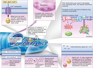
The role of mitochondrial dysfunction in disease remains unacknowledged because of some fascinating features of mitochondria. Mitochondria are an interesting mix of vulnerable and resilient. Mitochondrial DNA (mtDNA) and mitochondrial genes are more vulnerable than nuclear DNA and nuclear genes to damage caused by chemical toxicants (like pharmaceuticals and environmental pollutants) because mitochondrial genes “sit on a single circular chromosome (unlike the linear chromosomes of the nucleus) and are ‘naked’ – they’re not wrapped up in histone proteins.” (Lane p. 15) Histone proteins protect nuclear DNA and because mtDNA isn’t wrapped in histone proteins, it is vulnerable. This vulnerability means that mtDNA is easily damaged. This slide describes additional factors that affect mitochondrial vulnerability to environmental pollutants:

Factors that affect mitochondrial vulnerability to environmental toxicants
Mitochondria as a Target of Environmental Toxicants. Permission to print graphic provided by Joel N. Meyer.

Despite its vulnerability, mtDNA is, at the same time, quite hearty and resilient. MtDNA can take a punch, and a threshold of damage must be crossed over before a disease state will ensue. In Mechanisms of Pathogenesis in Drug Hepatotoxicity Putting the Stress on Mitochondria it is noted that, “damage to mitochondria often reflects successive chemical insults, such that no immediate cause for functional changes or pathological alterations can be established. There is indeed experimental evidence that prolonged injury to mitochondria, such as that which typifies oxidative injury to mitochondrial DNA or to components of the electron transport chain (ETC), has to cross a certain threshold (or a number of thresholds) before cell damage or cell death becomes manifest.” The researchers go on to note that, “This non-linear response can be explained upon consideration that the molecules that subserve mitochondrial function (e.g., mitochondrial DNA, mRNA, and ETC proteins) are present in excess of amounts required for normal cell function. This reserve (or buffering) capacity acts as a protective mechanism; however, at a certain stage of damage, the supply of biomolecules needed to support wild-type mitochondrial function becomes compromised.” (6)

Pharmaceutical Safety and Mitochondria: No Testing Required

To put it simply, because of the tolerance threshold that mitochondria have to damage, the damage done to mitochondria will not show up as a disease until the threshold is crossed. This makes the testing of the deleterious effects of pharmaceuticals and environmental toxins on mitochondria difficult. The damage done by the chemical toxin doesn’t show up until multiple exposures to mitochondrial damaging toxins have been experienced (and it likely doesn’t need to be the same toxin – different mitochondrial damaging toxins can erode the mitochondria’s tolerance threshold). Also, mitochondria display an “initial adaptive response was followed by a toxic response” (6) to damaging toxins.

The mitochondrial tolerance threshold for damage would need to be taken into consideration when testing drugs or environmental pollutants for their adverse effects on mitochondria, IF drugs and pollutants were tested for their effects on mitochondria at all. Unfortunately, “mitochondrial toxicity testing is still not required by the US FDA for drug approval.” (7) The authors of Mitochondria as a Target of Environmental Toxicants note that, “growing literature indicates that mitochondria are also targeted by environmental pollutants” but the EPA does not require testing of environmental pollutants for their affects on mitochondria either.

Studies have shown that bactericidal antibiotics (including fluorouquinolones) (8), statins (9), chemotherapy drugs (3), acetaminophen (6), metformin (a diabetes drug) (10), and others, damage mitochondria. The environmental pollutants that have been shown to damage mitochondria include rotenone, cyanide, lipopolysaccharide, PAH quinones, arsenic, and others (3).

Though it’s not excusable, it’s understandable that the FDA and EPA have not historically required testing of pharmaceuticals or environmental pollutants for their effects on mitochondria. Until very recently, much of what is currently known about mitochondria was not yet discovered. The link between multi-symptom chronic illnesses (including autism) and mitochondrial dysfunction and damage (4) was not yet known when the vast majority of the drugs that are on the market were going through their initial testing and review. What is known now about the important role of mitochondria in epigenetic signaling was not known until recently – and almost all laymen and probably plenty of scientists still don’t realize how much the molecules generated in our mitochondria affect our genes. All of the drugs and environmental pollutants that are on the market have been put on the market without their effects on mitochondria being studied, or even noted by the regulatory agencies that are entrusted with protecting our health and safety. The ignorance of everyone involved would be less consequential if people weren’t so sick. In addition to being connected to the mysterious diseases of modernity, mitochondrial damage is also implicated in the following disorders: “schizophrenia, bipolar disease, dementia, Alzheimer’s disease, epilepsy, migraine headaches, strokes, neuropathic pain, Parkinson’s disease, ataxia, transient ischemic attack, cardiomyopathy, coronary artery disease, chronic fatigue syndrome, fibromyalgia, retinitis pigmentosa, diabetes, hepatitis C, and primary biliary cirrhosis” (7) as well as cancer (11).

The Paradigm Shift

We’re in an interesting and strange situation where medicine hasn’t caught up to science and science hasn’t caught up to medicine. By this I mean that mitochondria damaging chemicals were created long before we knew the importance of our mitochondria, but now that scientists are realizing the importance of our mitochondria, the damaging pharmaceutical culprits are so entrenched in medicine that they can’t be extricated. For example, nalidixic acid, the precursor to fluoroquinolones (mitochondria damaging antibiotics) (8), was first created in the 1960s, long before what we currently know about mitochondria and the effects of mitochondrial damage was discovered. Now that the effects of depleting mtDNA on human health has been discovered, the myriad of strange health symptoms observed in patients who have taken fluoroquinolones can be explained. Mitochondrial damage can cause multi-symptom chronic illness (4). We know this now. However, fluoroquinolones are so widely used (20+ million annual prescriptions in America alone), and so widely regarded as safe, that it would be difficult, if not impossible, to restrict their use now – even though they have been found to cause mitochondrial damage and oxidative stress (8). It’s time for disease paradigms to shift to note the importance of mitochondria in human health. After all, chronic diseases, many of which are related to mitochondrial function, are the leading cause of death in the U.S.

Mitochondria are important. It’s time we started paying attention to them. It’s time for disease models to shift. It’s time for iatrogenic mitochondrial dysfunction to be recognized as a cause of chronic diseases. The chronic diseases are happening, whether we recognize the role of mitochondrial damage, and the role of pharmaceutical and environmental pollutants in damaging mitochondria, or not. Ignorance isn’t bliss – people are sick. With recognition of the importance of mitochondrial health, maybe we can prevent others from getting sick in the future.

Information about Fluoroquinolone Toxicity

Information about the author, and adverse reactions to fluoroquinolone antibiotics (Cipro/ciprofloxacin, Levaquin/levofloxacin, Avelox/moxifloxacin and Floxin/ofloxacin) can be found on Lisa Bloomquist’s site, www.floxiehope.com.

Participate in Research

Hormones MatterTM is conducting research on the side effects and adverse events associated with the fluoroquinolone antibiotics, Cipro, Levaquin, Avelox and others: The Fluoroquinolone Antibiotics Side Effects Study. The study is anonymous, takes 20-30 minutes to complete and is open to anyone who has used a fluoroquinolone antibiotic. Please complete the study and help us understand the scope of fluoroquinolone reactions.

Hormones MatterTM conducts other crowdsourced surveys on medication reactions. To take one of our other surveys, click here.

To sign up for our newsletter and receive weekly updates on the latest research news, click here.

What Else Can I Do To Help?

Hormones MatterTM is completely unfunded at this juncture and we rely entirely on crowdsourcing and volunteers to conduct the research and produce quality health education materials for the public. If you’d like help us improve healthcare with better data, get involved. Become an advocate, spread the word about our site, our research and our mission. Suggest a study. Share a study. Join our team. Write for us. Partner with us. Help us grow. For more information contact us at: info@hormonesmatter.com.

To support Hormones Matter and our research projects – Crowdfund Us.

References

Lane, Nick (2005). “Power, Sex, Suicide: Mitochondria and the Meaning of Life” Oxford University Press Inc., New York.

  1. Journal of Cell Biology, “Signal Transduction by Reactive Oxygen Species
  2. Cell, “Mitochondria: In Sickness and in Health
  3. Toxicological Sciences, “Mitochondria as a Target of Environmental Toxicants
  4. Nature Preceedings, “Oxidative Stress and Mitochondrial Injury in Chronic Multisymptom Conditions: From Gulf War Illness to Autism Spectrum Disorder
  5. Biochimica et Biophysica Acta (BBA) – Gene Regulatory Mechanisms, “Mitochondrial DNA Damage and its Consequences for Mitochondrial Gene Expression
  6. Molecular Interventions, “Mechanisms of Pathogenesis in Drug Hepatotoxicity Putting the Stress on Mitochondria
  7. Molecular Nutrition & Food Research, “Medication-Induced Mitochondrial Damage and Disease
  8. Science Translational Medicine, “Bactericidal Antibiotics Induce Mitochondrial Dysfunction and Oxidative Damage in Mammalian Cells”
  9. NIH Public Access, “Statin Adverse Effects: A Review of the Literature and Evidence for a Mitochondrial Mechanisms
  10. Biochemical Journal, “Metformin inhibits mitochondrial permeability transition and cell death: a pharmacological in vitro
  11. Contemporary Oncology, “Oxidative Damage and Carcinogenesis