thiamine - Page 4

From Mother to Daughter: The Legacy of Undiagnosed Vitamin Deficiencies

14148 views

This is a story of a mother with undiagnosed vitamin B deficiencies who gave birth to a daughter who was also born with undiagnosed vitamin B deficiencies. In the eyes of conventional doctors and labs, there was not much wrong with us, but we knew that life was harder than it should be. We lived managing debilitating dizziness, daily migraines, fibromyalgia pain, chronic fatigue, allergies, hormonal changes, anxiety, and depression. Until we discovered that we were both hypermobile with histamine issues, hypoglycemic, and had many vitamin B deficiencies. The biggest challenge was for my daughter to start taking thiamine (vitamin B1). Her heart rate was all over the place and she had such a bad paradoxical reaction to thiamine that we believe she had been living with undiagnosed beriberi along with POTS.

Mom’s Health Marked by Asthma, Anxiety, Migraines, and a Difficult Pregnancy

All I remember as a child is being afraid to talk in school even if I knew the answer to a question. I had allergies and could not exercise due to asthma. During college, I had to read over and over the same thing because I could not concentrate. I worked extremely hard because the fear of failure was too much to bear. I started to have hormonal imbalances and missing periods. I successfully finished college and moved away to another state. That is when migraines started. Later, I became pregnant with my first child and started having blood clots. Anxiety and depression would come and go with hormonal changes.

When I was pregnant with my second child, my daughter, I was sick every morning with nausea.  After 6 months of pregnancy, I had gained only 6 pounds. Ultrasounds showed that the baby was growing normally, but I was losing weight. At that point, I also could see blood clots on my leg. I was placed on bed rest. By the 8th month, my water broke and my daughter was born. She was jaundiced and placed under UV light for a week. I also stayed in the hospital for a week dehydrated, with blood clots, and with the “baby blues”. We left the hospital after a week, and she had a “normal” development. However, you could see that she was a baby that would not go with anyone, not even the people close to us, indicating some anxiety.

Daughter’s Early Health Issues: Selective Mutism, Asthma, Concentration Issues

When my daughter turned four years old, we moved out of state and that is when she stopped talking outside the house. I later found out that it is called selective mutism, a form of severe social anxiety. She started seeing a school counselor to try to help with her anxiety and self-esteem issues. I brought a girl scout group to my house so that she could start having friends and talk to others in her area of comfort. She also developed asthma and needed nebulizer/albuterol treatments frequently and daily QVAR for prevention. She was given Singulair, but it made her very depressed. Her grades in all classes were all over, from A to D.  She would spend the whole time after school trying to complete homework, but she couldn’t. Her teacher told me that she really did not have that much homework. I would ask her to watch the dog eating and to take her outside as soon as the dog finished but she would be wandering around the kitchen and could not pay attention to the dog. Her neurologist gave her Strattera and that helped a little. Her EGG also showed some abnormal activity. The doctor recommended anti-seizure medicine and said that she was probably having mal-petit seizures. I refused medication based on how she reacted to Singulair and because the doctors were using words like “probably” and “just in case”. I kept an eye on her and noticed when she ate ice cream and got asthma. I had her stop sugars and dairy.  Soon after that, a teacher called me, excited to tell me that my daughter was talking at school. She also was able to stop all asthma medication except for 2 weeks every year when seasonal allergies would hit. At this point, it had been already four years since she stopped talking outside our house. She started excelling in all classes and we were able to stop Strattera. However, the continuous anxiety remained.

The Teenage Years: Continuous Migraine, More Medications, and No Answers

At 16 years old, she got a cold that turned into asthma with a continuous headache that just would not go away. She started waking up every day with a migraine, depressed with no energy. We had to wait three months to see a pediatric neurologist. Meanwhile, I would take her to my chiropractor early in the morning, give her an Excedrin, and she would go to school whenever she felt better. She began drinking at least 2 cups of coffee every day to help with the pain. Sometimes she would go to school at 11am, sometimes at 1pm. Even if there was just one class left, she would go to school. At this point, she felt that she wouldn’t have a future.

When we finally went to the neurologist, he recommended amitriptyline. I had been on amitriptyline and woke up one day not knowing which year or season was, but I was told that the issue was the high dose given to me (125mg), after decades of it increasing it every year. I agreed as long as it was a low dose.  Amitriptyline lessened the continuous headache, but it was not really gone, and she still needed some Excedrin. She started daily aspirin as well. She was just getting by day to day trying to manage her pain and mood and trying to have a normal teenage life.

Increasing Weakness When Outdoors: Untangling Root Causes

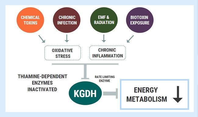
She became very weak whenever we would go to the beach or to a park. We would have to drag her indoors and give her water. On some occasions, she would say that she could not see. Somehow, she successfully managed to graduate from high school. We started seeing functional doctors. We found that she had some variants related to mitochondria dysfunction, but we really didn’t know how to address this. We also found out that she had Hashimoto’s and antibodies against intrinsic factors, which was indicative of pernicious anemia. We knew right there, that she had issues that conventional doctors had missed.

We also did a Dutch test and found that all of her hormones were high. The functional doctors suggested sublingual B12, folinic acid, and a B complex. She said the vitamins made her feel awake for the first time. However, chronic fatigue was still a major struggle for her. Eventually, she had to stop folinic acid because it made her depressed and unmotivated. Meanwhile, she managed her anxiety with herbs, but it was a real struggle.  She also continued to have asthma requiring albuterol every fall season. She chose a very challenging career in cell biology with biochemistry. She went through college with many cups of coffee just to control migraines, have energy, and be alert.

Discovering Her POTS Symptoms

The summer of 2019, before her senior year of college, the nurse checked her vitals as part of her new summer internship. The nurse thought the pulse monitor was broken because her heart rate was 120 sitting down. After a few minutes, it went down to 99, so the nurse dismissed it. When she told me that, I started paying attention to her heart rate. We went to her physician and neurologist and in both instances, her heart rate was 100, just sitting down waiting for the doctor. I asked if it was normal, and they said that it was in the upper range but not a concern. I was still concerned and made an appointment with a cardiologist but also bought her an iwatch. She noticed right away how her standing heart rate would be over 100, and by only taking a few steps, her heart rate would go even higher and she would become fatigued and even dizzy. From the heart rate monitor on her iwatch, we could see how quickly her heart rate would climb upon standing and then slow a bit when sitting.

That is when I remember that I have read about POTS and hypermobile people. I remember that when she was a child, the neurologist had said that she was hypermobile, but never said that it could be a problem for her. It just seemed like a fun thing to have. I started asking in health groups and someone mentioned that her medications could also cause high heart rate. I searched and amitriptyline did have that side effect.  That is when my daughter showed me that her resting heart rate was in the 90s and it would fluctuate from 29 to 205 without exercising. When we went to the cardiologist and explained all of this, he said that he did not even know how to diagnose POTS because it is rare. He did testing and said that the heart was fine but there was some inefficiency due to some valve leaking but that it usually does not cause symptoms. I asked about amitriptyline and he confirmed that it could raise heart rate.  At that point, she stopped amitriptyline and her maximum heart rate was 180 instead of 205.

She went back to her last year of college when Covid hit. She came back home and we could see the lack of energy and how much doing any little thing or stress would crash her for days. Since I needed glutathione for chemical sensitivities, I decided to see if it would help her. Glutathione with co-factors helped her recover, instead of crashing for days, she would recover the next day. That is when she told me that every time she walked to school, she felt that she would pass out. When she gets up in the morning, she ends up lying on the floor because of dizziness. Despite her dizziness, daily muscle pain, daily migraines, and chronic fatigue, she had big dreams. She just kept pushing through day by day, with coffee, herbs, and whatever it took, but she knew that something had to change. She successfully graduated in May, Magna Cum Laude, and she had a couple of months to deal with her health before she would leave to start her graduate studies and research job. That is when I found people that knew about Dr. Marrs’ work and thiamine, and her life finally changed.

Introducing Thiamine and Other Micronutrients: Navigating the Paradox

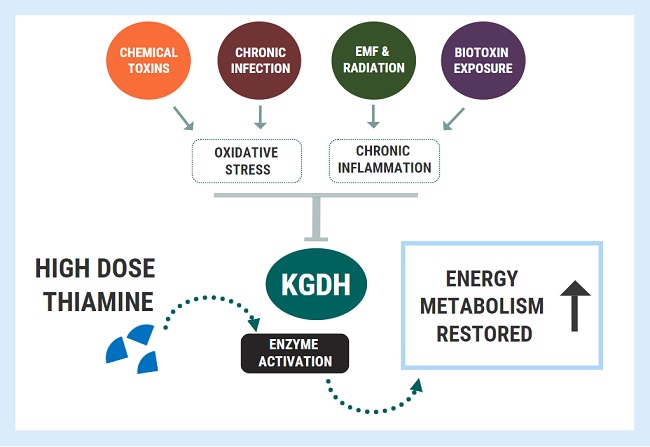
A functional doctor recommended magnesium and niacin for her migraines and they significantly helped. This gave the functional doctor the idea to try tocotrienols. High doses of tocotrienols worked better for reducing her migraine pain than amitriptyline and aspirin combined. Then she started taking high doses of B6. This helped her muscle pain and improved her mobility. Despite being hypermobile, easy stretches gave her intense muscle cramps prior to starting B6. Guided by very knowledgeable researchers belonging to Dr. Marrs’ Facebook group, Understanding Mitochondrial Nutrients, we started Allithiamine. The first thing she said was “wait, the sun does not hurt?”.  I asked her what she meant.  She explained that all her life, being in the sun gave her pain in her eyes and forehead and that she couldn’t understand why people wanted to be outside. No wonder she never wanted to go outside. She also said her migraines were gone. We have waited 4 years to hear that!

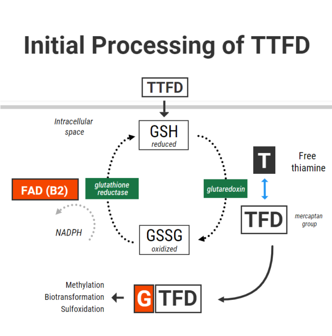
After just a couple of days, she started having a lot of nausea and lower-intensity migraines returned.  The researchers knew right away that she needed more potassium. She started to eat apricots, coconut water, or orange juice every time she had nausea and it helped. However, it was happening every hour so we decided to try a different Thiamine. We tried half Lipothiamine and Benfotiamine but she didn’t feel as much benefit and still gave her issues. We went back to 1/10 of Allithiamine. Chatting with the researchers, one asked if she also experienced blinding episodes. Yes! Finally, someone that knew about that! They recommended B2 and we started it. That’s when we discovered that her pain in the sun and dizziness were caused by a B2 deficiency. She continued waking up with crashes needing potassium every hour. She did not sleep that week. The researchers suggested taking cofactors including the rest of the B vitamins, phosphate salts, phospholipids, and beef organs. Beef organs and phospholipids helped with energy and bloating, phosphate salts helped with nausea and irritability.

Then researchers suggested that she needed to stabilize sugars and have more meat. That is when we realized that she had some type of hypoglycemia. We had noticed that she would get very tired and got shaky hands if she didn’t eat. Functional doctors had mentioned that she may have reactive hypoglycemia since she had a fasting glucose of 70. She started having more meat to stabilize her sugars and removed all packaged foods, sugars, grains, and starches. She started having just fresh meat, veggies, rice, beans, nuts, and berries. She felt that she was so much better with beef that she started using it for potassium between meals and bedtime.

She was able to increase allithiamine little by little. She would mix a little bit with orange juice since it tasted so awful. Little by little, she started having fewer crashes and feeling better. It took a month for her to be able to tolerate one capsule of Allithiamine. She was sleeping more but not the whole night. That is when our functional doctor suggested supporting adrenals. That really helped but then she began having stomach pain and nausea after eating beef and developed frequent diarrhea. Chicken always increased her hunger and reduced her energy compared to beef and but now she was afraid of having beef. She stopped all sources of beef and phospholipids.

We consulted a very good functional doctor. She did Nutraeval and confirmed that all her B vitamins were low or deficient and recommended TUDCA and Calcium D Glucarate along with trying lamb and bison first. Both helped in reducing bloating/nausea and she was able to start eating lamb and bison along with reintroducing a minimal amount of carbs. Soon after, she was eating beef again with no pain.  After starting TUDCA, her bilirubin levels were normal for the first time in her life. We continued to work with the functional doctor to fix other deficiencies.

Recovery from Multiple Nutrient Deficiencies and the Prospect of a Normal Life

After Allithiamine and vitamin B2, we worked with our functional doctor to balance the remaining B vitamins. She is now able to go out in the sun without bothering her eyes and without passing out. She gained weight after starting the B vitamins and began looking healthier, compared to how skinny and underdeveloped she looked before. She also learned how to manage electrolytes. She sometimes needs more sodium, but other times needs more potassium. She feels sick when electrolytes get out of balance. Although she still had some continuous pressure in her head, she no longer needs any amitriptyline, aspirin, or Excedrin for pain. One thing that remained problematic was folate deficiency. She still became depressed with folinic acid, so she tried methylfolate instead. She felt so unmotivated that preferred not to have it, but she realized that it was key to something that she struggled with all her life: anxiety. She figured that she could have methylfolate every other day, so that she could have less anxiety.

Now, for the first time, she began to have a normal life. She can now exercise daily without dizziness and her heart rate skyrocketing.  Her heart rate in general is more normal, doesn’t go down to 29 or up to 205. She had not had any asthma requiring albuterol.  She started driving without having to deal with anxiety and panic attacks.  She was able to walk to her office without fainting.  She now can now live alone dealing with the stress of having a full-time job, graduate classes, cooking her food, and exercise every day! She is not cured completely but for a person that once thought she couldn’t have a future, she is doing pretty darned good!

We Need Your Help

More people than ever are reading Hormones Matter, a testament to the need for independent voices in health and medicine. We are not funded and accept limited advertising. Unlike many health sites, we don’t force you to purchase a subscription. We believe health information should be open to all. If you read Hormones Matter, and like it, please help support it. Contribute now.

Yes, I would like to support Hormones Matter.

This article was published originally on July 22, 2021. 

Health Requires Energy. Energy Requires Nutrients.

11431 views

A Fundamental Issue

I was horrified to watch the “60 Minutes” program Sunday, April 12th, 2020 on television that dealt with the colossal number of Americans suffering from obesity, chronic fatigue and diabetes, both types I and II. About half of the program dealt with the essential consumption of natural food, reminding us that Hippocrates, 400 BCE said “let food be your medicine and let medicine be your food”. This must have been said thousands of times but everything in our modern civilization is totally destructive to the whole idea. I have seen so many hundreds, if not thousands, of people whose illness was caused by themselves. It was treatable by making sure that each individual understood the fundamental issue. So before I illustrate a typical illness situation within my experience, I will try to state what I mean by describing what I consider to be the “fundamental issue”.

“We behave according to what we eat”.

I have stated this so many times but unfortunately the American medical profession is the major inhibitor to its clinical success. When a suffering patient with many symptoms arising from what I call “dietary mayhem”, goes to his or her physician, they simply do not recognize the clinical expression of the popular high calorie malnutrition. The many symptoms are usually referred to as psychosomatic and the unfortunate patient is told that “it is all in your head”. I have witnessed this so many times; I cannot understand why the physicians don’t pay a little more attention to what the patient is trying to tell them. Often the patient has discovered the real cause of the problem but find that her words are considered to be the voice of ignorance and delusion.

Food and Energy

Our food consists of fuel that must be burned (oxidized) to liberate energy. In any text this element of the food is described as “calories”. The energy quality of our food intake is measured in kilocalories and a single one is defined as “the energy needed to raise the temperature of 1 kg of water through 1°C”. Notice the use of the word energy, the result of oxidation. Now, as everyone knows, vitamins and minerals, known as non-caloric nutrients, are vital to the release of energy from the caloric elements. To understand how this combination of chemicals works, there must be a ratio of calories to the noncaloric elements. That is why high calorie foods without vitamins or minerals are known as empty calories. It is the consumption of empty calories across America that has given rise to the idea of high calorie malnutrition. I have actually seen a written statement that this is an oxymoron. “How can excess of calories be considered a form of malnutrition?” It seems that few people understand this vital ratio and they seem to think that as long as you are consuming calories, you will flourish. Also, the food industry fills the grocery store with cartons of temptation and seems to have no regard for the well-being of its consumers. They keep using the term “all natural” so much, it becomes meaningless.

A Typical Case of Energy Deficiency

I was a pediatrician at Cleveland Clinic and one of my interests was sudden infant death (SIDS). So one day I was having lunch with one of the surgeons who practised ear nose and throat surgery. He told me that he had been called to the medical ICU because a woman had stopped breathing and he had performed a tracheostomy. He was intrigued by the reason for this disaster and, knowing my interest, he suggested that I should take a look. Pediatricians are assumed to be familiar with diseases of children but ignorant of adult disease and I knew that I was  not welcome. I found a 50-year-old woman who was grossly edematous and unconscious. Without considering the technical details, I proved that she had the vitamin B1 deficiency disease beriberi. With injections of thiamine she became conscious and the edema disappeared. During her recovery she developed a progressive anemia, thought to be evidence of internal bleeding, but all the tests were negative. I took some urine from her and subjected it to a special type of test. It showed that she was deficient in folate, another B vitamin. It is important to note that she did not develop folate deficiency until she began her recovery from thiamine, it was masked by cellular energy deficiency. When she began to receive folate there was an immediate recovery from the anemia but she had been given at least one injection of thiamine by then.

She was discharged from hospital, wheelchair bound, taking both thiamine and folate. When she returned as an outpatient, I found that she had a skin rash and that her legs were, if anything, weaker. It had long been known that anemia would develop from either folate or B12 deficiency, but the folate deficient variety required B12 supplementation as well as folate. If B12 was not provided, the patient would develop paralysis of the legs and I had forgotten this. Also, it is not well-known that vitamin B12 deficiency can cause a skin rash. I gave her an injection of B12 and the rash disappeared. However, for a few days she had muscle aches and fever that I did not understand at that time. Looking back I would now assume that this was what we call “paradox” on Hormones Matter. To those that may not have read about this it is the temporary worsening effect by introducing an essential nutrient to someone who has long been deficient in that nutrient. One of the things that had probably been a serious indictment on self cause was that she was a chronic cigarette smoker, a well-known habit that damages oxidation.

Energy Metabolism

Can we extrapolate from this case any general ideas about how medical treatment should advance? Perhaps the general opinion would be that this is a rare and unusual case, an outlier from the “usual and customary diagnosis”. But if we consider the facts; long-term cigarette smoking, dietary indiscretion and genetic risks appear to be quite common. I treated a 12-year old girl  with a conventional diagnosis of Juvenile Rheumatoid Arthritis, using a  nutritional supplement. Without discussing the technicalities of laboratory evidence, it was clear that defective energy metabolism was the underlying cause. The combination of genetic risk, failure to adapt to any form of stress (infection, trauma, chronic useless brain activity etc) and inadequate energy metabolism are the three factors that either collectively or singly lead to breakdown of health. As Selye predicted, energy for adaptation is the essential ingredient.

We Need Your Help

More people than ever are reading Hormones Matter, a testament to the need for independent voices in health and medicine. We are not funded and accept limited advertising. Unlike many health sites, we don’t force you to purchase a subscription. We believe health information should be open to all. If you read Hormones Matter, like it, please help support it. Contribute now.

Yes, I would like to support Hormones Matter. 

Image by Inns-Web from Pixabay.

This article was published originally on April 22, 2020.

Paradoxical Reactions With TTFD: The Glutathione Connection

41987 views

Some individuals experience negative reactions and the worsening of symptoms when they begin thiamine repletion therapies using the more biologically available formulations like thiamine tetrahydrofurfuryl disulfide (TTFD). Dr. Lonsdale calls these paradoxical reactions. In this article, I examine the chemistry beyond these reactions and provide some hypotheses regarding why they happen and how to mitigate them.

TTFD Basics

In order to understand why some individuals react negatively to TTFD supplementation, it is essential to understand the basics behind TTFDs molecular configuration and how it is processed by cells. The primary difference between ordinary thiamine and TTFD is an extra chemical group called a mercaptan group. The mercaptan is derived from allicin, a compound found in garlic, and is connected to the thiamine molecule via a special sulfur-sulfur bond called a disulfide bond. Importantly, it is this unique chemical group that accounts for TTFD’s ability to traverse membranes in the body without the need for a transport system.

Upon ingestion, TTFD is mostly absorbed into the blood from the gastrointestinal tract in whole form as TTFD. As it travels through the blood, it can penetrate the brain and other organs without cellular transporters. One of the main sites of absorption is the red blood cells. Upon penetration of the red blood cell membrane, TTFD must first be processed or “broken apart” before it can release the thiamine contained within its chemical structure. After thiamine is released into the cell, the ancillary mercaptan group must also be processed and/or detoxified through alternative pathways. It is therefore theoretically plausible that errors involved in the processing of TTFD could contribute toward negative side effects or reactions to this nutrient.

How TTFD Is Processed Inside the Cell: The Glutathione Connection

For TTFD to “release” its thiamine, its disulfide bond must gain electrons from another donor molecule. In chemical terms, this process is referred to as reduction. Once this reduction occurs, thiamine is freed and can then go on to participate in cellular biochemical reactions.

Of the few molecules which have been shown to reduce TTFD, glutathione performs this function most effectively. As the cell’s primary antioxidant, glutathione is responsible for donating electrons to neutralize reactive oxygen species, and can either be found in its reduced form or its oxidized form. Once a reduced glutathione molecule (GSH) has donated its electron, it bridges with another to molecule to form oxidized glutathione (GSSG). GSSG is then recycled back to two GSH molecules through accepting electrons from NADPH via the enzyme glutathione reductase (vitamin B2 as FAD dependent). TTFD and Glutathione

When TTFD enters cells, GSH in red blood cells chemically reduces TTFD via a process called “disulfide exchange” (presumably using a protein called glutaredoxin). Reduced glutathione becomes oxidized glutathione and TTFD “releases” thiamine to producing free thiamine inside the cell with an extra TFD mercaptan group left over.

The initial phase of processing TTFD requires that cells have enough reduced glutathione. Furthermore, the more GSH you have – the faster the rate of this reaction. So in simple terms, to obtain thiamine from TTFD the cells “use up” their reduced glutathione.

I recently had correspondence with one individual who only gained tolerance of TTFD after supplementing with 200mcg of selenium in the form of sodium selenite. Selenium supplementation in different forms has been shown to increase red blood cell GSH levels by up to 35%. This is thought to occur due to selenium’s ability increase glutathione synthesis through upregulating the enzyme gamma-glutamylcysteine synthetase. I suspect that poor glutathione status might be one of the reasons for benefit from selenium.

Having enough glutathione is clearly very important, but recycling it is also essential to maintain a pool of glutathione in its reduced form. Unfortunately TTFD can place a burden on this system, and this was demonstrated in one old study from Japan which showed that TTFD administration rapidly lowered red blood cell GSH. Interestingly enough, that same experiment showed that GSH levels were restored within 5-10 minutes. This restoration was accomplished by the vitamin B2 (as FAD)-dependent enzyme glutathione reductase, which donates electrons to GSSG with the reducing power of NADPH to recycle it back to two GSH.

What this basically means is that cells require a robust antioxidant system to properly process TTFD and return back to their original state. First, cells need enough of the antioxidant GSH to cleave thiamine. Second, cells also need to be able to recycle the oxidized glutathione back to its reduced state.

Poor Glutathione Status and Difficulty With TTFD

Immediately, we see two potential issues that could arise from TTFD supplementation which might provide a better understanding of why some people may not tolerate this molecule.

In someone who has poor glutathione (GSH) status, they might theoretically be less able to cleave thiamine from TTFD. There are many reasons why someone may have poor glutathione status:

  • Low precursors (cysteine, glutamate, glycine)
  • Chronic oxidative burden and/or inflammation
  • Deficiencies in the nutrients required to generate, process, or utilize glutathione (B6 or selenium)
Alternatively, an individual may have enough resources to make glutathione, but if they cannot recycle it through the necessary machinery (i.e glutathione reductase), then taking a substance which depletes their GSH (like TTFD) might further contribute towards their oxidative burden.

A total and/or functional riboflavin deficiency is the probably the most common culprit responsible for poor glutathione reductase activity. The glutathione reductase enzyme also requires adequate reducing power from NADPH to drive the enzymatic reaction. NADPH is derived from niacin (vitamin B3) and is generated in the pentose phosphate pathway which, ironically, requires the thiamine-dependent enzyme transketolase.

In the context of poor enzyme activity, without the reducing powder to drive GSSG back to GSH, the oxidized form of glutathione can theoretically drift towards the path of generating a free radical called the glutathione radical. This alone could further contributes to oxidative stress and cell damage.

Below is a hypothetical scenario to demonstrate my point:

  1. An individual suffers from long-term thiamine deficiency and has suboptimal riboflavin status
  2. Thiamine deficiency leads to lower activity of transketolase
  3. Low transketolase activity produces a lack of NADPH
  4. A lack of NADPH and a lack of FAD means that glutathione reductase is unable to efficiently recycle glutathione, which produces an imbalance between reduced/oxidized glutathione.
  5. Intracellular GSH is further lowered by taking high dose TTFD, and there is not enough enzyme activity to recycle it back
  6. Oxidative stress is made worse

In the above scenario, taking a high dose of TTFD may not be appropriate. Rather, restoring NADPH levels through supplementing with ordinary thiamine and supporting the glutathione system via other measures might be advised before starting with TTFD. Optimal riboflavin status is also necessary for the above processes to run smoothly.

Older research in Japan showed that TTFD supplementation could lead to a secondary B2 deficiency through increased urinary excretion. The increased need for glutathione reductase could at least also contribute to this effect. When taking TTFD, it has downstream effects on other nutrients. Hence, these supporting nutrients should also be taken in conjunction when someone is supplementing TTFD in high doses.

Some basic laboratory measurements of glutathione status include:

  • Whole blood glutathione (low)
  • Gamma-glutamyl-transpeptidase (high)
  • Urinary pyroglutamic acid (high)

Furthermore, there are several functional markers which can be measured to assess riboflavin status, including direct measurement of red blood cell glutathione reductase activity:

  • Urinary glutaric acid (high)
  • Whole blood B2
  • Urinary adipic, suberic, ethylmalonic acids (high)
  • Urinary succinic acid (high) can also be suggestive along with a few other organic acids
  • Erythrocyte glutathione reductase activity (low)

To summarize, the initial cellular processing of TTFD requires adequate levels of reduced glutathione. Glutathione becomes oxidized, and so TTFD has can have a depleting effect on GSH and increase the requirement for recycling. If there is insufficient active B2 (as FAD) or NADPH levels, glutathione is not likely to be recycled sufficiently and may lead to GSSG radical formation.

It is therefore possible that the glutathione-depleting effect of TTFD could be responsible for some of the side effects associated with supplementation. This is probably most applicable in individuals with poor glutathione recycling and underlying oxidative stress. Therefore, nutrient therapies that may support this initial phase of TTFD metabolism include:

  • Selenium (improve GSH levels)
  • Riboflavin (improve GSSG-GSH recycling)
  • Niacin (increase NADPH)
  • Ordinary thiamine (increase NAPH via PPP)
  • NAC, glycine and/or glutathione TAKEN HOURS AWAY from TTFD (GSH precursors)

We Need Your Help

More people than ever are reading Hormones Matter, a testament to the need for independent voices in health and medicine. We are not funded and accept limited advertising. Unlike many health sites, we don’t force you to purchase a subscription. We believe health information should be open to all. If you read Hormones Matter, like it, please help support it. Contribute now.

Yes, I would like to support Hormones Matter.   

Image by Layers from Pixabay.
This article was published originally on October 26, 2020. 

Are You Attractive? Connections Between Low Thiamine and Mosquito Bites

6066 views

A new study shows that you can be “highly attractive” if you have enough carboxylic acid on your skin. Highly attractive to mosquitos, that is.

Mosquitoes are an ideal vector for pathogen contraction and transmission between hosts. These pathogens include viruses such as Dengue, Yellow Fever, Zika, West Nile Virus, and Chikungunya. Parasites that cause malaria in tropical and subtropical countries are also transmitted by mosquito bites. It is therefore logical that increased exposure, or attractiveness, to mosquitos will increase the probability of contracting such pathogens. Why then do some individuals seem to be targeted by mosquitoes more than others?

A new study attempted to shed light on this age-old question of why some people are greater mosquito magnets than others. Indeed, some are victims of relentless mosquito bites and could in fact function as a “mosquito adsorbent” – a courtesy for those around them on a humid day at the park. It turns out that mosquitoes utilize their sense of smell to find food, as expected, but that those with higher levels of certain metabolites get bit more frequently than others. What is interesting is that some genes in mosquitoes allow this sensing to occur. To determine how all of this worked, researchers induced olfactory deficits by knocking out genes in mosquitoes. This caused less odor sensing to occur, but surprisingly mosquitoes were still able to distinguish between highly and weakly attractive individuals. This all indicates that attractiveness to mosquitoes is likely an amalgamation of mechanisms involving genetics and metabolomics.

The specific metabolites analyzed in this study are pentadecanoic, heptadecanoic, and nonadecanoic acids – all grouped as carboxylic acids. These are considered odd number fatty acids (ONFA), a minor group of the fatty acid repertoire in humans. Pentadecanoic and heptadecanoic acids are the most abundant ONFA, composed of 15 and 17 carbon chains, respectively. In contrast to other fatty acids, ONFA are thought to predominantly accumulate in the body from foods (e.g., dairy, meats). However, they can be synthesized in the body from straight chain fatty acids, through the mechanisms of alpha-oxidation.

The Thiamine Connection

It has long been rumored that thiamine could prevent, or at least reduce, mosquito bites. Is there research supporting this claim?

The literature is not clear, but does seem to indicate an indirect connection between thiamine (Vitamin B1) deficiency, or inadequacy, and attractiveness to mosquitos. For example, a research group studying yellow fever mosquitoes, showed that mixed with lactic acid, the carboxylic acid strongly synergizes to elicit mosquito attraction. The authors state:

Addition of lactic acid markedly increased the degree of attractiveness of formerly less attractive human odour samples and they were preferred over those which were originally the most attractive.

Indeed, lactic acid alone, acts as a sufficient mosquito attractant, but is relatively weak compared to the synergism with carboxylic acids. Another older study utilized a bioassay to identify attractants from human skin extracts. Here the mosquitoes were allowed to fly upwind toward odor sources (human skin wash extracts). They found that lactic acid was a major constituent of skin extract and that it was a necessary component for mosquito attraction.

What can cause lactic acid to build up (either in blood or tissues)? One mechanism would be the shunting of pyruvate into the lactate pathway. This can be measured in blood when someone has low thiamine. Indeed, in some conditions, the skin has been shown to carry higher amounts of lactic acid compared to blood levels, due to increased anaerobic glycolysis. Here we would expect lactic and carboxylic acids to suffuse below or above the surface of skin where it can be detected by mosquitos. In addition, a surfeit of lactic acid build up, in an overt thiamine deficiency, may not be required. This is because the mosquitos’ high sense of smell can pick small amount of odors, even when major olfactory-related genes are knocked out.

Contrary Research

On the other hand, a potential argument against the effectiveness of thiamine in repelling mosquitoes comes from a 2005 study. The authors gave individuals over the counter B vitamins in two experiments:

  • Experiment 1: B-complex containing 50 mg of B1(thiamine)/B2/B3/B5/B6, 400 µg Folate, 50 µg B12, 50 µg Biotin, and 500 mg Vitamin C. Vitamin C by itself was used as a control since the B-complex contained Vitamin C. Subjects (14 males, and 9 females) were utilized in a cross over experiments, meaning that they would start either placebo or the treatment for 7 days and then switch over to treatment or placebo, respectively. In this scheme each individual would act as his/her own paired experiment, statistically.
  • Experiment 2: Used 100 mg B1, with Vitamin C as a control. Subjects (10 males, and 7 females) were studied in the same manner to experiment 1, with 100 mg B1.

The authors did not find statistically significant differences between the groups, and concluded that B vitamins were not helpful in repelling mosquitos.

Possible Problems With the Research

There may be some caveats to the protocol used in this research, that likely masked the true effects of thiamine supplementation. First, the authors may have been limited, but should have used a non-Vitamin C formulation. Although somewhat inconsistent in the scientific literature, Vitamin C supplementation can reduce blood lactate levels in certain settings, “increase in free radical production and lactate levels….were offset by vitamin C supplementation”. This, in theory, would reduce lactate in skin and therefore the strong synergistic effects of lactic with carboxylic acids would be minimized. Second, and more importantly, the method only evaluated volatile components that can be transferred from the skin into the glass bottle used to attract mosquitoes. Lactic acid is a low volatility organic compound. The other positive studies discussed above had instead extracted skin organic compounds, including lactic acid, utilizing ethanol. Overall, the 2005 study was well designed for the questions asked and provided basic preliminary experiments. Unfortunately, the design of the study negated the effects of one key compound, lactic acid, due to volatility-based evaluation. Hence, a follow up study should be performed with these concepts in mind:

  1. Premeasurement on skin extracts using organic acid extraction methods (ethanol or other compounds).
  2. Treatment with higher dose thiamine. An example would be 0.5 – 1 gram of thiamine-HCl, for 1 to 4 weeks.
  3. Mosquito attraction should be performed concomitantly with measurements of thiamine status (ETKA test). Similarly, measurements of lactic acid in skin extracts of treated and non-treated individuals should be performed in order to correlate quantity with phenotype.

The expectation is that individuals who are thiamine deficient would have increased lactic acid in their skin extracts and therefore would serve as a mosquito magnet.


Does Thiamine Status Play a Role in Mosquito Attraction?

The above studies combined elucidate some of the mechanisms in play in individuals who are highly attractive to mosquitos. These include interplay between genetics of mosquitos and organic compounds present in human skin. Individual differences in mosquito attraction may also be attributed in part to temperature, moisture, visual cues, and body odor. Out of these possibilities, body odor plays a major role in mediating these interactions. Specifically, both lactic and carboxylic acids play a synergistic role in attracting mosquitos, and therefore increase the odds of contracting disease. Mechanisms that result in increased lactic acid production such as exercise, and potentially low thiamine metabolism, are likely a risk factor for mosquito bites. Overall, the role for lactic acid in mosquito attraction seems to stir the arrows of mosquito-magnet theory, in-part, towards a mild/subclinical thiamine deficiency, or at least an abnormal metabolic activity. As suggested above, more studies should be performed in order to directly evaluate the effects of thiamine on lactic acid content of human skin in conjunction with effects on attracting mosquitos. In the meant time though, if you are one of those people who seems to attract mosquitos, consider looking into thiamine.

We Need Your Help

More people than ever are reading Hormones Matter, a testament to the need for independent voices in health and medicine. We are not funded and accept limited advertising. Unlike many health sites, we don’t force you to purchase a subscription. We believe health information should be open to all. If you read Hormones Matter, like it, please help support it. Contribute now.

Yes, I would like to support Hormones Matter. 

Image by Welcome to all and thank you for your visit ! ツ from Pixabay.

This article was published originally on February 27, 2023. 


Medication and Vaccine Adverse Reactions and the Orexin – Hypocretin Neurons

11656 views

A paper published in Science Translational Medicine, provides preliminary evidence that the H1N1 Flu Vaccine Pandemrix can evoke immune system mediated damage to the orexin – hypocretin neurons and induce narcolepsy in individuals with a particular genetic variant. The orexin – hypocretin neurons were only recently discovered in the mid 1990s, by two separate research groups, hence, the two names for the same molecule. For this paper, we’ll be utilizing the orexin nomenclature.

Initially, the orexin neurons were thought to be involved only in feeding behavior, as damage elicited hypophagia in animals. Soon it was learned that more severe damage to the orexin neurons induced narcolepsy and the orexin system became a key focus in narcolepsy related research. With time, however, it became quite clear that these neurons were involved in regulating a myriad of hormone and neurotransmitter systems and their consequent behaviors. Narcolepsy or rather the ability to sustain wakefulness, is but one of the many functions regulated by the orexin system.

In a previous paper, I touched briefly on the possibility that the orexin neurons might be damaged and have diminished functionality in individuals suffering from post Gardasil side effects. In particular, I suspected these neurons were indicated in post-Gardasil hypersomnia, a derivative of narcolepsy. That may be only the tip of the iceberg. As I soon learned, the hypocretin/orexin neurons are brain energy sensors and may be involved in array of post medication or vaccine adverse reactions. Indeed, they may be central to the ensuing state of sickness behaviors that emanate once an organism becomes overwhelmed.

The Orexin – Hypocretin Basics

Orexin nuclei are located in the lateral hypothalamus, the section of the hypothalamus that is most known for regulating feeding, arousal and motivation. The hypothalamus is the master regulator for all hormone systems and hormone related activity including feeding, sleeping, reproduction, fight, flight, energy usage – basically every aspect of human and animal survival. It sits at the interface between the central nervous system functioning and the endocrine system functioning.

From the lateral hypothalamus, orexin neurons project across the entire brain with its two receptors (OXA and OXB) differentially distributed throughout the central nervous system and even in the body, including in the kidney, adrenals, thyroid, testis, ovaries and small intestine. The orexin neurons also modulate local networks of adjacent neurons within the hypothalamus that in turn influence a myriad of behaviors.

The most densely innervated brain regions include the thalamus, the locus coeruleus, dorsal raphe nucleus, accounting for the hormone’s role in arousal, feeding and energy management. At the most basic level, release of the orexin induces wakefulness. When orexin neurons are turned on and firing appropriately, arousal is maintained. When orexin neurons are turned off, diminished or dysfunctional, melatonin, the sleep promoting hormone, is turned on. The two work in concert to manage wakefulness and sleep.

Orexin receptors are also located in the amygdala, the ventral tegmental area (VTA) and throughout the limbic system, accounting for its role in emotion and the reward system. Orexin directly activates dopamine in the VTA. The VTA is the reward, addiction, and in many ways, the pleasure center of the brain. All drugs of addiction, all pleasurable activities, activate dopamine in the VTA. Through the release of dopamine, here and elsewhere, orexin modulates the motivation to sustain pleasurable activities. When orexin is diminished, not only does dopamine diminish, but the motivation to sustain behaviors decreases and dysphoria increases.

That’s not all. Orexin influences the release of many other neurotransmitters and hormones, several of which are co-located on the orexin neurons themselves. For example, the neuropeptide dynorphin is co-located on orexin neurons. Dynorphin is an endogenous opioid involved in the perception of pain and analgesia. It has dual actions that can both elicit analgesia or pain depending upon dose and length of exposure. Stress activates dynorphin. Dynorphin then inhibits orexin firing by as much as 50%. Illness is a stressor, a vaccine is a stressor, either could activate dynorphin and inhibit orexin. After the initial activation of dynorphin, and the ensuing decrease in orexin, the presence of chronic stressors and chronic pain could begin a continuous feedback loop of diminished arousal, and increasing pain.

Other Neurochemical Connections

  • Consistent with orexin’s role in arousal, orexin neurons contain glutamate vesicles. Glutamate is the brain’s primary excitatory neurotransmitter. Drugs that increase glutamate, also increase orexin. Drugs that block glutamate, via its NMDA receptor, decrease orexin. Common migraine medications block glutamate and thereby may also diminish orexin.
  • Serotonin and norepinephrine decrease hypocretin/orexin firing (suggesting if one is concerned with hypersomnia, anti-depressants might not be a good option).
  • As one might expect, orexin neurons are inhibited by GABAα agonists – sedatives. From a women’s health perspective, consider that cycling hormones would also affect orexin neurons through the GABAα pathway. Progesterone is a GABAα agonist – a sedative, while DHEA and its sulfated partner DHEAS are GABAα antagonists, anxiolytics that block GABAα, reduce sedation, and thereby increase anxiety and wakefulness. There may be a cyclical nature to orexin firing that has yet to be investigated.
  • The hypocretin/orexin neurons also influence galanin, a GI and CNS hormone that seems to inhibit the activity of a variety of other neurons in those regions.

These are but a few of the brain systems that the orexin neurons touch in some way or another. Damage to this system would have serious health consequences by initiating a cascade of biochemical changes within the brain and body. Many of which, we have yet to fully understand.

How Might the Orexin Neurons Become Inhibited?

Quite easily, apparently. In addition to the orexin’s vast interconnected pathways with a myriad of neurotransmitters and neuropeptides, the orexin neurons act as energy and activity sensors with some unique intracellular mechanics that make them especially sensitive to the changing dynamics of the extracellular milieu. Disruptions in ATP, glucose and temperature, elicit reactions in orexin functioning.

Orexin neurons require as much as 5-6X the amount of intracellular ATP to maintain firing, and to maintain a state of wakefulness or arousal. This extreme sensitivity to reduced ATP makes the orexin neurons uniquely positioned to sense and monitor brain energy resources, early, before ATP levels become critical in other areas of the brain. The orexin neurons cease firing when ATP stores become low, thereby allowing the reallocation energy, perhaps to those cells required for survival, breathing and heart rate. As Hans Selye observed many decades ago, one of the first, and indeed, most consistent of the sickness behaviors, no matter the disease, is lethargy, fatigue and sleepiness. Orexin is at the center of this behavior.

Orexin neurons react to extracellular glucose levels, though perhaps not as one might expect. When extracellular glucose levels are high, orexin neurons stop firing via what is called an inward rectifying potassium (K+) channel that is ATP dependent. That means that when extracellular glucose is high, intracellular ATP is allocated to open K+ channels and flood the cell with the inhibitory K+ ions. K+ hyperpolarizes the cell, prohibiting it from firing. This mechanism reminds me of Dr. Peter Attia’s talk about the nature of Type 2 Diabetes and our approach to treatment. He proposes that the body’s metabolic response – the conservation of energy – to Type 2 Diabetes is not something aberrant but is exactly as it should be with a disease state. We’re just not treating the correct disease state.

Another way we can shut down the orexin neurons is via increased temperature. The orexin neurons are very sensitive thermosensors. Increased temperatures shut down orexin firing via the inward K+ flow. Again, this is consistent with sickness behaviors and the reallocation of resources.

Orexin – Hypocretin Neurons in Migraine and Seizures

Diminished orexin has been linked to migraine and seizure activity. With migraines specifically, orexin may contribute to the early warning, hours to days, of impending cortical disruption via changes in feeding and sleep patterns that often precede migraine onset. Orexin may also be linked to the pre-migraine aura mediated by changes in brain electrical activity that prelude the migraine pain itself by minutes, called cortical spreading depression or more appropriately, cortical spreading depolarization – the massive spreading change in ion balance of the neurons. Initially the wave is excitatory, neurons are firing, but that is soon followed by a period of neural silence. Finally, orexin is also connected to the vasodilation of the trigeminal nerve, the nerve responsible for migraine pain. These findings have led some to call orexin a migraine generator.

Diminished cerebral spinal concentrations of oxerin have been found in patients generalized tonic-clonic seizures. Conversely, in rodent studies, injections of orexin elicit seizure activity. Despite the somewhat contradictory findings in seizure activity versus migraine activity, it is likely that the orexin system is involved both disease processes.

Pulling it all Together: Orexins Monitor and Mark Disruptions in Brain Homeostasis

Here’s where it gets really interesting. Although some have argued orexin, particularly diminished orexin functioning, is the cause and culprit of disruptions in brain homeostasis, leading to narcolepsy, excessive sleepiness, migraine, seizures and other diseases, I think this system represents merely a marker of a disease process. I think the orexin system is the stopgap, the final barrier of disrupted cellular energetics, of mitochondrial function. Mitochondrial ATP is the key.

When we consider orexin’s role in migraine, in particular, we see clearly how environmental changes (diet, stress, illness, medication/toxin exposure) can lead to changes in the extracellular milieu where orexins reside. The orexin sensors adjust to these changes, mostly by reducing neural firing in attempt to counteract damages. The reduction in orexin then elicits the premonitory phases of the impending brain disruptions, sleep and hypophagia – the sickness behaviors. If it progresses, the massive waves of electrical disruption ensue, and migraine, perhaps even seizures are evoked. When the extracellular environment become chronically disrupted, so too does the diminishment of orexin activity, thereby initiating a perpetuating loop of dysregulated brain activity. We can hypothesize that similar progressions exist with disease processes marked by aberrant electrical activity, such as epilepsy.

We know that mitochondrial dysfunction is often generated by genetic polymorphisms and can predispose individuals to an array of seemingly unrelated conditions like migraine and fibromyalgia, dysautonomias and cognitive deficits. At the root of the dysfunction is a error of some sort in mitochondrial energy processing – ATP.

What has become increasingly clear, is that the production of cellular energy, can be disrupted environmentally, by diet, illness and exposures, if co-factors necessary for the production ATP like thiamine are diminished. It is via diminished ATP production, that I think some medications and vaccines evoke adverse reactions in some individuals. The orexin system, because it is so exquisitely sensitive to changes in cellular energy, is our warning system; first by subtle changes in neurochemistry, then by changes in arousal and feeding behavior, and finally, by an all-out reallocation of resources – excessive sleeping. If ATP remains deficient chronically, and an individual is so disposed, then the cortical misfiring we see in migraine and seizure ensues, along with autonomic dysregulation and the syndromes associated therewith. It is not the orexin – hypocretin system that is at root of many of these diseases, but rather, the causes are deeper yet and reside with mitochondrial health.

We Need Your Help

More people than ever are reading Hormones Matter, a testament to the need for independent voices in health and medicine. We are not funded and accept limited advertising. Unlike many health sites, we don’t force you to purchase a subscription. We believe health information should be open to all. If you read Hormones Matter, like it, please help support it. Contribute now.

Yes, I would like to support Hormones Matter. 

Image created using Canva AI.

This article was published originally on January 29, 2014.

Beyond Deficiency: Using Thiamine as a Metabolic Stimulant

17770 views

Throughout the past few years, I have been prescribing thiamine more and more often for individuals with a range of different health conditions. I have witnessed major symptomatic improvement in some people who displayed none of the key risk factors for thiamine deficiency, and many times had been following clean, whole-food dietary regimes which contained levels of thiamine beyond what is suggested by the RDA. I began asking myself:

Why does thiamine, in sustained high doses, work so well for such a wide variety of diseases? Is it merely addressing a deficiency or is there something else going on here?

I have since come to the conclusion that one does not need to be deficient in the nutritional sense to benefit from this type of therapy; that high-dose thiamine is not simply working by correcting nutritional deficiency. Rather, thiamine is functioning as a metabolic stimulant to restore oxidative energy metabolism in cells that have been inhibited by factors unrelated to nutritional status.

Overwhelming toxicity and chronic oxidative stress have the capacity to inactivate thiamine-dependent enzymes involved in the generation of cellular energy, producing biochemical changes which are similar to clinical thiamine deficiency. This could basically be referred to as “functional” thiamine deficiency. In a functional deficiency, dietary thiamine intake is somewhat irrelevant, because the concentrations obtained via the diet are simply not sufficient to overcome enzymatic inactivation.

Instead, high concentrations of thiamine are often necessary to overcome the “metabolic block” and restore the deranged metabolism back to normal. Dr. Derrick Lonsdale has discussed this concept on many occasions and laid out the theory in his various writings. In this article, I will explain the rationale behind high-dose thiamine therapy as a tool for bypassing these metabolic blocks and examine how this can be a useful therapy for chronic health conditions.

Understanding Enzymes

To appreciate thiamine’s potential utility in mega-doses, we should first look at the very basic function of enzymes. Enzymes are a type of protein that the body uses as a catalyst to facilitate or “speed up” the rate of biochemical reactions.

Enzymes are responsible for driving the reactions involved in practically every known function of the human body, including building things up, breaking things down, modifying or changing molecules, and converting one molecule into another. Vitamins and minerals act as necessary cofactors or “helpers” for specific enzymes to work as they should. In the hypothetical diagram below, the enzyme responsible for converting substrate “A” into product “B” can only fulfill the task once it has bound its cofactor/coenzyme.

Enzyme activity

The ability of an enzyme to bind with its respective cofactor is referred to as the coenzyme affinity (km). A simple way to conceptualized this is to think of the enzyme like a magnet. Enzymes with high affinity for their coenzyme/cofactor exert a strong magnetic pull and can bind very readily with their coenzyme.

With high coenzyme affinity and more binding, the activity of the enzyme speeds up and the rate of reaction (A->B) increases. In contrast, enzymes with low coenzyme affinity exert a much weaker “magnetic” pull, meaning that they are less able to bind with the cofactor/coenzyme. Less cofactor binding means that the rate of reaction decreases.

Genetic Enzyme Defects and Nutrients

A variety of inherited, genetic conditions feature the production of defective enzymes with poor cofactor affinity. For these unfortunate individuals, the concentrations of nutrients found in food are simply not sufficient to overcome the genetically determined lack of affinity.

nutrient cofactors enzymes

A successful strategy used for these conditions is the administration of pharmacologic/mega-doses of the nutrient cofactor. By saturating the cell, one can bypass the low affinity and restore enzyme function back to its normal state. Extremely high doses are often required to achieve this effect and this therapy must be maintained lifelong.

  • Thiamine-responsive maple syrup urine disease: A genetic defect in the branched chain ketoacid dehydrogenase enzyme results in remarkably low affinity for its coenzyme TPP. Continued high doses are necessary restore the function of this enzyme complex.
  • Thiamine responsive Leigh’s disease: Inherited mutation in the gene encoding Pyruvate Dehydrogenase, with a decreased affinity for its TPP cofactor. Treated with pharmacological doses of thiamine to stimulate defective enzyme activity.
  • B12-responsive Methylmalonic acidaemia: Genetic defect encoding the methylmalonyl-CoA mutase enzyme, causing low affinity for adenosylcobalamin cofactor and a pathological accumulation of methylmalonic acid. This condition can be treated with megadoses of B12.
  • Biotin-responsive holocarboxylase synthetase deficiency: Genetic mutation renders biotin-responsive carboxylase enzymes much less able to bind with biotin cofactor due to markedly decreased affinity. Supraphysiologic doses can restore normal enzyme function.
  • B6-responsive homocysteinuria: A rare defect in the cystathionine-B-synthase enzyme reduces affinity for its coenzyme pyridoxal-5-phosphate. This leads to the toxic buildup of homocysteine. Mega-doses of vitamin B6 can return enzyme activity back to normal.

It is worth noting that these genuine genetic defects are extremely rare and are not applicable to the large majority of people. Nevertheless, similar principles can also be applied when an enzyme has been inactivated by other factors.

Prolonged Oxidative Stress, Inflammation, and Enzyme Activity

The activity of different enzymes is tightly regulated depending on metabolic requirements, energy intake, and numerous other conditions within the cell. In simplified terms, if cells need to break something down, build something up, slow a process down, speed a process up, the activity of the enzymes involved in those pathways will reflect that. Enzyme activation/inhibition is a necessary part of normal cell physiology. However, the activity of specific enzymes can also be affected by other factors including toxins. There are certain enzymes involved in energy metabolism which are particularly susceptible to inactivation by free radicals and oxidative damage. Short-term, this is most likely beneficial, but under conditions of chronic oxidative stress, such as that found in chronic disease, enzyme inactivation can become pathological.

A key enzyme involved in mitochondrial energy metabolism called alpha ketoglutarate dehydrogenase (KGDH). Several nutrients serve as cofactors for this enzyme complex, with thiamine taking center stage. KGDH is a rate-limiting step in in the TCA cycle, meaning that when this enzyme slows down, every other downstream step also slows down. Whilst a deficiency of any of the necessary cofactors will reduce the activity of this enzyme, it is also exquisitely sensitive to oxidative stress. KGDH appears to be more sensitive to disturbed homeostatic factors than other enzymes, playing the role of a metabolic redox sensor, capable of switching oxidative phosphorylation “on” or “off” depending on the cellular redox state and requirement for energy. Reactive oxygen species will selectively inactivate the KGDH complex and slow down oxidative energy metabolism. This inhibition is functionally beneficial for cells in the short-term as an attempt to avoid energy overload and oxidation. Not only is KGDH a target of oxidative inactivation, but it is also a significant generator of oxidative free radicals. Here, it plays a regulatory role which clearly serves essential functions in maintaining cell homeostasis.

Under long-term conditions of oxidative stress, chronic KGDH inhibition is thought to be a driving factor underlying many neurodegenerative diseases. In chronic fatigue syndrome, recent metabolomic analysis found that one of the few metabolites (out of 800+) elevated with statistical significance was alpha-ketoglutarate, which is perhaps also consistent with chronic KGDH inhibition. Several toxic and inflammatory factors have also been shown to inhibit KGDH. Immune cells in the brain called microglial are involved in neuroinflammation and can be activated by a variety of stressors including toxins, trauma, and infectious insult (think Lyme, or lipopolysaccharide coming from a leaky blood brain barrier). Microglia produce myeloperoxidase and downstream products including hypochlorous acid and mono‐N‐chloramine – all of which are powerful inhibitors of KGDH. Heavy metals including aluminum and arsenic, along with fungal mycotoxins inhibit thiamine-dependent enzymes including KGDH and pyruvate dehydrogenase (PDHC).

Activated microglia caused by inflammation in the brain generate excess amounts of nitric oxide and its free radical peroxynitrite, both of which further inactivate KGDH. Polyunsaturated fats lining neuronal membranes are prime targets for oxidative damage in the brain, yielding a toxic byproduct called hydroxynonenal (HNE). Once more, HNE was shown to inactivate both KGDH and PDHC, whereas other mitochondrial enzymes were unaffected.

Endogenous neurotoxins such as and isoquinolone derivatives (breakdown products of catecholamine neurotransmitters) have been associated with Parkinson’s disease, and also inactivate KGDH. These metabolites include oxidized derivatives of dopamine and norepinephrine. Other KDHC and PDHC inhibitors include the breakdown products of halogenated toxic chemicals such as Tetrafluoroethylene (TFEC).

KDGH enzyme modulation
Oxidative stress and chronic inflammation are the hallmarks of chronic disease and both factors appear to inhibit/inactivate KGDH. As the rate-limiting step in oxidative phosphorylation, the chronic inhibition of this enzyme can spell devastating consequences for cellular energy turnover. A person could be obtaining a great amount of thiamine through their diet, but the underlying inhibition of these enzymes will produce the exact same outcomes as a dietary deficiency. In other words, these changes will induce a functional deficiency.

Mega-Dose Thiamine to the Rescue

When enzyme inhibition becomes pathological, we can apply similar principles as outlined above with nutrient-responsive genetic conditions. We can use high doses to bypass or overcome the metabolic blocks caused by enzyme inhibition. This concept was wonderfully illustrated in a study titled: Thiamine preserves mitochondrial function in a rat model of traumatic brain injury, preventing inactivation of the 2-oxoglutarate dehydrogenase complex.

For this study, researchers investigated the effects of traumatic brain energy (TBI) on energy metabolism, using several groups of rats who were not deficient in thiamine. They showed that the oxidative stress associated with TBI inactivated the KGDH enzyme, causing great reductions in energy synthesis, which was coupled with brain damage. Administering massive doses of thiamine to the rats before TBI was able to completely protect the KGDH enzyme. The thiamine-treated group maintained normal activity of KGDH, mitochondrial respiration, and ATP despite being exposed to the injury. Furthermore, the restoration/protection of KGDH might have also conferred some degree of cytoprotection by combating inflammation, which was demonstrated by reduced inflammatory gene expression at three days post-TBI.

KDGH and thiamine

What this study demonstrated was that very high doses of the cofactor could provide protection against an insult which was not related to deficiency. In fact, similar results have been shown in several other studies:

  • Thiamine administration protected neurons against inflammation-induced impairments in neurogenesis caused by exposure to radiation, both in vitro and in vivo. Thiamine treatment also significantly increased lifespan. Attenuation of these inflammatory effects are thought to be due to increased stimulation of KGDH activity.
  • A more recent study also looked at traumatic brain injury (TBI) with a focus on glutamate neuroexcitoxicity. They showed that excess nitric oxide and peroxynitrite found in neuroinflammation led to the inactivation of KGDH. KGDH inhibition reduced glutamate uptake into the Kreb’s cycle, producing glutamate excitotoxicity and neuronal cell death. Once again, extra levels of thiamine reversed this issue by stimulating KGDH, increasing glutamate clearance and protecting the cells against injury. The authors concluded:

Thus, the impairment of OGDHC [KGDH] plays a key role in the glutamate mediated neurotoxicity in neurons during TBI; pharmacological activation of OGDHC may thus be of neuroprotective potential. 

Interesting choice of words, huh? They are basically telling us that the pharmacological use of thiamine might be helpful in conditions where KGDH is inactivated, and enzymatic stimulation can be protective against glutamate neuroexcitoxicity. For the reader’s reference, here are a quick list of conditions which are thought to involve neuroexcitoxicity as part of the disease-process:

Spinal cord injury leads to significant neuroinflammation similar to that found in TBI, with excess nitric oxide production and deficits in brain glutathione levels (an intracellular antioxidant). In one study: thiamine in high doses ameliorated excess nitric oxide levels and maintained brain levels of glutathione. The authors hypothesized that this was related to changes in precursor amino acid availability. However, this is likely also related to the stimulation of transketolase (TKT) activity (a thiamine-dependent enzyme involved in replenishing reduced glutathione). Under conditions of oxidative burden and increased requirement for glutathione recycling, there is a need for increased TKT activity and thiamine.

High doses of thiamine will stimulate the transketolase enzyme to maintain glutathione levels. This was shown in a different study using metabolomic analysis in cardiac ischemia, which found increased levels of ribulose-5-phosphate suggestive of increase TKT activity. Indeed, both thiamine and benfotiamine were found to increase the genetic expression and activity of the transketolase enzyme to counteractive oxidative damage and cell injury in diabetic vascular endothelial dysfunction. High doses of thiamine can also restore activity of the pyruvate dehydrogenase enzyme complex in the face of inactivation. Cardiac arrest was shown to markedly depress PDHC activity through inactivation.

In rats, high-dose thiamine post-cardiac arrest restored pyruvate dehydrogenase activity in brain, mitochondrial respiration, improved neurological function, reduced brain injury, and improved survival at 10 days. The quantity of the enzyme did not change, showing that thiamine worked by stimulating PDHC activity at high doses, thereby preventing injury-induced inactivation of this enzyme complex.

Pre-treatment with thiamine pyrophosphate protected against cardiac ischemia by maintaining mitochondrial function, ATP concentrations, and inhibiting mitochondrial fission.

Furthermore, copper toxicity was shown to inactivate the PDHC , produce mitochondrial dysfunction and neurological damage in rats. High doses of thiamine protected against the inhibition of Pyruvate dehydrogenase, markedly extended life span and protected against neuronal death.

The Use of Mega-Dose Thiamine in Clinical Practice

The late Italian neurologist A. Constantini published several case studies on the use of mega doses of thiamine for different conditions and saw impressive results. In one of the case reports on fibromyalgia, two patients saw an abrupt and immediate improvement only when they reached 1,800mg per day. At lower doses, improvements were negligible. High dose thiamine produced appreciable improvements in fatigue in 15 MS patients. Likewise, high doses were shown to produce remarkable and rapid improvement in the neurological condition essential tremor. Severe chronic fatigue in IBD patients with normal thiamine lab tests was reversed in most patients with megadoses.

Thiamine injections completely reversed gait abnormalities and motor failure in two patients with Freidrick’s Ataxia. Importantly, Constantini and colleagues concluded:

From this clinical observation, it is reasonable to infer that a thiamine deficiency due to enzymatic abnormalities could cause a selective neuronal damage in the centers that are typically affected by this disease.

Furthermore, in a case report of two patients, dystonia was reverse with thiamine administration. I have also seen this occur in several children with autism and/or neurodevelopmental abnormalities. Another case report detailed high-dose thiamine injection in patients with Parkinson’s disease, all of which had “normal” plasma thiamine levels, meaning that they were not classically diagnosed as having deficiency. The patients experienced between 30 and 77% improvement in motor coordination. We have seen from the research above that the neurotoxic metabolites which are thought to drive Parkinson’s also have the strong capacity to inhibit thiamine-dependent enzymes. It is therefore no wonder why thiamine can have such as tremendous impact on this condition.

Constantini and colleagues completed a larger study with 50 patients two years later, and found that 100mg thiamine injection twice per week produced massive improvement in both motor and non-motor symptoms, with some patients experiencing complete clinical remission.

Are these results simply addressing a deficiency or is something else going on here?

The daily recommended dietary intake of thiamine is merely 1 – 1.5 mg per day. Surely, if the benefits were simply due to nutritional repletion then we would see benefits at similar levels, or even 10x that amount? Except we do not. Rather, most people are required to consume 100 to 1000 times the daily recommended intake to see restoration of metabolism and symptomatic improvement. This is what I see in clinical practice on a frequent basis, and this is also what has been demonstrated in the case literature.

Beyond Treating A Deficit

The sheer amount of the nutrient necessary for clinical improvement is not consistent with simply addressing a deficit. Nutritional repletion is by no means an adequate explanation for this magnitude of effect. It IS consistent with stimulating enzyme activity to overcome inactivation, however.
Constantini hit the nail on the head with one quote from another paper:

We may suppose that symptoms decrease when the energetic metabolism and other thiamine-dependent processes return to physiologic levels. Our aim was not to correct a systemic deficit of thiamine, but rather to increase the activity of enzymes involved in cell production energy in selective brain regions.

Indeed, Constantini understood that thiamine could be used as metabolic enhancement to stimulate the enzymes involved in energy metabolism which had otherwise been inhibited by other factors. This is where we are dealing with a “functional deficiency” which can only be addressed by supraphysiologic concentrations to saturate the cell for improved bioenergetics. As I said mentioned previously, Dr. Derrick Lonsdale has highlighted on many occasions how thiamine’s effective is due to its pharmacological action, rather than nutritional repletion.

Rather than remaining hyper-focused on correcting a deficit, we should be using this molecule to improve bioenergetics regardless of nutrient status. This means that someone does not necessarily need to be nutritionally deficient to benefit from thiamine supplementation at high doses.

The Non-Enzyme Functions of Thiamine

It is worth noting here that there are a few other variables which I have not discussed thus far. Outside of the context of genuine inherited genetic defects, there are numerous polymorphisms in genes related to thiamine transport and metabolism. These polymorphisms can influence enzyme activity, albeit to a lesser extent, and can predispose one to developing a deficiency. Nonetheless, this does not alter the fundamental principles laid out in this article. It is also important to understand that the clinical improvements demonstrated are not just due to thiamine’s role as a cofactor to drive biochemical reactions to their completion. Rather, this nutrient exerts numerous non-coenzyme functions including allosteric regulation of other enzymes in energy metabolism, direct anti-oxidant and anti-inflammatory actions. It has been shown to influence the transcription of genes involved in modulating and dampening inflammation and oxidative stress upstream. Thiamine and benfotiamine supplements exhibit “anti-stress” properties in the brain, protecting against stress induced suppression of hippocampal neurogenesis. These effects stem from anti-oxidant, rather than coenzyme roles. A review of thiamine’s non-enzyme actions can be found here.

Thiamine as a Front Line Therapy

At this point, I hope that the reader can appreciate some of the potentially beneficial applications of thiamine therapy in high doses. Since this nutrient exhibits extremely low toxicity, is relatively cheap and easy to access, I believe that it should be considered as a front-line therapy, in conjunction with other interventions, for disorders involving mitochondrial dysfunction and chronic oxidative stress. This especially applies to neurological diseases. Whilst many people do not require pharmacological doses, there are many who DO benefit from this. I have seen it on many occasions, and I am sure that I will continue to do so in the future. As it currently stands, the therapeutic potential of this nutrient is untapped. 

Disclaimer

I should note, before beginning any type of treatment, consult your physician. The work above represents the current state of research and observations from my own clinical practice. It should not constitute medical advice. Please be aware that although this vitamin is non-toxic and one cannot overdose, some individuals with longstanding health issues exhibit negative reactions upon taking even small doses of thiamine. These reactions are often associated changes in electrolyte homeostasis, other nutrient deficiencies and/or can be associated with the formulation of thiamine administered. There are a number of articles on these reactions on this site under the search terms, paradoxical reaction, refeeding syndrome, and more recently, calcium management and heart function.

We Need Your Help

More people than ever are reading Hormones Matter, a testament to the need for independent voices in health and medicine. We are not funded and accept limited advertising. Unlike many health sites, we don’t force you to purchase a subscription. We believe health information should be open to all. If you read Hormones Matter, like it, please help support it. Contribute now.

Yes, I would like to support Hormones Matter. 

Photo by Jack Carter on Unsplash.

This article was published originally on EONutrition on November 17, 2020 and edited and republished here with permission. 

Atomic Imprint: A Legacy of Chronic Illness

8140 views

In a sense, my complicated health history began a decade before I was born. In 1951, on a chilly pre-dawn morning in Nevada, my father-to-be crouched in a trench with his Army comrades and shielded his eyes with his hands. Moments later, an atomic blast was detonated with a light so brilliant that he could see the bones in his hands through his eyelids, like an x-ray. The soldiers were marched to ground zero within an hour, exposing them to massive amounts of radiation. My father suffered many physical issues and died of chronic lymphocytic leukemia at 61 – a far younger age than usual with this disease.

Many of the soldiers exposed to atomic tests and military radiation cleanup efforts paid dearly with their health, and the legacy was passed on to their offspring in the form of miscarriages, stillbirths, deformities, retardation, childhood cancers, and chronic health issues. I never wanted children, in part because I was concerned that my own genes were affected by my father’s radiation exposure.

Early Markers of Ill Health

Physically, I didn’t feel right as a child. I had mononucleosis as a baby and needed a prednisone shot to get well. I was sick often and lacked stamina. I had mono again in high school and relapsed in college.

I fared well as a young adult, but then hit a wall in my mid-30s when I suddenly became chronically ill with digestive issues, insomnia, brain fog, and fatigue. A hair test revealed off-the-charts mercury poisoning, so I had ten fillings replaced and detoxed. All my hormone levels crashed, so I went on bioidentical hormone replacement therapy for a time. I recovered quickly but adrenal and thyroid hormone support were still necessary. I even fared poorly with the ACTH cortisol stimulation test to assess for adrenal insufficiency (“adrenal disease” beyond so-called “adrenal fatigue”).

In 2001, a DEXA scan revealed I had osteopenia at just 40 years old and I tested positive for elevated gliadin antibodies, a marker for celiac disease, the likely cause of the bone thinning. I went gluten-free and began lifting weights – thankfully, my bone density resolved. I shifted away from a vegetarian diet and gained muscle mass and energy.

Over the next several years, I had bouts of “gut infections,” resolving them with herbal antimicrobials. About a decade ago, the dysbiosis flares became more frequent and difficult to resolve. I tested positive again for mercury. This time I did the Cutler frequent-dose-chelation protocol and reduced my mercury burden to within normal levels according to hair tests.

A Labyrinth of Health Issues

My health issues were becoming more numerous, complex, and difficult to manage as I grew older. Besides the persistent sleep and digestion issues, I often had fatigue, pain, bladder pain, urinary frequency, restless legs, migraines, Raynaud’s, chilblains, and more. Managing all these symptoms was a real juggling act and rare was the day that I felt right.

As I searched for answers, I turned to genetic testing, starting with Amy Yasko’s DNA Nutrigenomic panel in 2012 and then 23andMe in 2013 to learn which “SNPs” (single nucleotide polymorphisms) I have. A Yasko-oriented practitioner helped me navigate the complexities of the nutrigenomics approach – that is, using nutrition with genetic issues.

I learned that genes drive enzymes that do all the myriad tasks to run our bodies (which don’t just function automatically), and that certain vitamins and minerals are required to assist the enzymes, as specific “cofactors.” Genetic SNPs require even more nutritional support than is normal to help enzymes function better. So my focus shifted toward using basic vitamins and minerals to support my genetic impairments. I now understood that I needed extra B12, folate, glutathione, and more. I began following Ben Lynch’s work in elucidating the MTHFR genetic issue, as I had MTHFR A1298C.

Also in 2013, given my struggle with diarrhea, I was diagnosed with microscopic colitis via a biopsy with colonoscopy. In 2014, I learned about small intestinal bacterial overgrowth (SIBO), which gave me a more specific understanding of my “gut infections,” and tested positive for methane SIBO. I worked with a SIBO-oriented practitioner on specific herbal treatments with some short-lived success.

At the end of 2014, I learned that I have Ehlers Danlos Syndrome (EDS, Hypermobile Type), confirmed by a specialist. I came to understand that my “bendiness” likely had implications in terms of chronic illness, and I saw my bunion and carpal tunnel surgeries in a new context, as part of this syndrome.

Even with these breakthroughs in understanding, I still relentlessly searched deeper for root causes.

Genetic Kinetics

In 2018, Ben Lynch published Dirty Genes, focusing on a number of common yet impactful SNPs.

I learned that I had NEARLY ALL of these SNPs – NEARLY ALL as “doubles” and even a “deletion.” (Deletions are worse than doubles; doubles are worse than singles.) Researching further, I had doubles in many related genes with added interactive impacts. Typically people might have just a few of these SNPs.

Understanding my “dirty gene” SNPs revealed that I could be deficient in methylation, detoxification, choline synthesis, nitric oxide synthesis, neurotransmitter processing, and histamine processing. Each of these SNPs could potentially impact sleep, digestion, and much more in numerous ways. Now I potentially had a myriad of root causes.

Lynch warns people to clean up their health act before supplementing the cofactors, whereas I’d cleaned mine up years prior. Sadly, I found only limited improvements in adding his nutritional protocol. Suffice it to say I felt rather overwhelmed and disheartened.

But at the same time, I gained vital and necessary insights. I now understood why I had mercury poisoning twice: detox impairments. I understood why I had Raynaud’s, chilblains, and poor circulation: nitric oxide impairments. My migraines could be histamine overload. I needed high levels of choline for the PEMT gene to prevent fatty liver disease and SAMe for the COMT gene. Much was yet still unexplained. So I relentlessly soldiered on, following every lead, clue, and a new piece of information.

Later in 2018, a friend who also has EDS encouraged me to learn about Mast Cell Activation Syndrome (MCAS), as many with EDS also have this condition. A few weeks later, I had a three-day flare of many issues, which prompted me to delve into the MCAS world, which was just as complex as the genetic approach. In working with an MCAS specialist, I honed in on three supplements, quercetin, palmitoylethanolamide, and luteolin, to help stabilize mast cells, which improved my bladder pain, bone pain, migraines, fatigue, and generalized pain. This was the culmination of months of research and work. All of this points to further genetic involvement, even though I lack specifics.

Downward Spiral

Twenty-nineteen brought further insights. I integrated circadian rhythm entrainment work. I tried a low-sulfur diet, suspecting hydrogen sulfide SIBO, which made me feel worse; and I began taking dietary oxalates somewhat more seriously after testing positive on a Great Plains OAT test. I did glyphosate and toxicity testing, which provided a picture of my toxic load. Testing also indicated high oxidative stress and mitochondrial issues (very interrelated). Hair Tissue Mineral Analysis (HTMA) testing, with the assistance of a specialist, helped me understand my mineral status and to begin rebalancing and repleting.

In 2020, I took a hiatus from all this effort, during which time I turned my attention towards personal matters, but 2021 has been a doozy in redoubling my health efforts. My digestion had worsened, so I focused on this area. I learned about sucrase-isomaltase deficiency, a lack of certain enzymes to digest sucrose and starch. I hadn’t tolerated sugar and starch for years, and I found I had a SNP for this condition. In January, a zero-carb trial diet helped me feel much better, so I continued. I tested positive for hydrogen sulfide SIBO, and I wrestled with this “whole-other-SIBO-beast” – in February trying again the low-sulfur diet and again feeling worse. Combining the zero-starch and low-sulfur diets left few options. Despite all my best efforts, I experienced a downward spiral with a loss of appetite, nausea, and vomiting every few days.

Discovering Thiamine

Around this time, I read an article about low thiamine (Vitamin B1) lowering intracellular potassium – I had been trying unsuccessfully to raise my potassium level in my HTMA work. I began following author Elliot Overton’s articles and videos on thiamine deficiency and oxalates. I was finally persuaded to take oxalates seriously. I then read the definitive book “Thiamine Deficiency Disease, Dysautonomia, and High Calorie Malnutrition” by Drs. Derrick Lonsdale and Chandler Marrs. I learned how B1 was key in many processes involving energy, digestion, and much more. I found that I had multiple SNPs in the B1-dependent transketolase gene, which is pivotal in several pathways. I gained some understanding of how all this related to some of my other genetic impairments, and why I might need high dose thiamine to overcome some issues.

All this was quite a revelation for me. It fit perfectly with my emphasis on vitamins and minerals to assist genes…but why hadn’t I learned of B1’s significance sooner?

In early March, I began my thiamine odyssey with 100 mg of thiamine HCL, upping the dose every couple of days. At 300mg HCL, I added 50 mg of TTFD, a more potent and bioavailable form of B1, then continued to up the TTFD dose every few days.

Similar to my experience with other vitamins, I was able to proceed rather quickly in dose increases. Many other people are not so fortunate and must go much more slowly. I already had in place most of thiamine’s cofactors (such as glutathione, other B vitamins, and methylation support) – so perhaps this helped me proceed more readily. Without these cofactors, peoples’ thiamine efforts often fail.

Magnesium is one of the most important thiamine cofactors, and for me, the most challenging. My gut cannot handle it, so I must apply it transdermally two or more times a day. At times, I had what I interpreted as low magnesium symptoms: racing and skipping heart, but these resolved as I continued.

Additionally, one must be prepared for “paradoxical reactions.” Worse-before-better symptoms hit me the day after thiamine dose increases: gut pain, sour stomach, headache, fatigue, and soreness.

My symptoms improved as I increased the dosing. When I added 180 mg of benfotiamine early on, my bit of peripheral neuropathy immediately cleared. This form of B1 helps nerve issues. As I increased my thiamine dosing, the nausea abated, my appetite came roaring back, and gastritis disappeared. Diarrhea, fatigue, and restless legs improved. I was able to jog again. My digestion improved without trying to address the SIBO and inflammation directly; the strict keto and low oxalate diets may have also helped.

In June, I attained a whopping TTFD dose of 1500 mg but did not experience further resolution beyond 1200mg, so I dropped back down. At 1200mg for a month, a Genova NutrEval test revealed that I was not keeping pace with TTFD’s needed cofactors, especially glutathione and its substrates. Not too surprising, given my malabsorption issues and my already high need for these nutrients. I dropped the TTFD to 300 mg, but quickly experienced fatigue. I’m now at 750 mg, which is still a large dose, and clearly, there is more to my situation than thiamine can address. I still have diarrhea and insomnia, and continue working to address these.

The Next Chapter

With TTFD, its cofactors, and my new gains in place, I’ve turned my attention towards a duo of genetic deletions that I have in GPX1 (glutathione peroxidase 1, one of Lynch’s dirty genes) and CAT (catalase). Both of these enzymes break down hydrogen peroxide (H2O2), a byproduct of numerous bodily processes. This unfortunate double-whammy causes me a build-up of damaging H2O2 and lipid peroxides – in other words, oxidative stress, a major factor in mitochondrial impairment, many diseases, and aging. This might be one of my biggest and yet-unaddressed issues, and I am digging deep into the published medical literature. This new chapter is currently unfolding.

I believe these two deletions are related to my father’s radiation exposure, for reasons beyond the scope of this article. But what about all the other SNPs? Many questions remain unanswered.

All my gains have been so hard-won, involving much research, effort, and supplementation. Yet what other options do I have, besides playing whack-a-mole and spiraling downward? Looking back, my improvements have been substantial, given the multitude of issues I’ve had to deal with. Perhaps now at 60, my life can start to open again to more than just self-care.

I hate to think of where I would be now, had I never come across the thiamine deficiency issue. I believe a number of factors had driven my thiamine status dangerously low earlier this year, such as malabsorption, oxidative stress, and hydrogen sulfide SIBO. I’m forever grateful to Lonsdale, Marrs, and Overton for their invaluable thiamine work that helped guide me back from the brink, and to the numerous doctors and practitioners who have helped me get this far. Perhaps my story can help others struggling with chronic health issues.

We Need Your Help

More people than ever are reading Hormones Matter, a testament to the need for independent voices in health and medicine. We are not funded and accept limited advertising. Unlike many health sites, we don’t force you to purchase a subscription. We believe health information should be open to all. If you read Hormones Matter, like it, please help support it. Contribute now.

Yes, I would like to support Hormones Matter.

This article was published originally on September 23, 2021.

The Exquisite Simplicity of Health and Illness: Mitochondria and Energy

10652 views

For years I have struggled to get people to understand the relative simplicity of what causes us to get sick. Our medical model implies that each disease has a specific cause, and therefore, has a specific treatment. If you look seriously at what makes us tick, there are several obvious factors involved. Yes, we are provided with a “blueprint”, given in code called DNA, by our parents. Since the discovery of DNA, medical research has emphasized almost to exclusion of other factors, that genetics is the primary research area. The most amazing recent finding is that our cellular genes (the blueprint) can be manipulated by our diet and lifestyle.

Diet and Stress

Even though the great Hans Selye studied the effects of physical stress on animals, we have neglected it in relationship to human health. He said that humans were suffering from what he called the diseases of adaptation. What he meant by that was that any form of “stress” has to be met by an adaptation that requires a huge amount of energy. The brain causes the body to go into a defensive mode when we are attacked by a microorganism and it should not be surprising that it requires energy. Sometimes a severe form of stress is associated with fever that should be regarded as an automated defensive action. In fact, I knew of a patient in whom the cause of her persistent fever could not be determined by standard laboratory methods. It was written off as “psychosomatic”, because of personality factors.

The idea, however, seems to me to be a reduction to absurdity based on collective ignorance of the underlying mechanism. The symptoms that we develop are caused by all the actions that make up the defensive mode and we call that the disease. For example, fever is part of the defense because it renders the attacking organism less efficient. Hence, the attacking organism is a “stressor”. Perhaps prolonged mental stress can produce fever in a metabolically abnormal brain because of causative misinterpretation by the brain.

It has long been time-honored that we bring the temperature down artificially as part of the treatment for infection, thus losing an important part of the defense. It wasn’t the flu virus that caused Reye’s syndrome, a disease that caused the death of many children. It was the aspirin given by the mothers to bring their child’s temperature down.

Energy Deficiency and Mitochondria

When you read a telegram giving you bad news, when you ride a bicycle, when you run cross country or shovel snow, we take it for granted that the energy will be forthcoming, that is if we think about it at all. Energy deficiency in the heart muscle could easily explain the “drop-dead” phenomenon occasionally experienced by elderly people in the winter when shoveling snow, usually written off as a heart attack from coronary disease that could easily be part of the event. Could that death have been prevented by analyzing the state of nutrition for that individual?

Another great discovery is that we have a separate set of genes that preside over the functions of our mitochondria. These are the organelles within each of our cells that produce the energy that enables us to function. Sick mitochondria produce sick people, because energy consumed must be met by energy synthesized. We now know that mitochondria have their own genes completely separate from the “blueprint” genes. Mitochondrial genes are passed to the children by the mother. When damaged mitochondrial genes are passed on to children, it becomes a form of maternal inheritance. An obvious question is whether the damage to genes can be caused in adult life from malnutrition or whether the damaged genes passed on to the children are invariably inherited from grandma.

Energy synthesis depends upon an exquisitely complicated set of nutrients that are derived from what we eat, so nutrition becomes the third factor. It is therefore very likely that an element of each of these factors is always involved. Yes, it is true that a genetic mistake may be the primary cause, but a lot of genetic mistakes are really risk factors that begin to produce a given disease in relationship to “stress” and “nutrition”, both of which always play a part.

We now know that the induction of the first symptoms of beriberi, a well-known vitamin deficiency disease that has dogged mankind for centuries, can be fully initiated by sunlight exposure in a person with marginal deficiency. There may be mild symptoms attributed to other “more acceptable” causes or even no symptoms of vitamin deficiency prior to sunlight exposure. In the early investigation of beriberi, the appearance of symptoms in many individuals at the same time misled the investigators who concluded that it was due to a mysterious infection. We now have reason to believe that ultraviolet light imposes a “stress” in an individual whose metabolism is marginal, thus initiating the true underlying cause.

Healing Comes Naturally If We Let It

The human body, as we all recognize, is beautifully designed and healing is a natural phenomenon built into our system. The body knows exactly what to do, but like stress factors, healing requires energy. So, it seems to make absolute sense that we cannot possibly produce healing by the use of compounds that are completely foreign to our cellular system. Shouldn’t we be using methods that assist the healing process by stimulating mitochondria to produce the necessary energy? Surely, the only possible assistance must be through the use of nutrients. At present, we know that there are well over 40 separate non-caloric nutrients that we must get from our food to maintain health and this may not be a full complement.

Feeding the Body Fuel to Heal: Of Vitamins and Minerals

I give this as a forerunner to news that I came across quite recently. I am reasonably sure that it will be known by people who love American sports. Everyone knows the name of Bernie Kosar, the great quarterback of the Cleveland Browns back in the good old days. Bernie understood the highs and lows of football. He had hundreds of concussions, broken bones and torn ligaments over 8 ½ seasons. In retirement he suffered pounding headaches, sleepless nights, anxiety and increased weight. Speech slurring made people think that he was drunk. Amazingly, his family didn’t believe that he had genuine symptoms and thought that he was merely trying to gain attention. The slurred speech was thought to be due to alcohol, the weight gain from overeating. After his retirement, apparently he spent some time in Florida and he learned there of a physician who was using intravenous vitamins to treat the kind of symptoms of which he complained. He tried it and immediately began to feel better. In fact he was so impressed that when he came north to live in Ohio he looked for a physician who could continue this treatment. He was directed to a doctor Pesek, founding holistic physician and CEO of Vital Health in Cleveland, Ohio. Dr.Pesek uses holistic superfoods and megadose vitamins to treat his patients. Kosar gets two or three intravenous infusions of vitamins a month. His headaches have decreased, his sleep is improved and he has lost 60 pounds in weight. This is loss of accumulated water in the tissues, a signature of  mitochondrial disease, not loss of fat. In fact he is so impressed that he is going to bring it to the notice of the NFL concussion settlement. He wishes that he had started it earlier. He says that “he knows of guys who are older and some who are younger than me and it goes south quickly”.

Healing the Brain

Because the methodology is “out of the box”, it is likely that a common explanation would be the so-called placebo effect. But that effect has to have a mechanism and perhaps the approach with nutrients actually stimulates this effect. What we know about brain injury is that the damage upsets the normal balance of metabolism. It causes a release of oxygen radicals, a phenomenon that can be likened to the production of sparks in a fire. The damage is cumulative, eventually giving rise to the kind of symptoms experienced by Kosar and also by Mohammed Ali, who went on to suffer from Parkinson’s disease. Neglect the early symptoms, almost always mistaken for psychosomatic disease, and the damage slowly accumulates, eventually becoming irreversible and untreatable. I suggest that this is represented as one of the many neurodegenerative diseases such as Alzheimer’s or Parkinson’s. Under the present medical model, it might easily be assumed that intravenous vitamins are a specific treatment for the effects of concussion and should be reserved for that. The point is that there are many avenues to metabolic imbalance. For example, if type I diabetes was determined by a genetic effect, why do the symptoms not appear for many years?  If genes are solely responsible, diabetes should be present at birth. The answer is that other factors come into play including malnutrition and aging. In fact, in the state of genius, it might be that even the best possible diet does not provide sufficient energy, perhaps explaining the long-term illnesses of the historical figures, Mozart and Charles Darwin, both of whom suffered lifelong from symptoms that have often been regarded by historians mostly as psychosomatic.

We Need Your Help

More people than ever are reading Hormones Matter, a testament to the need for independent voices in health and medicine. We are not funded and accept limited advertising. Unlike many health sites, we don’t force you to purchase a subscription. We believe health information should be open to all. If you read Hormones Matter, like it, please help support it. Contribute now.

Yes, I would like to support Hormones Matter. 

Image by PDPics from Pixabay.

This article was first published on July 31, 2017.

1 2 3 4 5 6 12