wernicke's

Maternal Thiamine Deficiency and Fetal Brain Damage

8029 views

Over the last several months Dr. Lonsdale and I have been working on a book about thiamine deficiency and dysautonomia. Last week I wrote about the presumed connection between the Zika virus and microcephaly where I hinted at a thiamine connection. One might say, that I have thiamine on the brain and that would be a fair assumption. The old adage, ‘if one has a hammer, everything becomes a nail‘, may apply. I may be focusing too much on thiamine and its role on mitochondrial health. Alternatively, it could be that thiamine is just that important. After all, it sits atop at least two of the four energy producing pathways that give us ATP and is deeply embedded within the remainder of the oxidation process. The consequences of impaired oxidative metabolism in the brain are vast and include a range of disease processes like Alzheimer’s disease, amyotrophic lateral sclerosis (Lou Gerhig’s disease), Parkinson’s disease, multiple sclerosis, alcoholic brain disease, and stroke.  Without thiamine, the mitochondrial factories stop producing energy or ATP and without ATP, stuff slows and then dies. So yes, thiamine is critical to health.

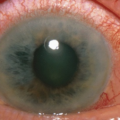
It is not difficult to imagine what happens to energy levels when thiamine concentrations diminish even slightly in an adult. An unrelenting fatigue is one of the early symptoms of struggling mitochondria and thiamine deficiency. More fundamentally, however, all the organs tasked with maintaining life, demand energy. When energy stores diminish, those organ systems struggle. The organ systems requiring the most energy, like the brain and the heart, are hit hardest. Maintain a slight deficiency chronically and damage ensues. In Cuba, for example, trade embargo policies resulted widespread thiamine deficiency in the population, which in turn initiated an epidemic of neuropathy – nerve damage. Over 50,000 Cubans were reported to have developed optic neuropathy, deafness, myelopathy, and sensory neuropathy related to embargo imposed dietary changes. In contrast to the more insidious damage initiated by chronically low thiamine concentrations, severe and acute thiamine deficiency is life-threatening, especially in children, but also in pregnant women.

With low maternal thiamine concentrations, the effects on fetal development, especially fetal brain development that requires enormous amounts of energy, are likely to be devastating. And indeed, they are. But we don’t study that very often, even in rats. Do a search on the subject and there is not much research out there. Sure, some researchers have investigated maternal thiamine deficiency in fetal alcohol syndrome (FAS), postulating thiamine might be the mechanism by which FAS develops, but that is about it. Given how critical it is to fetal development, I expected more research.

It is not just alcoholics who are at risk of thiamine deficiency. An increasing percentage of Western populations are likely thiamine deficient. Thiamine depletion occurs with numerous medications and vaccines via multiple mechanisms, many of which are just beginning to be understood. Conventional farming practices use herbicides and pesticides that block vitamin B absorption and so even diets presumed healthy may not be as nutrient dense as in the past. Poor absorption from altered gut microbiomes may be another common mechanism for thiamine deficiency and emerging evidence finds that Type 1 and Type 2 diabetics excrete significantly more thiamine than non diabetics, making them thiamine deficient as well. Not studying this more broadly is leaving millions of folks to suffer with entirely preventable disease processes. During pregnancy, however, this lack of recognition and research is just downright negligent, especially when we consider fetal brain development.

Thiamine During Pregnancy

Thiamine is absolutely critical for both maternal health and fetal development. Women with hyperemesis gravidarum, excessive vomiting during pregnancy, are at a particularly high risk for thiamine deficiency and though there is increasing awareness of maternal Wernicke’s encephalopathy during pregnancy, a condition typically associated with thiamine deficient alcoholics, the full scope of damage associated with maternal thiamine deficiency is insufficiently understood. There is little to no appreciation of the long term effects on maternal health and even less recognition of how the deficiency impacts fetal development in either the short or long term.

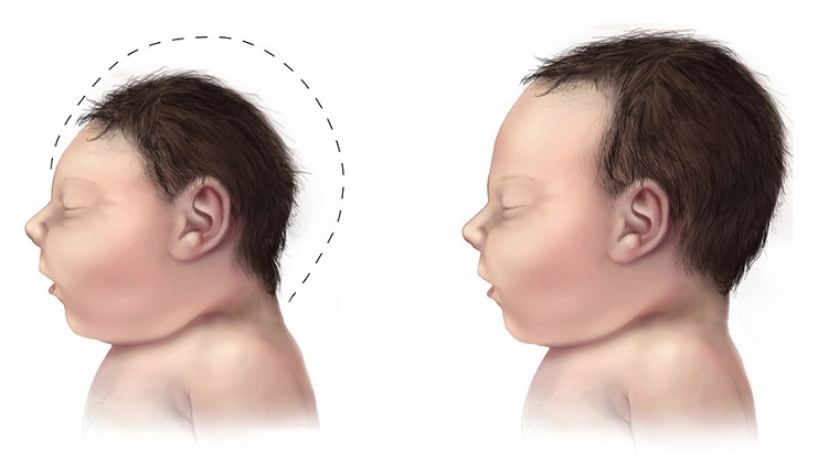
Provided mom survives a thiamine deficient pregnancy, what happens to the growing fetus? In 37% of the cases of severe maternal thiamine deficiency, spontaneous fetal loss occurs. If thiamine is critical for mitochondrial energy production, and fetal development requires exorbitant amounts of mitochondrial energy, what happens if one of the key components to that energy production process is lacking? All sorts of things, it turns out, including microcephaly. Beyond a rare congenital defect in thiamine transport believed to affect only consanguineous Amish, there are very few studies that have considered the effects of epigenetic and more functional maternal or fetal thiamine deficits. We know from the Amish cases, that when the fetal thiamine transporters are impaired, microcephaly ensues. Is it so hard to imagine that we might impair those transporters epigenetically or reduce maternal thiamine concentrations functionally by dietary choices, medications or environmental toxicants that leach nutrients and/or by malabsorption?  And yet, as I dig into this, I find only a few studies that have addressed maternal thiamine and fetal brain development. Here they are.

Maternal Thiamine Deficiency and Fetal Brain Damage

A 2005 study from researchers in West Africa showed that the pups from thiamine deficient dams, had significantly smaller brains by weight. Digging deeper, they found far fewer neurons in the hippocampus, the region of the brain responsible for memory consolidation and retrieval, than the pups from thiamine sufficient diets. Brain damage in the offspring could be induced by maternal thiamine deficiency either leading up to, during, or after pregnancy (while lactating) but varied in scope, severity, and pattern. The most significant damage occurred when the dams were deficient during pregnancy.

In the offspring from perinatal thiamine deficiency, hippocampal volume was reduced by almost a third due to neural cell death.  The neurons that survived were smaller than normal and misshapen. The hippocampus is critical to memory. Hippocampal damage in human adults causes all manner of amnesias and aphasias (speaking and language comprehension deficits) and is found in neurodegenerative disorders like Alzheimer’s disease.

The neurons affected most by the thiamine deficiency, the CA1 neurons, are especially susceptible to oxidative damage and insult. Thiamine is integral to brain oxidation and so this makes sense. What we have to remember though, is that in a fully developed human brain, oxidative damage to the CA1 region is associated with hippocampal ischemia, limbic encephalitis, status epilepticus, and transient global amnesia – very serious conditions. To a developing brain, requiring vast amounts of energy to grow, the consequences of hippocampal deficits are largely under-recognized except again in fetal alcohol syndrome.

Another animal study looked at the effects of maternal thiamine deficiency to the cerebellum of the offspring. The cerebellum is the region of the brain responsible for balance and coordinated motor movements. Here again, the damage was severe with a significant reduction of size, loss of neuron viability, and conduction. There have been a smattering of studies across the decades (here, and here, for example) looking at thiamine deficiency and brain damage in non-pregnant rats, but that’s about it.

Not much else is out there.

From these few animal studies, the work on Amish microcephaly and the work connecting neurodegenerative disorders to thiamine deficiency, we can surmise that thiamine is essential to brain development. More specifically, in pregnancies where thiamine concentrations are low, cerebral development of the offspring will be impaired in some pretty significant ways. Namely, the number and size of neurons is reduced, and as a consequent, total brain volume is reduced. If the deficiency is severe enough, microcephaly is possible and has been identified in the two of studies mentioned above. I think this is what is happening in Brazil. That is, a combination of seemingly unrelated factors, coalesce to produce fetal thiamine deficiency which results in microcephaly and other sorts of brain damage. The questions that remain include:

  1. By what mechanisms specifically is thiamine deficiency produced?
  2. What are the risks for maternal thiamine deficiency in other regions?

One of the most direct routes to thiamine deficiency during pregnancy is hyperemesis gravidarum, excessive vomiting. Case studies abound where it is often not recognized until the mother is in critical condition. It is considered a rare complication, but is it? Unless and until those questions are answered more fully and physicians recognize maternal thiamine deficiency as a potential problem, women and children will continue to be at risk for what are entirely preventable complications of pregnancy.

We Need Your Help

More people than ever are reading Hormones Matter, a testament to the need for independent voices in health and medicine. We are not funded and accept limited advertising. Unlike many health sites, we don’t force you to purchase a subscription. We believe health information should be open to all. If you read Hormones Matter, like it, please help support it. Contribute now.

Yes, I would like to support Hormones Matter. 

Image credit: Centers for Disease Control and Prevention, Public domain, via Wikimedia Commons

This article was first published on June 16, 2016. 

Metronidazole Toxicity, Thiamine Deficiency, and Wernicke’s Encephalopathy

9302 views

Dr. Marrs and I recently had a book published with the title of “Thiamine Deficiency Disease, Dysautonomia and High Calorie Malnutrition”. I was therefore extremely interested to read one of the book reviews published on Amazon books by a patient who had suffered from what has become recognized as metronidazole toxicity encephalopathy (brain disease). Subsequently, she shared her full story with us. It is published here.

A Comparison of Flagyl Induced Encephalopathy and Wernicke’s Encephalopathy

This drug, sold as Flagyl, is prescribed to treat infections caused by anaerobic bacteria and protozoa. Uncommonly, it causes central nervous system toxicity that has the same damage configuration in the brain as Wernicke encephalopathy (WE). WE is the condition that occurs in the brain in people with severe thiamine deficiency. A manuscript in the medical literature was entitled “Metronidazole-Induced and Wernicke Encephalopathy: Two Different Entities Sharing the Same Metabolic Pathway?”  There seems to be little or no effort to explain it as a cause and effect relationship rather than seeing the two situations existing with different causes.

Debilitating Symptoms Post Flagyl

The reviewer states that she has, for two years, suffered with what she describes as debilitating symptoms due to a reaction from Flagyl. She states how these symptoms mysteriously wax and wane. On some days she barely notices them, but of extreme importance, she adds that a mild illness or physical exertion will cause the symptoms to reappear, forcing her back to bed for a variable number of days. When she was taking the drug, she lost his ability to walk and speak, but experienced a dozen other symptoms that persisted. These include difficulty in swallowing and breathing, constipation, severe anxiety, insomnia, depression, heart palpitations, chest pressure and several other unspecified problems. She mentions that she has a borderline enlarged heart.

I must point out here that these additional symptoms are surprisingly common in patients attending physicians in America, often classified as psychosomatic. It is the anxiety, insomnia and depression that guides physicians to thinking that the entire list of “inexplicable” symptoms is psychosomatic. Even the use of the word borderline for heart enlargement indicates that the physician could not identify the total symptomology. If thiamine deficiency had been considered as a diagnosis, the “borderline enlargement” would have fit because it is a cardinal sign of this deficiency.

The patient had evidently done some research on her own and had discovered that metronidazole is a thiamine antagonist. She also reported that in the medical literature metronidazole toxicity is constantly being compared with WE. When she came across our book, she had evidently experienced a flare up of symptoms and began to take supplements of thiamine and magnesium. She stated that the flare up calmed down much more rapidly than usual, enabling her to return to work and function in social activities.

She discovered that doctors do not believe in adverse drug reactions and will not treat the condition since they will not acknowledge that it exists. As a result, she has started a support group for people who suffer from this toxicity. Starting with three affected individuals, there are now over 100, all of whom have the same symptoms. Interestingly, they even warn new members how their doctors will react, preparing them for the reality of being dismissed by the medical community.

Thiamine Deficiency and Psychosomatic Disease

As mentioned in our book repeatedly, the multiple symptoms described by this reviewer are common in the offices of American physicians. “Real” diseases, according to the present medical model, are supported “by laboratory confirmation”. Because vitamin deficiency is generally considered to be nonexistent in America, it is only a physician, open to the possibility, who will entertain the laboratory studies required. The cause of many abnormal current laboratory studies performed on behalf of a sick patient is often obscure. None are capable of identifying vitamin deficiency. When positive as a result of the biochemical changes induced in the body by the deficiency, they are ascribed to other conditions that are acceptable to the present medical model. Hence, the diagnosis of psychosomatic disease often rescues the physician from a failure to recognize his own ignorance. It has always seemed to me that blaming the patient for imaginary symptoms without thinking of the brain as an electrochemical machine represents a glaring deficiency in diagnostic perspective. Unfortunately, nutrition has for long been a neglected area of medical teaching and there are extraordinarily few physicians in practice who recognize its vital importance. What is even more important is the recognition that many drugs are capable of precipitating something as bizarre as thiamine deficiency.

Stress and Illness

I mentioned above that it was extraordinarily important for the reviewer to recognize that flare ups of symptoms occur following a mild illness or physical activity. To explain this, I turn to the teachings of Hans Selye, one of the early researchers in the effect of stress as a causative agent in precipitating disease. Stress must be defined as any environmental factor that imposes a necessity for an animal to adapt (resist). Just like a car that climbs a hill, energy requirement must increase to meet the demand. Mental stress is often more energy requiring than physical stress, explaining the breakdown of health that may follow divorce proceedings. Selye had recognized in animal studies that virtually any form of physical or mental stress imposed the requirement of some form of energy in the ability of the animal to adapt. This was pure genius because energy metabolism was largely unknown in Selye’s time. Modern biochemistry has unraveled a lot of its complexities and thiamine stands out as an absolute necessity in the production of energy. Adapting means that the animal being physically stressed in many different ways would be capable of maintaining a state of health. Failure to meet the increased energy demand was marked by many observed performance and biochemical changes commensurate with those seen in sick humans. One of Selye’s students had reproduced in thiamine deficient animals exactly the same manifestations as those caused by physical stressors.

Diseases of Adaptation or Maladaptation?

The conclusion must be obvious. Any form of mental or physical stress induces a complex reaction in the organism that requires a large amount of energy to run the necessary adaptive machinery. In fact, Selye had concluded that illnesses in human beings were what he described as “the diseases of adaptation”. Since it is a failure to furnish the necessary energy, I suggest a refinement by calling them “the diseases of maladaptation”.

A healthy diet is designed to meet the calorie and non calorie nutrients that fully enable the body to synthesize energy. If this capability fails to meet the demand, even under extreme environmental conditions that permit life to continue, disease follows. The weakness may be genetic or nutritional in character or the stress overwhelming. For everyday life, all three factors are in play continuously.

Please Mr. or Mrs. Reader. Talk about this to your friends and help it to go viral!

We Need Your Help

More people than ever are reading Hormones Matter, a testament to the need for independent voices in health and medicine. We are not funded and accept limited advertising. Unlike many health sites, we don’t force you to purchase a subscription. We believe health information should be open to all. If you read Hormones Matter, like it, please help support it. Contribute now.

Yes, I would like to support Hormones Matter. 

Image by Steve Norris from Pixabay.

Cognitive Testing Post Adverse Reaction: A Lost Opportunity

6095 views

In the not too distant past, before sophisticated brain imaging tests became available, it was the job of the neuropsychologist to assess brain function and brain damage based upon an array of cognitive and behavioral tests. These tests measured the functional capacity of different brain regions. They were entirely behavioral and performance based and could, with a fair degree of accuracy, identify whether and where a brain injury was located and the extent of the damage. Results from these tests could then indicate a need for surgical intervention and/or suggest a prognosis and therapeutic options; options that generally involved a cognitive therapy of sorts to retrain or regain lost capacities.

And then, technology caught up; brain imaging became possible and physicians no longer needed the neurocognitive assessment as a diagnostic, but only for rehabilitation purposes once the brain damage was identified. Non-invasive brain imaging was a remarkable technological advancement. How much better and more accurate diagnoses and interventions could be if physicians were able to see the damage in advance, and indeed, at every phase of treatment. No need to delineate the subtle behavioral signs linked to brain injury in order to diagnose, just scan the brain to rule in or rule out trauma and deal with the deficits after the fact.

Functional Cognitive Testing – A Missed Opportunity

I can’t help but wondering though, if we’ve lost something important by switching so completely to visually based diagnoses. For example, what if the damage is at the molecular level and unable to be detected via imaging, or even via current laboratory markers? How do we even know which lab markers to look for if we don’t ascertain that there are in fact decrements in functioning? Do we even recognize brain injuries as extant if they are not visible by current imaging or laboratory techniques?  I have a sense that we don’t. Cognitive deficits, especially those occurring in previously healthy individuals, following an illness, medication, vaccine or even post pregnancy, may be disregarded along with further diagnostic and therapeutic possibilities when the indices of injury exclude assessing functional capacity.

I was reminded of this recently from a patient story. She, and others like her, experienced a loss of reading comprehension post-fluoroquinolone reaction. Medication and vaccine induced cognitive disruptions are not uncommon. In elderly populations they are quite well documented. In the younger adult populations, however, the research is sketchy at best. In the case I mentioned, the patient was a previously healthy, active young woman. After taking a course of fluoroquinolone antibiotics, and in addition to a myriad of other side effects, she reports losing her ability to comprehend text; something that would be quite disabling in our current text-based world.

I lost a lot of my reading comprehension while I was floxed. I could still officially read – if you gave me a short memo that said, “buy milk,” or something like that, I could read it. But reading a novel or complex materials for work became really difficult. I lost track of the content of the beginning of a paragraph by the time I reached the end of the paragraph. I struggled to understand things that I used to be able to read with ease.

Another fluoroquinolone patient describes her deficit:

I remember going into a restaurant a few months after being floxed. I sat down, looked at the menu, and couldn’t understand a single thing. I couldn’t make sense of anything. It was as though trying to read a foreign language. I put it down, and wanted to stand up and start screaming, and breaking glasses and dishes.

Read any of the fluoroquinolone social media and these observations are not uncommon. Similarly, decrements in cognitive function have been reported in our research on the side effects of the HPV vaccine, during and after Lupron treatments, and even with oral contraceptives.

What I find both most interesting and most troubling is that the loss of attentional capacity, loss of short term memory and loss of language comprehension following the administration of a medication or vaccine may be indicative of a broader health issue; one that should be investigated further. No doubt in many patients these deficits were not explored, at least not functionally, as imaging tests are often negative. That is a shame. Functional cognitive assessments, like those common in clinical practice in the past, and yet still in academic research, would more finely delineate the patterns of medication induced cognitive disorders. These tests could tell us the brain regions susceptible to the medication-induced events in the absence imaging or lab markers. In fact, these tests might help us design more appropriate lab markers. More importantly, functional neurocognitive testing could provide clues about the patient’s overall health. Let me explain.

Linking Cognitive Performance to Overall Health

Each of the medications I mentioned above have distinctly different pharmacological mechanisms of action; so different, one might wonder why I would even consider looking for commonalities in their adverse reaction patterns. Initially, I didn’t. But then the data from our research began flowing in, and along with the data, patient stories began arriving. Slowly, pattern similarities began emerging; similarities that I could not explain by solely looking at the drug’s specific mechanisms of action. There had to be an underlying factor or factors that somehow connected these medications and vaccine reactions. What were they? And per the current topic at hand, how might have functional neurocognitive assessments inspired or expedited our understanding? Not all of the pieces to the puzzle are clear, but here are the clues thus far.

Clue 1. Three of the medications we study negatively affect the thyroid (Lupron, Fluoroquinolones and Gardasil). Thyroid influence on central nervous system functioning, cognitive and behavioral performance is well known.

Clue 2. Thyroid damage is linked to cerebellar ataxia, acute and chronic, via white matter demyelination. Cerebellar ataxia has been noted post fluoroquinolone, post Gardasil and post Lupron.

Clue 3. Thyroid damage is linked to peripheral demyelination. Again, all three medications include demyelination syndromes as part of their reaction profiles.

Thyroid dysfunction alone, without any other intervening variables could explain the cognitive and many of the neurological symptoms we were seeing, but was it sufficient to explain all of them? Probably not, there must something else at play. What could it be?

Clue 4. Each of these drugs are linked to mitochondrial damage (mitochondria are an unrecognized target for many pharmaceuticals and environmental agents). These drugs increase the production of reactive oxygen species (ROS) and decrease cellular energetics via changes in mitochondrial functioning. Mitochondrial damage evokes multi-system, seemingly disparate illnesses, much like what we are seeing. Cerebral mitochondrial dysfunction can cause serious cognitive and behavioral symptoms.

Clue 5. Thyroid and mitochondrial health are reciprocally connected. Damage the thyroid and mitochondrial functioning diminishes. Damage the mitochondria and thyroid functioning diminishes. We have two factors that are inherently related.

Thyroid and Mitochondrial Functioning

What factor could initiate a thyroid – mitochondrial cascade and connect completely dissimilar drugs to these reactions; reactions which are often complex, affect multiple physiological systems, but are also integrally dependent upon proper thyroid and/or mitochondrial function (because of their reciprocal relationship)?  Could there be such a connection?  A few more clues.

Clue 6. A heartwrenching patient story: A Long and Complicated History Topped by Levaquin, highlights a particular set of neurological symptoms that every neuropsych student should immediately recognize.

Clue 7.   Patients from the post fluoroquinolone and the Gardasil groups have been identified clinically with thiamine deficiency. I suspect post Lupron patients may also have thiamine deficiencies, but none have been tested yet.

Clue 8. Both the fluoroquinolones and Gardasil increase thiaminase, an enzyme that blocks thiamine. Higher thiaminase means lower thiamine. Oral contraceptives are believed to increase thiaminase and so women using oral contraceptives in combination with a fluoroquinolone and/or the HPV vaccine Gardasil or Cervarix would be at higher risk for thiamine deficiencies.

Drug Induced Thiamine Deficiency, Cognitive Deficits – The Mechanism

It turns out, thiamine deficiency, or more specifically, a medication induced blockade of thiamine may be at the root of these adverse reactions. Thiamine is a co-factor in mitochondrial and cellular energy, the currency of which is adenosine triphosphate (ATP).  Without thiamine, the mitochondria become defunct, as do the cells in which they reside, and they eventually die. High energy organs like the brain, the heart and the GI tract are often affected dramatically. Similarly, given the reciprocal relationship between the thyroid and mitochondrial functioning and their combined influence on cerebral, cardiac and metabolic homeostasis, diminished drugs that attack the thyroid and diminish thiamine may be doubly dangerous.

In most recent work, thiamine deficient syndromes have been expanded to include five conditions, with fair degree of overlap between them.

  1. Gastrointestinal beriberi: abdominal pain, lactic acidosis, vomiting.
  2. Neuritic beriberi: sensorimotor polyneuropathy, peripheral neuropathy (likely multiple B vitamins involved).
  3. Dry beriberi: high output cardiac disruption without edema
  4. Wet beriberi: high output cardiac disruption with edema (dysautonomias, including POTS)
  5. Wernicke’s encephalopathy: mental status changes, ocular abnormalities, gait ataxia

Given the current nutritional trends with high intake of sugar, fats and processed foods, it is likely that when these medications directly block thiamine production, they do so against the backdrop of already suboptimal thiamine intake. When we consider that oral contraceptives block also block thiamine and that women are more likely to already suffer from low thyroid function, the effects of either the fluoroquinolones or Gardasil on the mitochondrial thiamine could be devastating. How many other medications or vaccines affect mitochondrial functioning and/or thyroid health? How many other medications or vaccines contain anti-thiamine components and diminish this critical mitochondrial co-factor?

Loss of Reading Comprehension and Other Missed Opportunities

Thiamine deficient cognitive decline is well characterized and includes the loss of language comprehension, in more severe cases, deficits in language production, cerebellar ataxia, tremors and as it progresses, seizures, coma, and death. All reversible with thiamine replacement. The cognitive deficits reported by patients, post medication or vaccine reaction, when observed alone but especially when taken in combination with the other tell tale signs of incipient thiamine deficiency, could have lead researchers or clinicians to these diagnoses. At the very least, it should have lead clinicians to thyroid dysfunction, but more often than not, this was not the case.

Cognitive deficits in previously high functioning individuals are reported regularly after medication or vaccine reactions. Almost to a tee, most are ignored once imaging tests rule out blatant injury, but they shouldn’t be. These deficits, when functionally assessed, would provide valuable clues regarding the regions of the brain most susceptible to medication or vaccine induced injuries; clues that could identify damage and disease processes well before detected by imaging tests. By dismissing patient complaints of cognitive deficits we lose valuable research, diagnostic, and therapeutic opportunities. And perhaps, even more importantly, when we segregate symptoms by organ or body part and fail to see the inherent connections among symptoms and physiological systems, we miss the opportunity to help patients heal.

We Need Your Help

Hormones Matter needs funding now. Our research funding was cut recently and because of our commitment to independent health research and journalism unbiased by commercial interests we allow minimal advertising on the site. That means all funding must come from you, our readers. Don’t let Hormones Matter die.

Yes, I’d like to support Hormones Matter.

Image created using Canva AI.

This post was published originally on Hormones Matter on May 21, 2014.

Thresholds and Tipping Points in Thiamine Deficiency Syndromes

13953 views

I recently stumbled upon on a research paper published in 1968. It was not that long ago in the overall course of modern medicine, perhaps even its heyday, when all things were still possible and before the complete fealty to pharmaceuticals arrived. To the youngsters and to those coming of age in the last 20 years, however, anything published pre 1990 is ancient history.  What could such old paper tell me about medicine that is new and useful? It turns out an awful lot.

Back in the day, research was a little simpler and more focused, not on finding out which drug could be fit to which symptoms, but on how things worked. Good experimental design, answered mechanical questions, like if we apply X to Y or if we remove X from Y what happens?

In this paper, Encephalopathy of Thiamine Deficiency: Studies of Intracerebral Mechanisms, the researchers identified a very important component about Vitamin B1/thiamine deficiency – the time course of the disease process. That is, with a diet deficient in thiamine, how long does it take before symptoms emerge, what is the corresponding level of deficiency in the brain, and at what point, after supplementation, does recovery begin; important questions clinically.

Vitamin B1 – Thiamine Deficiency

Remember, vitamin B1 or thiamine deficiency at its worst is linked to severe decrements neurological functioning, like Wernicke’s Encephalopathy that include noticeable ataxic and gait disturbances (loss of voluntary control of muscle movements, balance and walking difficulties), aphasias (language comprehension and/or production difficulties), and if it persists, Korsakoff’s Syndrome (severe memory deficits, confabulations and psychosis). Thiamine deficiency was originally observed in only chronic and severe alcoholics or with severe nutritional deficits as seen in famine. Fortification of food stuffs was thought to relieve much of the nutritional risks for deficit, especially in impoverished regions. More recent research, however, indicates that thiamine deficiency has reared its ugly head once again and this time in modern, non-impoverished, regions where the food supply is ample. How can that be?

Non-Alcoholic Wernicke’s Encephalopathies

Thiamine deficits can be mediated by a number of factors, including by less obvious nutritional deficits where food supply is abundant but nutrition is lacking (a diet of highly processed, carbohydrate and fat laden foods), with thiamine blocking factors found in medications/vaccines, environmental toxicants and some foods, after bariatric surgery and in disease processes like AIDS. Over the course of our research, thiamine deficiency has been observed in previously healthy, young, non-alcoholic patients, post medication or vaccine, along with symptoms of dysautonomia.

What has always struck me about the thiamine deficits we observe is the differential expression and time course of the symptoms. In some people, the reaction leading to thiamine deficit appears linear, progressive and rapid. In others, the symptoms appear to wax and wane and to evolve more slowly. How is that possible? Certainly, individual predispositions come into play. Some individuals may be somewhat thiamine deficient prior to the trigger that initiates the full expression of symptoms, while others have higher baseline stores. Additionally, anti-thiamine environmental exposures and other medical conditions/medications may also come into play.  In the literature, however, the progression of symptoms from bad to worse is almost always direct and rapid, perhaps mistakenly so. Indeed, Wernicke’s Encephalopathy is a medical emergency necessitating immediate IV thiamine.  How is it then, that we see more chronic, remit and relapse patterns of thiamine deficiency, even in some cases where thiamine concentrations are being managed medically?

Cerebral Thiamine Deficiency: Crossing the Black Line

It turns out, there is black line with regard to thiamine deficiency, that when crossed overt symptoms emerge, and a similar black line, that demarks recovery. It is possible then that barring a continuous blockade of thiamine, one can move above and below those lines and the corresponding symptoms may wax and wane. The paper from 1968, cited above, found those black lines, in rodents, but we can extrapolate to humans.

The research. The investigators took three groups of female rodents, a paired group of thiamine deprived and thiamine supplemented, along with a group fed ad lib (as desired) and assessed the time course and concentrations of cerebral thiamine deficiency relative to the initiation and progression of the observable neurological symptoms associated with Wernicke’s encephalopathy in rodents (ataxia, loss of righting, opisthotonos –rigid body arching). The experiment lasted about 6 weeks.

Neither the control group (thiamine supplemented) nor the ad lib group demonstrated neurological deficits at any time during the study. The thiamine deprived group, on the other hand, demonstrated symptoms that began with weight loss, progressive anorexia, hair loss (recall our observations about hair loss) and drowsiness at about 2.5 weeks into the experiment. Interestingly, no neurological signs of thiamine deficiency were seen at that time.

The results. At 4.5 weeks in, the researchers noted a rapid progression of symptoms and decline of health over the course of the next 5 days (the black line). These symptoms included: incoordination with walking, impairment of the righting reflex, reluctance to walk, walking backwards in circles, imbalance, rigid posturing and eventually a total loss of righting activity and severe drowsiness.

One can imagine, if a similar deprivation of thiamine were observed in humans, the corresponding symptoms might also include the initial hair loose and weight loss, perhaps noticeable, perhaps not depending upon the time frame and severity of the thiamine deficiency. It would also include incoordination and difficulty with walking, balance and voluntary movement, perhaps tremors, excessive fatigue or sleepiness and the myriad of neuro-cognitive disturbances noted in Wernicke’s syndrome.

In the cited experiment, one injection of thiamine reversed these symptoms to a nearly normal, or apparently normal neurological state within 24 hours.

Brain Thiamine Thresholds

Animals from each of the groups were sacrificed and examined at each of the stages of the experiment. Brain thiamine and other markers of thiamine metabolism were assayed to determine the cutoff levels of thiamine that demark symptoms and recovery.  This is really interesting and the beauty of this entire study.  Neurological symptoms become apparent when cerebral thiamine concentrations reach 20% of normal.  Recovery begins when those concentrations climb to 26% of normal. At least in rodents, one has to deplete 80% of the brain thiamine stores before overt neurological symptoms become apparent; 80% – that is a huge deficit.  Similarly, it doesn’t appear to take much to right that deficit, only a 6% increase in thiamine concentration set the course for improvement.

If we extrapolate to humans, where life span, genetic and environmental factors likely moderate the degree of thiamine stores and consumption, we still contemplate a rather large thiamine deficit needed before overt symptoms of Wernicke’s emerge. Similarly though, it is also evident that a rather small change in thiamine can have enormous effects on neurological functioning. In the case of the rodents, a mere 6% point change reversed the symptoms. One might suspect equivalent deficit/recovery thiamine parameters in humans.

Waxing and Waning Symptoms:  A Case for Persistent Thiamine Deficiency

If we consider the possible course of non-alcoholic thiamine deficiency, where no extraneous variables like bariatric surgery or thiamine deficient parenteral feeding are present and where dietary thiamine varies daily and is not held constant as it is during experimental conditions or during famine, we can begin to see how thiamine related neurological symptoms may wax and wane. Different exposures and triggers may decrease thiamine periodically, even to the point where overt neurological symptoms present. When those exposures are removed and barring deficiencies in metabolism and diet, symptoms may abate, at least temporarily, and until the next trigger or until the black line is crossed anew and thiamine deficiency becomes the medical emergency observed in overt Wernicke’s.

In contrast, the more persistent or chronic thiamine deficits that do not cross the 80% depletion cutoff (or the human equivalent), may also wax and wane and show all the core neurological symptoms expected in overt Wernicke’s though to a much lesser degree. Additionally, as we have speculated, persistent thiamine deficiency might disable mitochondrial functioning in such a way that the patient presents with a myriad of seemingly unrelated symptoms, that are not typically attributed to thiamine deficiency, such as cardiac dysregulation, gastroparesis, autonomic instability, demyelinating syndromes and hormone irregularities, especially thyroid, but also reproductive hormones. These too may be related to thiamine deficiencies. Although, we cannot and should not rule out other causes as well, sub-optimal thiamine may be involved with a host of complex disease states and medication adverse reactions where neurological symptoms are present. Thiamine deficiency should be tested for and ruled out before more invasive therapeutic options are contemplated.

We Need Your Help

More people than ever are reading Hormones Matter, a testament to the need for independent voices in health and medicine. We are not funded and accept limited advertising. Unlike many health sites, we don’t force you to purchase a subscription. We believe health information should be open to all. If you read Hormones Matter, like it, please help support it. Contribute now.

Yes, I would like to support Hormones Matter. 

A Long and Complicated History Topped by Levaquin: Please Help

6832 views

Here is my story from the beginning. Well, not my beginning, but the beginning of what seems to be a downward spiral health wise. Please help us figure this out.

Two Pregnancies and Cervical Cancer

In 1998 at the age of 22, I became pregnant with my first born son. A normal pregnancy and natural delivery. Upon my six week check up after delivery, they found abnormalities in my pap smear. With further investigation, I was diagnosed with cervical cancer. The doctor said it was as if the wallpaper was cancer but the sheet rock and wood was not affected. I had a LEEP procedure that removed the damaged area with a good portion of my cervix. I was advised that if I wanted more children, I should do so within the next two years, because any further complications would mean a hysterectomy.

In 2000, I became pregnant and delivered my second child, a daughter. The pregnancy was a little more complicated. They feared my cervix would not hold well enough to get her to full term. During the pregnancy, I was diagnosed with hypothyroidism, likely due to autoimmune dysfunction. As a child, I developed vitiligo, an autoimmune disorder of the skin.

The birth of my daughter was natural, although she came four weeks early. The doctors were still investigating my thyroid condition and eventually determined that I had a big goiter and thyroid nodules. The endocrinologist said that the nodules were too small to biopsy, and though he could not say positively that they were benign, he thought that they were. I was instructed to just continue with my thyroid replacement hormones.

Endometriosis and Partial Oophorectomy

In 2004, I experienced terrible pain in my pelvic area. All testing came back normal and the doctors originally dismissed my pain. It got so bad that I could not even sit down without terrible pain. The doctor took me in on a emergency basis and an internal ultrasound showed a mass in my pelvic region. My local doctors believed it to be cancer, as it showed all of the characteristics of malignancy. I was sent five hours away to a cancer specialist. They performed an open surgery to explore the area and remove the mass. Pathology showed it to be benign, so they removed my left ovary and tube. I was diagnosed with endometriosis.

Autoimmune Disease

In 2005, I became very ill. It started with what they believed to be an infection. Later, I was diagnosed with mononucleosis. I was told that my Epstein Barr numbers were through the roof. I literally could not get out of bed. My body hurt so bad that moving, other than to get to the bathroom, pretty much did me in for the day. I was placed on a medical leave at work. Blood testing revealed high levels of antinuclear antibody (ANA) in my blood. My local doctor thought that I may have Lupus. I was referred to a rheumatologist who diagnosed me with Fibromyalgia and Chronic Fatigue Syndrome. After six months, I returned to work.

Hysterectomy and Complications

In 2006, after years of suffering with terrible periods and a few more abnormalities on my pap smear, my OB decided it was time for a complete hysterectomy. My first night in the hospital seemed to be smooth, but in the morning things took a change. I was on a morphine pump for pain, and though I had no pain from my pelvic region, I was having pain in my chest and my left arm. The nurse said my oxygen level was extremely low. The next thing I remember was doctors running in everywhere. I was rushed to CT and then to ICU where I spent the next few days. To this day, I do not know what happened. I got the doctor’s reports. They concluded that it was either a pulmonary embolism or a coronary event. Although, at discharge the doctor told me he thought it was anxiety.

Thyroidectomy, Lung Mass and Fatty Liver

During this time and through following the thyroid diagnosis I always felt awful. No energy, extreme fatigue and weight gain of in total 70 pounds.

In 2007, I developed an illness in my stomach and bowels. The first diagnosis was gallstones. I had surgery to remove the stones. They kept me in the hospital overnight because of the incident the last time I had surgery. I had an endoscopy a few days prior to surgery, and they found I had ulcers and tested positive for H pylori. I got C diff from the hospital and had to deal with that on top of everything else. A colonoscopy revealed that I had ulcerative colitis.

In 2010, my goiter was growing to the point that swallowing and sometimes speaking became an issue. My endocrinologist felt that those symptoms, coupled with the nodules, meant it was time to remove the thyroid. I had a thyroidectomy that year with no complications other than severe fatigue and a struggle to get my levels right.

In 2011, a minor fall left me with a torn meniscus and knee surgery, really not important, I know.

In 2012, I was diagnosed with mono again and the symptoms of pain in my abdomen called for a CT. In receiving the results, I was told I had a 7mm nodule in my right lung and a fatty liver. My liver levels had been high for a few years. I was sent to a pulmonary doctor at the Lahey clinic in Massachusetts. He said that because of my young age and the fact that I had never smoked it was likely not cancer, but that we needed to recheck in six months. My six month checkup revealed another nodule and I returned to Lahey clinic for another consultation. He again said that it did not have some of the characteristics of malignancy and because it was small our best bet was to rescan in another six months.

The Current Nightmare – Enter Levaquin

So this brings me to my current nightmare, one that has continued for seven months. It began on Easter Sunday. I had been sick with what I believed to be pneumonia as my husband had just had it, and I seem to get whatever is going around. We visited the local Emergency rooms and I was diagnosed with pneumonia. They gave me a pill to take while in the ER room. I asked what it was, as my husband was given a Z pack. She told me Levaquin and I took it without question, as it meant nothing to me at the time. We waited for the discharge paperwork and left with a few different pills and and a prescription to continue Levaquin for 10 days.

By the time we reached our house, 20 minutes away, I was itching all over. Hives began to form and my face and ears were starting to swell. I went to a different ER that was 5 minutes away. The rushed me in and administered IV prednisone and Benadryl. I was put on oxygen. After about an hour, symptoms started to slow and I was released. As the reaction was going on, I felt like I was crazy. I think, or at least thought at the time, that it was from the itching. By the time we left the hospital, all I could think about was breathing.

I felt that if I did not concentrate on my breathing I would forget to breathe.

My discharge instructions were to continue with oral prednisone for 3 days and take Benadryl every 4 hours for the next 24 hours. Monday, I slept all day. That evening, I decided I could not take anymore Benadryl. When I came out of what felt like a drug induced coma, I was scared, very frightened actually.

I could barely speak and I did not want my husband to leave me.

I am a very independent person and me feeling like I needed him was not usual. I was very different and it alarmed my husband. He felt it was the prednisone and would not let me take anymore. I finally begged that he let me take another Benadryl to sleep, as I was scared and hating the way I was feeling and functioning. My head hurt so bad that I felt like it may explode.

Tuesday this continued and I could not get off the couch or speak clearly.

Wednesday we returned to the ER. I underwent a CT scan which came out normal and the ER doc felt it was migraines. He dismissed the fact that it could be the Levaquin, as it was only one pill. I was treated with migraine medicine and released.

At first I felt a little better, but some of the symptoms would not go away. I had a limp with pain and weakness on the right side of my body. My neck and shoulders hurt so bad that I could not lay down. The headache seemed unending. I laid around feeling not myself for days.

I recognized my kids but could not come up with their names. I started calling objects by different names, wrappers for socks, and looper for bra, talker for phone.

By Monday my husband brought me to the walk-in clinic, as my doctor was away and the ER had proved to not be of much help. We shared all of the pain and symptoms.

The doctor concluded that it was anxiety and gave me Tramadol for the pain.

The next day my husband brought me to my primary care physician and she was mortified by my condition. She sent us straight to the ER and said she would call them to let them know I was coming and my condition. I was still in terrible pain my head mainly and my right side. I was sent for an MRI that came back normal and underwent a lumbar puncture. It took the radiologist four tries to get the spinal tap and then she forgot to get the pressure.

I was admitted to the hospital for further testing. I had a magnetic resonance angiogram (MRA) and numerous blood tests. I had debilitating pain that left me feeling like I literally may die. I could not stand the light, the nurses had to hang blankets from the windows. The littlest noise hurt me horribly. My husband stayed by my side, as I was still nervous to have him leave me.

I was released a few days later with a slew of different migraine medicines and an appointment to see the neurologist.

The neurologist and her staff were not my favorite from the get go. The nurse asked if something was wrong with me, as I could barely speak and continued to grunt in pain. She changed my medicines and sent me for an EEG that same day. She called a few days later.

I was having seizures in my left temporal lobe.

She prescribed Keppra and left it at that with no follow up appointment or anything. She did mail me a paper about epilepsy. The Keppra did not work well for me. I became very nasty and most of my words were very colorful.

After two weeks, we went back to that neurologist and she was gradually going to reduce the Keppra and start me on Lamictal. The next week I went to see another neurologist that was four hours away.

She said that I had status epilepticus and sent me right to the ER for an infusion of Dilantin.

The next day I returned to be almost myself. I was talking better and acting more like myself.

This sickness has also changed my personality. I say silly things and giggle after everything I say, most of which is inappropriate. I act very childlike or like someone who has mental retardation.

After two days, I slipped back into my previous state. This continued for months the medicine was too low, requiring me to take more than twice the recommended amount, then too high. After two more EEGs both showing slowed brain waves on my left temporal lobe, I was sent to a big hospital to have a long term EEG. There they found the same slowing/episodes that happened 8 to 15 times a day.

I was taken off the Dilantin and started to become myself again. I lost over 20 pounds in month without trying. I was getting around and helping around the house. I was regaining interest in some of my previous hobbies and wanting to rejoin society. This continued for a little over a month. Then, I started feeling bad again.

My cognition remained improved, but my body and my head felt as they did in the beginning of this nightmare. One evening, at my nieces birthday party, I started having pains in my head.

My hearing became very acute. Everything was magnified in sound and my vision again became very blurred.

We left immediately, and by the time we were home, I could barely speak.

My jaw hurt and felt like it could hardly move. My head was aching so bad and my fear had returned.

I have regressed into my previous state and that has continued for two weeks now. I was referred to the neuro-ophthalmologist who said I have pappiledema severe in my right eye and mild to moderate in my left. My neuro thought that I may have a tumor somewhere in my body and my immune system, as a result, was attacking my brain. This is because the testing for paraneoplastic syndrome came back showing positive striational antibodies.

This week I had a PET/CT scan and an third spinal tap. The PET scan showed no abnormalities, although I was given the disc and there is a clear hot spot, at least to my untrained eye, but I guess I need to trust the experts. The spinal fluid was being sent to the Mayo Clinic for testing. For the past weeks, I have had terrible pain in the left half of my face, including my ear, my jaw and near my temple. I know it is not a sinus infection, as I get them regularly and can spot them in an instant. I am not sure if its an infection, but I am inclined to think it is another chapter in this book. I will list some of my symptoms, diagnosis and current medications.

Current Symptoms

  1. Headache, daily
  2. Blurred vision
  3. Magnified hearing
  4. Increased anxiety and fear nothing like before
  5. Right sided weakness
  6. Numbness in tingling in my extremities
  7. Memory impairment
  8. Cognitive deficits
  9. Fatigue
  10. Body Pain
  11. Weight Loss 20 pounds (yay)
  12. Eye pain
  13. Poor judgment
  14. Child like behavior
  15. Clumsiness
  16. Lack of coordination
  17. Lack of focus and inattention
  18. Restlessness
  19. Insomnia

Current Diagnoses

  1. Encephalitis
  2. Temporal Lobe seizures
  3. Status epilepticus
  4. Encephalopathy
  5. Papilledema
  6. Paraneoplastic Syndrome
  7. High Blood Pressure
  8. Acid Reflux
  9. Fibromyalgia
  10. Chronic Fatigue Syndrome / Mono

Current Medications

  1. Synthroid 200 mg
  2. Lamictal 125 mg 2 times daily
  3. Fluoxetine 40 mg
  4. Prtonix 40 mg
  5. Linsopril 10 mg
  6. Vivelle patch (estrogen 100 change twice weekly)

The blood pressure medications and estrogen are new in the last two months.

Please Help

I apologize for the length of this documentation. I want to sincerely thank you for any time and consideration you put into this. I certainly know that it is not your responsibility or obligation. I have two beautiful children and this has taken a severe toll on them. I have gone from a mom who was involved in every aspect of their lives, to a mom who is constantly afraid of causing them shame. In this, I have lost my job and an income, which means paying an incredible price for cobra insurance. I feel like we are up against a wall and running out of possibilities. This is no way for anyone to have to live. I am willing to entertain or try pretty much anything at this point. Thank you again, this means the world to me, just to gain some insight.

With Gratitude and Appreciation.

Participate in Research and Support Hormones Matter

Hormones MatterTM is conducting research on the side effects and adverse events associated with a number of common medications. Our fluoroquinolone study (Levaquin, Cipro, Avelox) will come online soon. To sign up for our newsletter and receive weekly updates on the latest research news and research participation opportunities click here.

To take one of our other Real Women. Real Data.TM surveys, click here.

To share your health story with our community, click here.

If you think what we do at Hormones Matter is worthwhile, please contribute to our research programs and our continued health research reporting. Hormones Matter is totally unfunded and can use your help to continue operations. Crowdfund Hormones Matter – Buy an Unsubscription.