

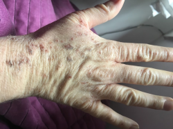
So many Gardasil and Cervarix injured report a host of skin related disorders that appear chronic and treatment resistant in nature. The diagnoses are often incomplete, sometimes contradictory, and more often than not, and no matter the diagnosis, the treatment involves the standard topical and/or injected corticosteroid to reduce the inflammation. If there is relief, it is temporary. I have to wonder what we are missing?
My untrained hunch, from the pictures sent to me is that many of these conditions involve undiagnosed vasculitis. In some cases, the rashes may also be related to undiagnosed Hashimoto’s and other autoimmune conditions that seem to be prevalent. To offer some assistance and encourage folks to press for diagnoses, I am listing possibilities to explore with your physicians.
Acute Urticaria or Hives
We all have had urticaria or hives at one time or another, the red, blistery, itchy, rash that appears after an allergic reaction to something. The rash is generally short-lived, less than six weeks, often recurring and remitting. It can appear with angioedema or swelling and usually responds to antihistamines or anti-inflammatory medications like corticosteroids.

Chronic Urticaria, Urticarial Vasculitis
When the rash last longer than six months, it is considered chronic urticaria and in many cases has a vascular and an autoimmune component. Urticarial vasculitis is a form of cutaneous vasculitis. The acute urticaria and urticarial vasculitis look similar to the naked eye but under a microscope look quite different and emerge from different causes. Non-vascular urticaria is an allergic reaction, whereas urticarial vasculitis represents an inflammation or an attack on the blood vessel wall and may be linked to systemic vasculitis and autoimmune diseases such as Lupus and Sjogren’s.
In comparison to the itchiness of acute urticaria, urticarial vasculitis itches more intensely, is painful and burns. Of interest, many patients with urticarial vasculitis also develop photo-sensitivity (sensitivity to light), joint pain and swollen lymph nodes, fever, abdominal pain, sometimes even difficulty breathing with lung and kidney problems. For color pictures of urticarial vasculitis (I could find no non-copyrighted pictures to post), click here. For pictures of cutaneous vasculitis, click here.
The lesions also may have petechiae or purpura (bleeding or bruising under the skin), common post Gardasil, documented in published case reports and among the many reasons, vasculitis should be ruled out when dealing with chronic rashes post HPV vaccine.

Urticarial Vasculitis and the Immune System
There are two types of urticarial vasculitis, normo-complement – meaning normal complement immune function (normal complement proteins found on blood testing) and hypo-complement or low immune system functioning based on a pattern of low and low plus normal complement blood proteins. Complement proteins are made in the liver and assist or complement the innate immune system by attacking pathogens. Complement deficiency predisposes one to infection and an increased risk of autoimmune diseases like Lupus and Sjogren’s Syndrome. Patients with urticarial vasculitis plus complement deficiency have more difficulty clearing the condition.
Urticarial vasculitis is mostly idiopathic or of unknown origins but is frequently associated with autoimmune diseases like Lupus and Sjogren’s, a viral or bacterial infection of the vessel wall, drug reactions, immunoglobulin disorders and some cancers.
Distinguishing Urticaria from Urticarial or Cutaneous Vasculitis Using the Cell Phone
Diagnosis of vasculitis requires a skin biopsy. Researchers wanting to increase the at-office diagnostic capability for skin related vasculitis found that viewing the rash at 10X magnification using a dermoscope allowed them to distinguish between regular urticaria and urticarial vasculitis. This was in 2004, before camera phones with high intensity magnification existed. I think we can use the same methods using the cell phone. This may give you an at home tool to use and take to your doctor’s office to encourage further testing.
The full report is published here: Surface microscopy for discriminating between common urticaria and urticarial vasculitis. Briefly, a section of the rash is covered in olive oil to eliminate the reflection and viewed under a dermoscope at 10X magnification. A cell phone, photographed at similar or greater magnification may work just as well. The photo examples are copyrighted, so I cannot publish them here, but if you suspect urticarial vasculitis, click the link above to view the sample images and the detailed instructions. Using this method, there are clearly visible differences, even to the untrained eye.
Eczema and Atopic Dermatitis
The catch-all skin disorder for rashes of unknown origin seems to be eczema or atopic dermatitis; an allergic reaction, characterized by dry scaly patches, that are itchy, sometimes oozy and often overly sensitive to internal allergens as well as external irritants. Individuals with atopic dermatitis or eczema often also have asthma, hay fever and food allergies. Treatment is focused on re-hydrating the skin and reducing inflammation.

Hashimoto’s and Celiac or Gluten Sensitivity Rashes
Hypothyroid and autoimmune thyroiditis or Hashimoto’s is common in this population. Anecdotal comments have indicated a degree of gluten sensitivity as well. A rash associated with these condition include the celiac rash, a blistery, itchy rash that appears when one eats foods with gluten. The rash can appear anywhere, but is usually confined to the buttocks, knees, elbows, back and scalp.

Lupus Rash
Again, anecdotal reports suggest Lupus may be common in this population. Lupus is an autoimmune condition that attacks multiple organs including the skin. Symptoms include joint pain, fatigue, lymph node swelling, and when attacking the skin, a specific set of rashes appear: the butterfly rash on the face, the cutaneous rash and discoid rashes on the legs. For a slideshow with images, click here.


Testing for Lupus includes a positive antinuclear antibody test and increased erythrocyte sedimentation rate.
These are among the rashes and vascular conditions that have crossed my desk in communication with post Gardasil/Cervarix patients and parents. I am not an expert in these conditions and so this offered only as suggestions for inquiry. If you suspect a vasculitis or any of the conditions listed here, please read the linked materials, do your own research and see your physician for diagnosis. I would also recommend sharing your stories and pictures with our broader audience so that others may offer input and/or learn from your experiences. These conditions, especially the vasculitis related, are rare and difficult to diagnose. Sharing stories will spread awareness and increase understanding for all.
We Need Your Help
More people than ever are reading Hormones Matter, a testament to the need for independent voices in health and medicine. We are not funded and accept limited advertising. Unlike many health sites, we don’t force you to purchase a subscription. We believe health information should be open to all. If you read Hormones Matter, like it, please help support it. Contribute now.


My daughter took an HPV vaccine and is now experiencing rash like symptoms like never before experienced with her.
How long after did she get a rash?
I am by no means anti-vax. I got the HPV shot when I got my covid booster and flu shot. I have never had a reaction to a vaccine. About 3 days after my legs and arms were covered in hives and my legs looks red and scaly from the calves down. I have went to many doctors and been told so many things and treated for so many things with no relief. I don’t know if it was this or something else I just know it started days after.
My 12 year old granddaughter developed eczema, 2 weeks after the Gardasil injection. She never had skin problems before. I hear that 7 other girls in her school also now have skin conditions.
My son is 12 and received Gardasil injection. A few days later developed extremely red rash on ears and now face that looks like eczema. So going to dermatologist now. Pediatrician did not even say anything about it being the vaccine when I questioned this. This article is informative. thank you!
This is quite educational. Thank you!
The more I read of these posts, the more I am convinced that the ultimate effect of Gardasil is in the disturbance of oxidative metabolism by damaging mitochondria. Since the limbic system of the brain is the most oxygen demanding tissue, it is only to be expected that the clinical issues will be related to this function. For example, the nervous system and the brain are created from the same cells that create the skin. This makes skin and brain function very intimately connected. Many years ago I treated a patient with lichen planus by hypnosis. This is a skin disease where the skin is covered with itchy scales. Every effort by a dermatologist for two years had been unsuccessful. I gave her a post hypnotic suggestion that the itchy scales which covered most of her body would simply fall off. As she undressed that evening to get into bed all the scales fell off. This has got nothing to do with psychology. Hypnosis is a strategy by which the unconscious brain can be manipulated. The limbic system is a computer that affects all the adaptive mechanisms of the entire body, including the skin which is the largest organ in the body. What I have learned in my many years of practice is that the limbic system becomes extremely irritable with a mild degree of hypoxic metabolism, as in damaged mitochondria. Thus, it is a signal from the brain to the histamine containing cells in the skin that causes the urticaria. Several posts on this web site have discussed the relationship of thiamine deficiency with post Gardasil POTS, a condition that comes under the general heading of dysautonomia. Nobody has suggested that this is the only way to produce the dysfunction associated with the vaccination. It does however provide a template for further discussion on oxidative stress. Thiamine deficiency leads to effects that are the exact copy of oxygen deficiency. The role of nutrition is an absolute essential to understanding the mechanisms of post-Gardasil disease.
Lisa, good points. I do wonder how many of these skin disorders are innate or relative to food/environmental allergens versus drug reactions. I suspect unless the reaction occurs immediately post medication use, most folks don’t consider it linked to the medication. In some cases, the effect takes a while to build up and may reflect an accumulation of stressors.
Hmmm…. I’m not trying to bring all of this back to fluoroquinolone toxicity, but it all may be related. One of my first symptoms of FQ toxicity was hives all over my body. Here is some information, taken from comments made on facebook with names removed, that is interesting food for thought –
But remember, when a cell is malfunctioning (do to a mutation caused by a toxin or radiation) the body deems it alien and begins and autoimmune response as a defense mechanism. Thus producing positive autoimmune antibodies in lab tests, but in actuality you don’t really have the disease, it is bad cells. For example I test positive for rheumatoid arthritis (RA +), but I don’t have RA, I have Fibrillan Connective Tissue destruction upon biopsy. But the doctor reads the lab report for RA+ and recommends anti-inflammatory steroids. Bad diagnosis, because the problem is not RA but Fibrillan and steroids will desolve the Fibrillan faster.
and
The cells in your tissue, organs, etc. are not functioning correctly, there is a mutation in there somewhere and the body is reacting to this weird cells as alien, thus producing an inflammatory process (which is painful). The only thing an MD has in his bag of tricks for that is 1. anti-inflammatory steroids or 2. Immune blockers. The steroids will work, but when you come off them the condition will get worse. The Immune Blockers are dangerous, especially for floxed people. I will NOT go on the Immune Blockers and risk lymphoma. Not with all the genes that are fractured in my gene profile.
The root cause of the myriad of symptoms experienced by those suffering from FQ toxicity, and possibly those suffering from other medication toxicities, is the fracturing of genes / the destruction of DNA. That’s the theory that makes the most sense to me at least.