Childhood Constipation: A True Crime Story
I suppose the normal reaction to the title of this article would be to wonder what childhood constipation could possibly have to do with true crime, but I’ll bet there are thousands of parents who will read it and think, “I hope this is going where I think it is.”
That’s because those parents know a doctor who told them to give their child MiraLAX. That is a bit of a spoiler, but I promise, knowing the answer to that part of the mystery only scratches the surface of a much bigger enigma.
Setting the Stage
Whether it’s curling up in front of the TV on a Friday night or listening to a popular podcast on the way to work, a growing audience eagerly seeks their next true crime binge. The attraction may be similar to that of a good Agatha Christie whodunit, but it’s the reality that resonates much deeper than any imagined character ever could – a real story being shared by the real people who lived the nightmare.
Told through the uneasy reflections of survivors, these stories often reveal an evil that we all hope to never personally encounter. Ultimately, we all want to believe that tomorrow will be even better than today. But, if we’re honest with ourselves, we know that not a single person ever woke up and planned to experience a life-changing tragedy.
That’s why the most haunting and most ubiquitous line in the true crime genre is:
“I never thought it could happen to my family.”
True Crime en Masse
Lately, thoughts have been swirling through my mind about what constitutes a “true crime story.” These stories are often sensational and extreme, but they don’t have to be. The villains often seem like an incarnation of the devil himself, but they don’t have to be. Sometimes the cases are solved only after an incredibly in-depth game of forensic connect-the-dots, but they don’t have to be.
What if we were to discover a true crime story where the villain is a system; the crime is frequently nothing more than obstinance and negligence; and the victims are too numerous to count?
Granted, it doesn’t fit the normal true crime template, and perhaps it doesn’t even sound like an interesting story. But, I think you will find that this story is indeed captivating and it is most certainly criminal… on a massive scale.
MiraLAX Intro
A few years ago, I was introduced to Mike Koehler by a doctor who had read my book about the dangers of birth control. She was interested in exploring some common medical practices that seem to be doing more harm than good, and believed Mike and I would have a lot in common.
It was a fruitful conversation, and to be honest, I had never even heard of MiraLAX before that day. I couldn’t believe that his son’s pediatrician had pushed him and his wife to give Bradley an adult dose indefinitely. MiraLAX is an over-the-counter laxative approved by the FDA for patients over 17 years old, not to be taken longer than a week. But then, I learned that this off-label, overprescribing of a drug that has never been approved for children isn’t unusual. In fact, it has become the standard of care for most pediatricians and pediatric GIs!
After taking some time to process this insanity, I wrote an article about Mike’s advocacy and the outlandish practice of giving MiraLAX to children. The response was overwhelming and ultimately inspired me to start interviewing more parents.
Entering Into the Fray
It’s hard to describe what it was like to speak with these families, but it was eerily familiar. It took me back to the days when I was working on my book and spoke with families who had lost daughters to complications from birth control.
If you’ve ever listened to a true crime podcast that touched your soul deeply, one where you carried the story with you as you went about your day – imagine that feeling times ten. Hearing their stories and connecting with them personally feels something like being dropped into the middle of an unnerving combination of an escape room and an interactive true crime experience. It’s sickening and incomprehensible – but once you’re in it, you can’t help but be compelled to find answers.
There’s always one clue that initially demands your attention, and in this mystery, behavioral issues jumped out as the first puzzle piece that appeared in nearly every MiraLAX story I’ve heard.
MiraLAX and Behavioral Issues
I think Mike Koehler would agree with me that his family is pretty fortunate. Their mild-mannered boy suddenly began to get into trouble frequently at school. He even demonstrated violent outbursts toward them. Everything in their lives began to spiral out of control. Like many parents in the same situation, they felt like they were losing their child.
Just as they began to connect their son’s behavioral issues to MiraLAX, the New York Times ran a front page article questioning the wisdom of MiraLAX as a childhood remedy. The timing was perfect because doctors had been gaslighting them, offering assurances (and chastisements) that the drug was perfectly safe for children. Some questioned whether they might simply be parents who refused to accept that they had raised a problem child.
In the end, they were able to reverse many of the ill effects Bradley suffered, and today, they help other parents do the same through a Facebook group that Mike runs, called Parents Against MiraLAX. You can hear more of his MiraLAX story in this short video excerpted from our interview.
Bad Behavior North of the Border
Another father who helps with the group lives in Canada and faced very similar problems after doctors recommended RestoraLAX for his son. He and his wife faced the same questions and doubts, the same gaslighting and fights with doctors, and the same hopeless Google searches after all else had failed. However, it took him a while to find Mike’s group because, as you may have noticed, the drug isn’t called MiraLAX in Canada.
One of the things that makes it more difficult to track how many adverse events this drug ultimately causes is that Bayer markets it under different names across the globe. Brands include MiraLAX, RestoraLAX, GlycoLAX, Movicol, Osmolax… Even generically, it can be referred to as polyethylene glycol, PEG, or PEG 3350 – not to mention other products such as those used when a hospital needs to do a bowel cleansing. These products combine PEG with electrolytes and are branded with names like GoLYTELY, NuLYTELY, Moviprep…
Normally, when companies try to build a strong brand, they focus on uniformity. Watch any drug commercial, and you will see consistency and uniformity executed to the hilt. They build their brand equity by tying everything back to the product. That’s why every article of clothing the actors wear, every prop they hold, and every graphic they display matches the color palette of the drug they are promoting. And, who among us hasn’t cringed after realizing we were singing the brand name of some drug from a catchy jingle?
That’s why, in this world of ultra-branding, it’s odd that Bayer would turn its back on consistency. Instead, they chose to go with a schizophrenic naming convention for their huge global constipation drug.
Despite the confusing naming convention, Chris did ultimately find the group of parents who came together to rally against PEG 3350. You can hear more about his RestoraLAX journey by clicking on this link.
MiraLAX, Allergies, and Kidney Stress
For any sleuth sifting through the evidence, the next signs would likely be allergies and kidney stress. A little boy named Ashton may embody these symptoms more than anyone. His mother, Nicole, told me that when she first saw his allergies getting worse, she never suspected it could be a side effect of the drug he was taking to help him poop. Who would?
She saw her son getting congested, sneezing, and coughing. Pollen counts were high. So, she gave him Benadryl. Unfortunately, Benadryl also contains polyethylene glycol. Which means, she could have unwittingly been making his allergies worse. His body had started developing antibodies to fight the invasion of PEG 3350 from the frequent doses of MiraLAX.
The assault on Ashton’s body escalated. He suffered extreme allergic reactions, hives, and even anaphylaxis – four times. His body was becoming allergic to nearly everything.
MiraLAX is basically liquid plastic. It is, as the name suggests, polymerized ethylene glycol (EG). If EG sounds familiar, that’s because it is the poisonous antifreeze that we use in vehicles.
As Ashton’s antibodies increased, he grew more allergic to household items that included other polymers. He literally became allergic to basketballs, bounce houses, soaps, shampoos, as well as clothes and sheets made of polyester.
And, because the MiraLAX was pulling water from his body into his digestive tract, the stress started to weigh on his kidneys. He developed dark eye circles, a telltale sign, and eventually began to pee blood, which lasted for eight months.
You can imagine Nicole’s passionate, if not frantic, desire to figure out what was happening to her son, and at every step of her pursuit, there were doctors ready to browbeat her into thinking MiraLAX had nothing to do with it.
Here is a clip from the interview where Nicole tells their MiraLAX horror story.
A Medical Threat
As with any true crime story, when you begin to peel the onion, many pungent layers manifest, each with its own distinct variation of rot. It becomes increasingly difficult to judge which layer stinks the most.
Obviously, nothing in the story will ever compare to the tragedy of children being harmed. However, nearly as infuriating is the way that the parents who love those children have been treated by the doctors responsible for injuring their child.
Nicole was one of several parents who mentioned that, at one point, she feared the doctors might report her to CPS. The first time I heard this it sounded a bit hyperbolic, but as more parents expressed the same concern, it took me back to an old notion that I used to ponder.
It starts with the consideration that allopathic doctors tend to like standardization. Their approach goes something like: “You have X symptoms, so I’m going to prescribe Y drug.” They generally don’t like challenges or mysteries. And, they really don’t like to be flustered.
When a child develops unusual symptoms, it puts the doctor in the uncomfortable position of not having answers for a particularly vulnerable patient. Once the discomfort reaches a certain level, the easiest solution becomes pointing the finger at the parents.
I have often wondered how much more common this type of scapegoating became after the movie, The Sixth Sense, which prominently featured Munchausen by proxy (MSBP). MSBP is a psychological disorder where a caregiver essentially abuses a person under their care by faking or exaggerating symptoms, and it’s kind of a perfect fit for the limited curiosity of the allopathic paradigm.
If your standard reaction to unexpected symptoms is, “I don’t know what’s causing that, so I’m just going to prescribe this drug to mask your symptoms,” then it isn’t a huge leap to land on, “I don’t know what’s going on with your child, so I’m just going to assume the problem is YOU.”
I’m not saying that this type of abuse doesn’t happen, only that it may be “over-diagnosed.” Perhaps one day, I will be able to write about parents who had to fight to get their children back, those who were locked up, and some who actually fled their home state in an effort to protect their family. Those parents are understandably reticent to share their stories publicly. However, if you doubt things could go to that extreme, I would like to suggest a documentary on Netflix (not related to MiraLAX) entitled, Take Care of Maya.
MiraLAX and Seizures
Some clues deserve attention because they are common. Others may be less common, but deserve attention because they stand out like a siren and flashing lights. Such is the case with MiraLAX induced seizures.
My research into MiraLAX represents a very small sample size, and yet, I have encountered several cases where the child suffered seizures. And, in nearly every one of these cases, it would be extremely easy to establish a case for cause and effect. However, the pediatricians and pediatric GIs who prescribe this drug day in and day out lack the intellectual curiosity to question what role PEG 3350 might be playing in their patients’ seizures as a newly acquired symptom. Sometimes the symptom leads to an epilepsy diagnosis and more drugs; other times it’s simply ignored.
Overall, only 4-10% of children in the U.S. will experience a seizure before the age of 16, and a large majority of those will be triggered by a high fever. That makes other seizures a pretty rare occurrence among children.
So, let me ask you, if you were a physician who frequently told parents to give their child MiraLAX, how many reported seizures would you need to hear about before you started questioning the cause?
Tragedy Beyond Criminal
In this article, we have talked about victims, their families, as well as the evidence and fallout from the crime, but you may have noticed one key element that’s missing. Where is the determined detective who says, “We weren’t going to stop until we found the bad guy”?
Sadly, that’s a role that hasn’t been sufficiently filled by any outsider. A few tenacious parents have attempted to fill that void with the Facebook group previously mentioned. Ideally, valiant scientists and medical researchers would have identified the dire situation years ago and would be working to stop more injuries. For a time, it looked like there was hope.
The New York Times article that Mike Koehler found back in 2015 featured some prominent players who appeared to be ready to fill the role of dogged detective. First, you had Dr. Kent Williams from Nationwide Children’s Hospital in Columbus, who seemed willing to acknowledge the red flag, if not wave it. He told the Times, “Every pediatric GI physician, I would guarantee you, has told a family this is a safe product… it may not be true.”
Then, you had the announcement that, in the previous year, the FDA awarded nearly $325,000 to Dr. Robert O. Heuckeroth and Dr. Ritu Verma at The Children’s Hospital of Philadelphia (CHOP) “to study whether PEG 3350 might be absorbed by the very young and whether use of the laxatives is linked to the development of psychiatric problems.”
Dr. Verma assured, “It’s a medicine that helps a fair number of children. We want to be sure it’s not harming them.”
That was then. This is now.
It’s been well over a decade since the CHOP study was first funded. Dr. Verma moved on from CHOP. Dr. Heuckeroth continued the study, which officially started in 2022 (eight years after the initial funding). The actual completion date is listed as August 9, 2025. (It was listed as May 2024 when I wrote my first article on MiraLAX). Like everything with the study, it feels like the researchers are hoping to play around with smoke and mirrors long enough that people will stop paying attention. We can keep our fingers crossed that they will eventually publish their findings. I’m guessing since it took eight years to even begin the study, it will take at least that long to form their conclusions.
And, other than being involved with a small study in 2018 that looked at the short term effects of MiraLAX on children, Dr. Williams has fallen silent.
A Fresh Set of Eyes
We sit at a crossroads. All the researchers who seemed to show interest have either disappeared or are severely dragging their feet.
For families who advocate for MiraLAX awareness, their greatest hope may be that the story is incomplete. Perhaps we are sitting at the end of Act 2. The case has grown cold, and before we can reach a resolution, a new sheriff will have to take the reigns and review the evidence. In short, this story still longs for a hero.
In the meantime, we should remember why families usually agree to share their stories with a true crime audience. They don’t want their tragedy to happen to anyone else.
So, much like we might share a tip with a parent to help them protect their tween from online predators after watching a true crime story, let’s spread the word about MiraLAX. If someone mentions their child is constipated, ask them if they’re doctor recommended MiraLAX and then ask if they’ve heard about the problems associated with it.
You could also be even more proactive. If someone mentions that their child is on the spectrum or has other special needs, ask them if they struggle with constipation because many of these children have metabolic differences that lead to a higher incidence of constipation. They also tend to have more doctor visits in general. So, there is a good chance that the parent you’re speaking with has already been sold a bill of goods by at least one doctor.
Simply by educating parents to watch for behavioral changes in their child if they start taking MiraLAX, we could actually influence a sea change. We could empower parents to push back against their doctor when he/she discounts the effects this drug is having on their child.
I’ve seen the beginning ripples first hand. Parents have been talking, Googling, sharing their experiences. They no longer have to go through that long phase of wondering what is happening to their child.
Unfortunately, most of the medical establishment is still denying there is a problem with giving this drug to children, a patient group that the FDA never approved. And, that is a true crime.
…Or, at least it should be. On a macro scale, the most frustrating part of this story may be that none of the actions mentioned in this article are actually prosecutable. No laws have been broken. Even negligence in the face of a mountain of evidence would be hard to establish because the medical establishment holds on to the “consensus” that prescribing this drug off-label to children is a safe practice. And, of course, they can keep saying that as long as it isn’t “proven” to be unsafe.
Perhaps we could start a new genre. Unfortunately, true unethical stories doesn’t have the same ring.
We Need Your Help
More people than ever are reading Hormones Matter, a testament to the need for independent voices in health and medicine. We are not funded and accept limited advertising. Unlike many health sites, we don’t force you to purchase a subscription. We believe health information should be open to all. If you read Hormones Matter, like it, please help support it. Contribute now.
Yes, I would like to support Hormones Matter.
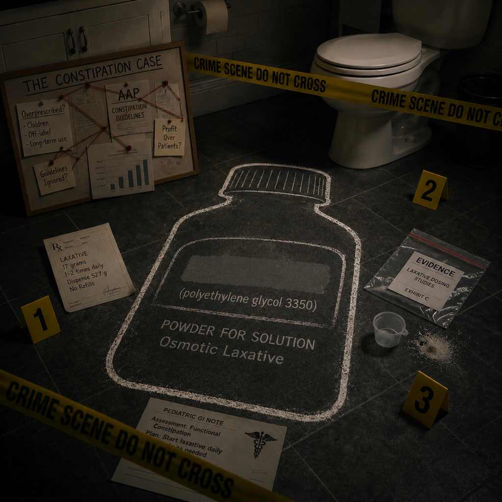
Graphic created using AI.


You might be interested in