ASD

ASD, Seizures, and Eosinophilic Esophagitis: Could They Be Thiamine Related?

3008 views

My 18 year old son has ASD and has had a seizure disorder since he was 6 years old. He has tried virtually all anti-epileptic drugs. Either the side effects were unbearable, they made his seizures worse, or had no effect on his seizures. He was diagnosed with Eosinophilic Esophagitis. He is underweight and of short stature, and always has been. Mitochondrial tests show that complex II is working at 26% capacity. He is also autistic. He has tested positive for folate receptor antibody.

Over the years he has done several rounds of antibiotics, including Flagyl, which I have since learned that it significantly depletes the body of thiamine. He has also taken several rounds of Diflucan, Azithromycin, Vancomycin, Augmentin, Amox for various issues including candida, clostridia, gram negative gut bacteria, etc.

He is currently on Lamictal and just started Briviact for seizures. The Briviact causes anger and aggression issues. He currently deals with OCD tendencies. He was recently found to have bone density of 2.8 standard deviations below normal. This falls in the range of osteoporosis, but he has not been diagnosed with it because of his age.

He eats fresh and a lot of dried fruit, meats, raw and cooked greens, white rice, lots of cooked veggies, eggs. He also takes Lipothiamine 100 mg/day, Magnesium 550 mg, a multi-vitamin, calcium, vitamin D, and K, all at the direction of his doctors.

Childbirth and Infancy

M was born on July 9th 2005 7lbs 9oz. He was full-term. I had high blood pressure at 41 weeks and labor was induced. He would not drop into position and he became distressed and so was delivered via cesarean while I was under general anesthesia.

He spent 4 days in the NICU because he aspirated meconium and would not latch to feed. While in the NICU, he was administered antibiotics. He was formula-fed, not breast-fed.

As an infant, the large size of his head was somewhat of a concern for the pediatrician. He was administered vaccinations according to the CDC guidelines for the first 12 months. He had infantile spasms off and on. He spiked a fever for every vaccination. Tylenol was administered. He received 3 doses of flu vaccine, accidentally, within 3 months.

He did not sleep well, and still doesn’t.

Initially, he was very precocious. As an infant, he would put puzzles together that were for much older children. He would complete sorting activities that were well beyond his age range. He did not babble and eye-contact was fleeting.

After his 18 month vaccination, he lost just about everything within 2 weeks. After these vaccinations, he couldn’t do his puzzles, bring food to his mouth, smile, couldn’t stand to be read to when he previously loved to be read to. He also developed a sensitivity to light and sound and cried a lot.

At 24 months, he was diagnosed with profound autism.

PANDAS/PANS and Eosinophilic Esophagitis

At age 10 years, he abruptly lost skills again and it was thought he had PANDAS/PANS as he had several strep infections treated with antibiotics. He did a several month long courses of Augmentin or Azithromycin to treat PANDAS/PANS. He had a severe trauma at age 11. He was horrifically abused by a school employee.

He has always been of short-stature nearing 5th percentile for height, and slightly overweight for his age, until age 14 when he started having symptoms of Eosinophilic Esophagitis. He was diagnosed with EoE at 15 and has struggled to keep his weight high enough as he dealt with the intense pain, fatigue, and esophagus issues with this condition. He is currently taking Dupixent for his Eosinophilic Esophagitis as the PPI and Budesonide slurry were not addressing the issues. So far Dupixent is allowing him to eat. His diet remains very restricted due to having so many trigger foods and he has almost no appetite.

He eats a lot of dried and fresh fruit. He loves greens, raw and cooked. He also eats meat, white rice noodles.  He eats mostly an organic diet. He does occasionally enjoy candy.

Seizures

He developed seizures at age 6. These were controlled for a while on Depakote, but the side effects of Depakote were too much for him and so we had to stop. His seizures are now not controlled. He has 1-2 tonic-clonic seizures per week, plus several staring spells all throughout the day. Recent EEG showed abnormal spikes and discharges in the frontal and temporal lobes. It indicated his seizures involved many places on his brain. Brain surgery was being considered for seizures at this time, but ruled out as an option due to the nature of his seizures.

He has failed several other seizure meds including Vimpat, Zonegran, Aptiom, Topamax, Onfi, and others. He is currently on Lamotrigine and Epidiolex for his seizures. He also takes trazadone and gabapentin for sleep, although these do not consistently help him sleep. He is so consumed by fatigue and can hardly get out of bed even to walk across the room. With tons of encouragement he can do brief periods of school work. The meds cause him to lose focus and become frustrated. He seems to almost always be lost in a fog and unable to participate in basic conversations without losing focus or becoming too exhausted to continue. Each seizure will cause him to be in bed for 2-3 days. He has fallen many times going into a seizure and is now afraid to leave the safety of his bedroom. He will come out, but rarely.

He has intermittent issues with nystagmus. He had a bad case of COVID 2 years ago, which caused clusters of seizures and constant nystagmus.

He has an exaggerated startle response.

Despite It All

M is a sweet young man. He is brilliant. He loves animals. He tells everyone he sees that he is so happy to see them. He is working with a local legislator on how to improve rights for non-speaking people, especially in the court room. He is completing all of his high school courses at home with straight A’s and he is a published poet.

He does not speak, but he communicates by pointing to letters on an alphabet board. This is a skill that took him years to learn. He communicates at an age-appropriate level or higher. He is working, slowly, toward a standard high school diploma.

Postscript

Based upon what I have learned from this website, I discussed thiamine with our physician. It turns out, she heard Dr. Lonsdale speak years ago. She recommended 50mg of Lipothiamine. The entire time he was taking it, he had no seizures. I was not sure that it was thiamine or the meds until we ran out for about a week. The seizures returned, but as soon as we resumed the Lipothiamine, they disappeared again. He has been taking it again and now it has been 2 weeks without seizures. I don’t want to get my hopes up, but it could definitely be a piece of the puzzle. Are there others out there with similar experiences?

We Need Your Help

More people than ever are reading Hormones Matter, a testament to the need for independent voices in health and medicine. We are not funded and accept limited advertising. Unlike many health sites, we don’t force you to purchase a subscription. We believe health information should be open to all. If you read Hormones Matter, like it, please help support it. Contribute now.

Yes, I would like to support Hormones Matter. 

Image by Gerd Altmann from Pixabay.
Food Aversions in Children with Autism

5806 views

Dietary Aversions and Preferences in Children with Autism

“They are a super picky eater. They won’t eat vegetables.”

These are some of the most common words I hear from the parents of my autistic patients. Of course, we all know that eating our veggies is healthy, but for a child on the spectrum what the bacteria would do with the vegetable fiber can have a massive impact on how their immune system and brain work. There are a few reasons they might dislike certain types of foods.

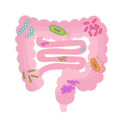
  • Altered gut bacteria. They may have too many bacteria in the small intestine due to poor bowel motility which then could cause pain and bloating.
  • Parietal lobe issues. It is also possible that a child on the spectrum could have their parietal lobes affected. This could lead to an aversion to the texture as well as the taste of certain foods. Since there is typically altered sensory perception with a child on the spectrum, including on the lips and tongue, this will impact how they perceive food textures and the taste of food itself.
  • The foods they like may be exorphins. Certain foods like breads with gluten, for example, can activate opioid receptor sites. That is, when the child, eats these foods, he or she gets a boost of nature’s endogenous painkillers. This makes the child addicted to the very foods that have the potential to be the most problematic. When a parent asks me what foods their child should avoid, I typically ask them what foods they seem addicted to and we have some of the answers.

Pieces to the Puzzle: Immune System and Microbiome Crosstalk

Although the research is ongoing, it is becoming more apparent that either the child with autism or his/her mother suffer from a pattern of immune activation that can lead to disrupted gut barrier. This, in turn, contributes to an alteration not only in the gut bacteria, but also, in how the immune system in the brain respond to immune insults. Since these bacteria are responsible for metabolizing the food we eat and extracting from those foods essential nutrients, that then are used for building neurotransmitters, and modulating immune function, the alteration in gut bacteria can lead to an inflammatory cascade and cycle, which then affects the proper development of the nervous system. As the diet becomes narrower, it brings further nutrient depletion, causes more of a microbial imbalance, and leads to a pro-inflammatory environment. Although most people with chronic disease have some form of inflammatory response, researchers have found that when a subset of immune cells called TH17 are hyperactive, either in the mom during pregnancy, or the child themselves, we see an inflammatory response so imbalanced that a neurodevelopmental disease is more likely to develop.

A Few More Pieces: Bacteria, Fiber, and Immune Modulation

When bacteria eat different types of fiber from dark leafy green vegetables or grains, they make compounds called short-chain fatty acids (SCFA). These SCFAs activate several parts of the enteric nervous system, which then communicate with the central nervous system. Specifically, one SCFA called butyrate seems to particularly important. Butyrate is a primary energy source for cells in the large intestine used to ‘fix the gut’, but it also has a great impact on the immune system and healing the gut lining. It does this by multiple mechanism, but mainly by reducing bad bacteria that can be driving the immune issues in the first place. Butyrate helps turn off pro-inflammatory signals as well as turn on anti-inflammatory signals and bring balance back to the immune system by helping with something called T-regulatory cells. T regulatory cells do exactly what the name suggests, they regulate the immune response. This is especially important given that the brain and GI tract are in constant communication, not only via the vagal nerve, but the sympathetic nerve fibers send signals directly to the brain. Given that the microbiome and inflammatory cytokines are an important part of this relay system, their involvement with something like Autism should not be ignored.

For those of us that have been doing this enough, it is blatantly apparent that there is immune system involvement, with some articles showing upwards of 70% of children on the spectrum have an autoimmune disease process going on in their brain. Hence, to help modulate the altered immune response, the bacterial by-products of fermenting fibers can go a long way in helping bring balance to the immune response, gut lining, and nervous system.

Returning to the picky eater problem in children with autism, it appears that because of altered bacterial balance and possibly also sensory issues, they may be drawn to the very foods that sustain an inflammatory response, and conversely, naturally avoid those foods that might dampen that response and rebalance the gut microbiome. The concept of oral tolerance is where the immune system is told to ignore foods that are being consumed so they don’t contribute to an inflammatory response. This can be a large catch-22 in the sense that the less diverse a child’s diet, typically the lower their oral tolerance of foods, so unfortunately, the immune system and microbiome get painted into a corner that can be difficult to get out of.

A Picture Emerges

Although these are only a few pieces of the puzzle, giving the gut and immune cells of a child with autism what they need can really help. Eventually, the goal is to have these kids lose their taste aversion and start eating veggies and other healthier foods. To accomplish this, we have to address the root causes of their food aversions. This means addressing proper magnesium levels of the brain and re-balancing gut microbiota. Once we see how all the pieces are connecting, we can paint the clearest picture possible. Since each child is unique, each picture is unique as well. The good news is, once a path to healing is identified in a child, the effect can snowball in the sense that the fewer food aversions a child has, the better the gut and immune system work and so the better the brain works, which means the more foods they will eat and enjoy. That is, the food/health/immunity cycle becomes more favorable.

We Need Your Help

More people than ever are reading Hormones Matter, a testament to the need for independent voices in health and medicine. We are not funded and accept limited advertising. Unlike many health sites, we don’t force you to purchase a subscription. We believe health information should be open to all. If you read Hormones Matter, like it, please help support it. Contribute now.

Yes, I would like to support Hormones Matter. 

Image by StockSnap from Pixabay.

This article was published originally on April 8, 2019. 

Energy Deficiency and ASD

6041 views

I was a pediatrician at Cleveland Clinic from 1962 to 1982 and the clinical research, much of which I have published, was done during those years. I can distinctly remember that a patient with autism was considered to be unusually rare at that time. It was a habit of the pediatric staff to meet regularly with the residents in order to discuss clinical cases, particularly those that were uncommon or rare. I remember a case of autism coming up for one of those conferences because of its rarity. No longer rare, there are many ways in which a child can present symptoms, so the disease is now referred to as autistic spectrum disorder (ASD). This may imply that a lot of different disorders are conceived as being within a spectrum, each with a different cause, or it may simply refer to the wide variation of symptoms.

I looked up the statistics on the Internet and found that ASD today affects one in 68 children (1% of the childhood population). Parents who have a child in the spectrum have a 2% to 18% chance of having a second child affected, implying a genetic risk. I took early retirement from Cleveland Clinic and joined a private practice specializing in nutrient therapy. I became active in the group of physicians who are members of the American College for Advancement in Medicine (ACAM, acam@acam.org). A very big fraction of my patient population during those years was many children with ASD. My clinical experience between 1962 and 1982 had strongly suggested that thiamin(e) deficiency was common in the United States, in spite of the general medical opinion that it never occurred. During those years I had worked with an associate, a doctor who worked in the laboratory. Because of my clinical need, he had researched the literature for ways and means of accurately measuring thiamine deficiency. He had initiated a blood test known as transketolase activity and I need to explain this briefly.

Understanding How Thiamine Works and How to Test for Deficiency

Transketolase is the name of an enzyme whose activity is completely dependent on the presence of thiamine and magnesium, both known as cofactors to the enzyme. There are a series of biochemical reactions in which this enzyme plays an integral part. Known as the hexose monophosphate shunt, this series of reactions occurs in red blood cells. By taking some blood from a patient, the activity of this enzyme can be measured in the laboratory and is reported as transketolase activity (TKA). What we discovered was that the TKA could be quite normal when the symptoms of the patient strongly suggested thiamine deficiency. In order to prove that the patient was deficient, there was a second part to the test. Thiamine pyrophosphate (TPP), the biologically active form of thiamine in the body, had to be added to the reaction container and the transketolase activity measured again. If the TKA accelerated, it showed that the enzyme was lacking the necessary cofactor, in spite of it being in the normal range of activity with the first analysis. It was reported out as a percentage increase of activity as compared with the baseline (thiamine pyrophosphate effect or TPPE).

My friend in the laboratory had done a series of control tests on people that were purportedly healthy. His results compared favorably with reports of the test in the literature and these healthy people could have a TPPE of up to 18%. Anything over 18% for the TPPE guaranteed deficiency of thiamine or magnesium, so both of them would have to be supplemented for the benefit of the patient. Unfortunately, laboratories in prestigious institutions presently only do the TKA and assume that the patient does not have thiamine deficiency if the TKA is in the normal range. From my experience, I am aware that they are missing the majority of patients who have this deficiency. The TKA is not a valid indicator of deficiency by itself. Having given considerable thought to this, I concluded that if the enzyme is equipped with an adequate supply of thiamine there should be no acceleration in its activity after the addition of TPP. The only strictly normal TPPE would be zero (i.e. no TKA acceleration). The percentage acceleration of TKA would reveal a gradual deficiency compatible with no clinical significance until a certain increase in TPPE was recorded as being symptomatic of deficiency. Therefore a TPPE of, say, 15%  might be more clinically significant in one individual, whereas 20% might be less significant in another individual. The use of the test requires the clinical experience and adequacy of knowledge of the observer.

Clinical and Laboratory Experience

Without thiamine and magnesium as cofactors, the several enzymes dependent on them begin to become less and less efficient. One of these enzymes is essential to energy synthesis and so thiamine deficiency, because it powers pretty well every cell in the body, can literally cause any disease, since energy is integral to all functions of the body. Depending on the cellular distribution of the deficiency, the clinical expression will vary and there is no typical repetitive clinical expression (phenotype) for thiamine deficiency. However, the brain and heart being the most metabolically active organs are therefore much more susceptible to this deficiency. Thus, brain and heart symptoms are the commonest forms of clinical expression. Thus, it can be assumed that any gross deviation of behavior is evidence of electro-chemically driven changes in brain metabolism related to energy availability.

Case Evidence

Many of the children that I saw with ASD had an increase in their TPPE. Most of them with an accelerated TPPE had a TKA that was in the low normal range. When they were treated with thiamine, many of them responded clinically and their TPPE decreased into the acceptable range, so the evidence was published . The case histories of a mother and her two children were reported . The mother was a recovered alcoholic and alcohol is frequently responsible for causing thiamine deficiency. Some alcoholics react to sugar, particularly if there is a family history of alcoholism that suggests genetic risk. She and both of her children had symptoms that were typical of autistic spectrum disorder, but also they all were recognized to experience the clinical effects of dysautonomia. A description of beriberi clearly indicates that thiamine deficiency disrupts the normal functions of the autonomic nervous system (dysautonomia). All of them had intermittently abnormal transketolase studies indicating abnormal thiamine pyrophosphate homeostasis. Both children had unusual concentrations of arsenic in the urine. All of them had symptomatic improvement with diet restriction and supplementary vitamin therapy, but they quickly relapsed after ingestion of sugar, milk, or wheat. All three individuals became aware that their symptoms were related to their dietary indiscretion, but were quite unable to resist their ingestion of the “foods” that they all knew to be responsible for their relapse. The presence of arsenic in the urine was in keeping with our experience in finding the presence of heavy metals in ASD. My colleagues and I were so impressed with thiamine deficiency as a major cause of ASD that we conducted a pilot study, using a derivative of thiamin known as thiamin tetrahydrofurfuryl disulfide (TTFD). Eight of 10 children showed clinical and biochemical improvement .

The negative relationship of the brain with sugar is mindful of a case that I reported in 2016 and whose story appears on this website. A 14-year-old boy was strongly addicted to sugar and throughout the first eight years of his life his repetitive symptoms had been classified elsewhere as psychosomatic in nature. At the age of eight years it had been discovered that he had the relatively recently described disease known as eosinophilic esophagitis. He also had the classical clinical picture of dysautonomia. It was hypothesized that some form of genetic risk, coupled with thiamine deficiency from out-of-control sugar ingestion, was the responsible combination. The vagus nerve, whose course runs from the brain to the spleen, (and beyond) presides over the control of inflammation. The medical literature has reported food allergy as the cause of eosinophilic esophagitis. The vagus nerve requires acetylcholine as its neurotransmitter, a chemical that is derived from the citric acid cycle (CAC), the cellular “machine” that synthesizes energy. In turn, the nerve is dependent on thiamine that is essential for the entry of glucose into the CAC. Lacking the suppressive function of the vagus on inflammation resulted in a failure to suppress it in the esophagus. Thus, it was conjectured that thiamine deficiency was the primary underlying cause of the esophagitis.

Insufficient Energy and ASD: The Three Circles of Health

Mitochondria are very sensitive to environmental stressors such as toxicants, medication, immune activation, and metabolic disturbances suggesting that mitochondria are involved as a cause of ASD.

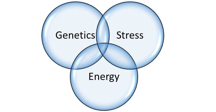
Figure 1. Three Circles of Health


From what has been written above, it must become clear to the reader that I am proposing a very different approach to the cause of ASD and disease in general. It is based on the use of Boolean algebra as represented in Figure 1. This is a statistical method of measuring the influence of variables by representing them as overlapping circles. The individual effect of one circle is represented by its area and its relationship with the other two circles by the area within the overlap. All three circles also overlap, representing the concept that disease might be 100% from one circle alone, by the combination of two defective circles, or by the variable combination of all three.

Genetics

The construction of the human body is based on a code in the form of DNA and represents a “blueprint”. Known as the genome, perfection in the code would create a perfect human body. However, we know that DNA has imperfections, but in most cases a human individual can get along quite well in spite of the imperfections.  Although an abnormal gene may be the single cause of disease, the late onset of most genetically determined conditions indicates that an additional factor is required to cause disease expression. This emphasizes the importance of the quality of nutrition in preserving health. For example, type 1 diabetes has a genetic risk, but is not usually expressed for many years, so another factor is necessary. That is why the disease is stress related, making its clinical appearance following something as simple as an infection, a divorce or an injury. Type 2 diabetes often has a genetic risk where either diet or stress, or both, may be initiators.

Epigenetics: Environment and Nutrition

Epigenetics is a relatively new science that seeks to explore how malnutrition and poor lifestyle can have a negative effect on our genes. There are several genetically determined diseases known as inborn errors of metabolism that make their appearance at birth. If any one of them is not recognized in the newborn infant, the result is often a mentally retarded individual. Some of them, perhaps the commonest being phenylketonuria (PKU) require a special diet initiated at birth in order to prevent the mental retardation. That is why in every state in the United States, a special laboratory has been set up to screen a blood test from every newborn infant, in spite of the rarity of the diseases. Sometimes, an infant is born with a gene that creates a risk for generating the disease, but it does not appear unless a secondary factor such as an infection or an injury (stressor) is experienced. There is a disease known as maple syrup urine disease (because the urine smells exactly like maple syrup) that can appear immediately at birth spontaneously or will only appear when the child is hit by some infection or trauma. There are some cases of this disease that respond to megadoses of the vitamin (thiamine/magnesium) that is normally necessary for the mechanism whose failure produces the disease. This has significant importance because a head injury or an infection might be blamed as the sole cause of a clinical problem, whereas the truth could be that the stress factor has initiated a metabolically determined disease previously unsuspected. If such a similar disease responds to the necessary associated vitamin, it can be said that the patient was successfully treated epigenetically.

Stress

In my view, this word is used too carelessly. It is most frequently used to describe the result of stress by saying that someone is “all stressed out”. So let us be clear that stress is some kind of force that a person has to meet and to which an adaptive response is required. It may be a mental force such as a divorce, a business deadline, or a physical assault such as an infection, or an injury. The three circles of health uses the philosophy proposed by Selye. This famous researcher used many forms of physical and mental injury (stress) on rats and studied the biological effects by examining their blood and tissues. He came to the conclusion that each animal went through a process of resistance that he called the General Adaptation Syndrome (GAS). The laboratory results that occurred if the GAS failed to restore health were strikingly similar to those registered in sick people and he proposed the idea that human diseases were the diseases of failure to resist or adapt to stress. What was completely revolutionary was that the GAS required some form of energy to power the machinery that enabled the animal to adapt.

In Selye’s time the biochemical mechanisms of energy synthesis were not well known. Today, these mechanisms are understood and our lives are spent in meeting the daily stresses to which we have to adapt. Our failure to meet the GAS may well be of course that the form of stress is overwhelming, such as a car collision or a virulent infection. However, what we meet on a daily basis is a mental or physical form of stress to which we have to adapt or from which we have to heal. The central factor is the mobilization of sufficient energy to meet the demands of defense. Therefore, the three circles of health predict that the cause of human disease agrees with the concept of the “diseases of adaptation” proposed by Selye.

Energy and the Ability to Adapt

Assuming that the adaptive machinery in the body is genetically adequate, all that is required is sufficient energy to drive it. The symptoms generated during the process of adaptation simply represent the locality of the energy deficiency and constitute a warning by their perception in the brain. They have to be interpreted for their underlying meaning rather than accepting them as the effects of a named disease such as Alzheimer’s or Parkinson’s. Since the brain is an electrochemical “machine”, the nature of the patient’s diet, as well as family history, are essential to beginning to unravel the problem. In the case of ASD, the mother’s diet during pregnancy is of vital importance. Occasionally, a seemingly irrelevant observation by a physician can be valid. I was riding in a car with a gentleman who had a group of infant’s shoes dangling behind the windscreen. I asked: ‘what was the significance?’ He responded by telling me that his first child died in infancy from a rare genetically determined disease. Subsequent children had survived and this was his method of keeping them in memory.

Nutrition as Medicine

When Homo sapiens arrived on the face of the earth, or even when his ancestors were present, the food was available and the general concept is that we were hunter gatherers. To find out what kind of food an animal should be taking, you have to look at the teeth. We have cutting teeth, canine teeth and grinding teeth, indicating that we are omnivores and can consume meat, vegetables and fruit. The necessary non-caloric nutrients (vitamins and essential minerals) were in the natural food. In the modern world, our food is far from natural and the high calorie content, particularly in the form of sweets, is overwhelming the cellular machinery that synthesizes energy. For this reason, many of the illnesses that haunt a physician’s office are merely a reflection of a mild to moderate energy deficiency in brain cells. If recognized for what they represent and treated with appropriate nutrition, the symptoms quickly disappear. If not, we can hypothesize that there may be permanent damage that is diagnosed as one of the neurodegenerative diseases.

The trouble with understanding this concept is that the distribution of the energy reducing mechanism, together with genetic risk, will vary from individual to individual. A child with a genetic risk, or, more commonly, a victim of poor maternal diet, is born a candidate for energy deficiency. That is why ASD can be the result of thiamine deficiency or any other mechanism that interrupts the flow of energy. However, the same deficiency can give rise to many other conditions presenting with an unpredictable array of symptoms. There is a great deal of evidence that food is burned (oxidized) in the cells of the body to create a form of chemical energy (ATP) that is transduced to electromagnetic energy. It is this energy that is used to drive physical and mental action. There is also evidence that a biological form of thiamine (thiamine triphosphate) is important in the electomagnetic transduction process, thus making thiamine uniquely indispensable. It is for this reason that I have spent many years emphasizing the life-giving properties of this extraordinary vitamin. I am not therefore surprised when I read that a constellation of symptoms called Parkinson’s disease has been reported to respond to treatment with thiamine.

We Need Your Help

More people than ever are reading Hormones Matter, a testament to the need for independent voices in health and medicine. We are not funded and accept limited advertising. Unlike many health sites, we don’t force you to purchase a subscription. We believe health information should be open to all. If you read Hormones Matter, like it, please help support it. Contribute now.

Yes, I would like to support Hormones Matter.

Autistic Spectrum Disorder, Mitochondria, and Nutrient Deficiencies

5484 views

Much has been written in the medical literature concerning the biochemical and potentially treatable nature of a group of conditions known as Autistic Spectrum Disorders (ASD). Then why are practicing physicians treating these children with medications rather than nutrients? The answer, of course, is that the disease model is being rapidly antiquated by a better knowledge of Mother Nature. This post discusses some of the important published information that indicates very strongly where the research should be.

Mitochondria

Hopefully, the reader of this post will be aware that mitochondria are the organelles that produce cellular energy. It has become clear that what the authors describe as a large subgroup of individuals with ASD demonstrate abnormalities in mitochondrial function and suffer gastrointestinal symptoms. These authors state that the majority of children with ASD do not have a primary genetic mutation. This raises the possibility that their mitochondrial disorder is acquired, or results from a combination of genetic risk interacting with a wide range of environmental triggers. “Spotty”energy deficiency in the brain may be the bottom line. Cellular distribution of the deficiency might vary from individual to individual, thus explaining the variety of symptoms. These researchers also reviewed the evidence that enteric bacteria play a part in this disease. The microbiome is the term used to describe the full complement of bacteria that live in the human intestine and play a huge part in maintaining health. The microbiome and its connection with disease was recently reviewed on Hormones Matter.

ASD and Malnutrition

One hundred and five children with ASD and 495 typically developing children of 6 to 9 years were compared in Valencia, Spain. The affected children failed to meet dietary recommendations for thiamine, riboflavin, vitamin C, and calcium. It was recognized that cultural patterns and environment may influence food intake. The majority of parents reported some concern regarding the feeding behavior of their ASD children in India. Relative to controls, the affected children had significantly lower daily intake of potassium, copper and folate although they did not differ in their calorie intake. We have pointed out in these posts that a high intake of empty calories with a normal blood concentration of thiamine is the equivalent of a normal calorie intake with dietary thiamine deficiency.

Autism rates in the United States are increasing at a rate of 15% a year. A recent study established the autism rates for each of the 50 states and calculated the percentage of infants who participate in the Women, Infants and Children program (WIC). The states with the highest WIC participation had significantly lower autism rates. Infants who were solely breast-fed, however, had diets that contained less thiamine, riboflavin, and vitamin D than the minimal daily requirements. The results suggest that autism may be nutritionally related to vitamin deficiency. However, the question remains as to whether the malnutrition was the effect of poor eating behavior resulting from brain changes due to autism, a typical “chicken and egg argument”. Which is cause and which is result?

An 11-year-old boy with autism developed liver dysfunction and became less responsive. His diet for several years was self-limited exclusively to a single “fast food”. He improved rapidly with the administration of thiamine but developed epileptic fits two weeks later that required the administration of vitamin B6. It has long been recognized that pharmacological doses of vitamin B6 can be used in some cases of seizure disorder, requiring the physician to be alert to the possibility.

Genetic Factors, Nutrition, and Autism

To illustrate that nutritional effects can be linked with genetic risk, we reported a recovered alcoholic mother who had two children with ASD. All three subjects had evidence of autonomic dysfunction that improved with dietary restriction but quickly relapsed after ingestion of sugar to which they were clearly addicted. Improvements and relapses were marked by an intermittently normal and abnormal laboratory test for thiamine deficiency. A genetic relationship between sugar consumption and substance abuse, including alcohol, has been demonstrated. I came across several cases where an individual with sugar addiction had a family history of alcoholism. In one instance a boy had developed autonomic dysfunction (Postural orthostatic tachycardia syndrome, POTS) following a vaccination with Gardasil. His test for thiamine deficiency was strongly positive. His father was known to have a classic case of brain thiamine deficiency (Wernicke encephalopathy) and a family history of alcoholism on his side of the family. This case suggests that the boy had a genetic risk for alcoholism from his father and that sugar ingestion might have increased that risk, creating a marginal, possibly mildly symptomatic or asymptomatic, state of thiamine deficiency before he received the vaccination. The question remains therefore whether his succumbing to POTS demanded a combination of all three factors, genetic risk, asymptomatic marginal thiamine deficiency with symptomatic precipitation by the “stress” imposed by the vaccination.

Oxidative Metabolism

The published research suggests that the primary cause of autism (and many other brain diseases) is an underlying defect in the energy requirement for the brain. Although we do not claim that thiamine deficiency is the only cause of this disease, it is strongly suspected that the cause in common is anything that interferes with energy metabolism, an idea that has already been published. Since thiamine deficiency appeared to be a common cause of mitochondrial dysfunction, we reviewed our medical records from ASD children that we had treated. Besides blood tests revealing thiamine deficiency, there was evidence of traces of heavy metal accumulation from hair analysis, suggesting that some children with ASD may have particular trouble excreting these heavy metals.

Genetic studies have not revealed dominant genetic errors common to all cases of ASD, although it is assumed to be a complex disorder due to mutations in hundreds of common variants. Insight has been published, offering evidence that perhaps many of these neurologically afflicted children could be successfully treated with micronutrients. Evidence has been published that, in infantile autism, the areas of brain damage that occur are the same as those resulting from alcohol abuse, well known to be associated with thiamine deficiency. It is of particular interest that concentrations in blood testing of inactive thiamine (thiamine and its monophosphate) were normal whereas the concentrations of active thiamine (thiamine pyrophosphate) in autistic children were significantly decreased.

Many people (including physicians) are not aware that thiamine ingested from the diet does not have any action in the body until it is “activated” (converted to its phosphate form). Failure in the body to activate it is of course equivalent to being diet deficient in the original vitamin. In 2002, I published a paper reporting that I had treated 10 children with ASD using a derivative of thiamine, now available as Lipothiamine. Although eight of 10 children had marked clinical improvement, it was only a pilot study to assess whether this substance had any value in treating this disease. I had intended to set up a major study in collusion with a number of university scientists but the particular circumstances we required were not permitted by the FDA and we were unable to go ahead. Two recent publications have provided further details concerning the role of thiamine in health and disease (Lonsdale, Marrs 2017. Lonsdale, 2018)

Conclusions

It seems to be clear that ASD is a group of disorders with overlapping symptoms represented by a mixture of genetic risk, response to the stresses of life, and poor nutrition. The symptomology may come from genetic risk alone, but it is argued that in most cases it is a variable mixture of all three factors.

We Need Your Help

More people than ever are reading Hormones Matter, a testament to the need for independent voices in health and medicine. We are not funded and accept limited advertising. Unlike many health sites, we don’t force you to purchase a subscription. We believe health information should be open to all. If you read Hormones Matter, like it, please help support it. Contribute now.

Yes, I would like to support Hormones Matter. 

Image by H. B. from Pixabay.

Potential Link between Oral Contraceptives and Autism

8295 views

It is estimated that 1 in 68 children are diagnosed with ASD in the United States. Increasing awareness and the rapidly growing number of cases of Autism Spectrum Disorder (ASD) have caused national alarm, compelling scientists to search for clues about the causes and contributing factors of ASDs. Many explanations for the rise in prevalence of ASD have been offered and yet, causal factors for ASD are still inadequately understood. Scientists agree that ASD is a complicated disorder thought to be due to interactions between genes and the environment, but as yet, there is no known cause that explains the increase in prevalence of autism and autism spectrum disorders.

Oral contraception use is one possible risk factor for the increase in prevalence that has been profoundly overlooked in the biomedical and epidemiologic literature. Interestingly, as the prevalence of ASD has risen over the last fifty years, so has the prevalence of the usage of oral contraceptives. Usage of oral contraceptives in the United States has increased from 1 million women in 1962 to almost 11 million women today. Because oral contraceptives were created to mimic natural human hormones and disrupt endogenous endocrine function to inhibit pregnancy, there is good reason for concern that the synthetic hormonal components may be causing the harmful neurodevelopmental effects that lead to the increase in ASDs.

Oral Contraceptives are Endocrine Disruptors

One of the compounds found in oral contraceptives is the synthetic estrogen called Ethinylestradiol (EE2). EE2 is a known endocrine disrupting compound (EDC) capable of causing harm to the endocrine system and to progeny. Studies show that EDCs have the potential to do harm by adversely affecting the sensitive hormonal pathways that regulate reproductive function in a variety of species including humans. The National Institute for Environmental Health Sciences (NIEHS) reports that EDCs may disturb the endocrine system and produce adverse developmental, reproductive, neurological, and immune effects in humans and wildlife. The NIEHS indicates that research also shows that the highest risk of endocrine disruption occurs during prenatal and early postnatal development. Humans might be exposed to EDCs through foods, beverages, pesticides, and cosmetics, but the case with EE2 is particularly striking because EE2 exposure in female humans occurs at a pharmacologically effective dose, administered every day, for extended periods of time.

Hormones and their signaling pathways are essential to normal functioning of all tissues and organs in invertebrate and vertebrate species. Normal communication of the endocrine system can be disrupted by exogenous substances like EDCs, which have the same attributes as endogenous hormones. EDCs possess the ability to be active at low concentrations and like endogenous hormones, they are able to bind to receptors at very low concentrations. Therefore, endocrine disruption can occur from low-dose exogenous hormone exposure or from hormonally active substances that interfere with receptors for other hormonally assisted processes. In addition, some EDCs are able to interact with multiple hormone receptors concurrently. They can work simultaneously to create additive or synergistic effects not observed with the individual compounds. EDCs can act on a number of physiological processes in a tissue specific manner. And, as with endogenous hormones, it is often not feasible to extrapolate low-dose effects from the high-dose effects of EDCs. Thus the mimicry of estradiol (E2) and the information that such compounds can cause harmful effects on reproduction and the endocrine system provide mechanistic evidence that EE2 found in oral contraceptives may adversely affect the oocyte or developing embryo.

Disrupting Hormones Chronically: Is This Safe?

Exposure timing is of interest and importance. When does exposure to the endocrine disruptor EE2 in oral contraceptives disrupt the endocrine system? Oral contraceptives were designed to disrupt the endocrine system throughout the month to keep a woman from becoming pregnant. During this disruption, what happens to follicles or the oocytes? As they are repeatedly exposed to the compound EE2, does this modify or change either or both of them? It is conceivable that with contraceptive EE2 exposure alteration in follicles or oocytes occurs, since data from animal models suggest that hormonal compounds do cause changes in follicular, embryonic, and fetal development. Does repeated exposure to the synthetic hormone EE2 cause harmful changes to human follicles and/or oocytes as well? If so, in this case, the adverse effects of disruption would happen even before fertilization occurs.

Becoming Pregnant while on Oral Contraceptives: Potential Dangers

Oral contraceptives are reported to be 99.9% effective if used properly. Less than 1 out of 100 women will get pregnant each year if they always take the pill each day as directed. Moreover, about 9 out of 100 women will get pregnant each year if they don’t always take the pill each day as directed. That means that out of the 11 million U.S. women using oral contraceptives, up to 100,000 may get pregnant while continuing to take EE2 after oocyte fertilization. Those embryos would then be directly exposed to pharmacologic doses of EE2. It is conceivable that exposure to EE2 could adversely affect the developing embryo. And, the time-frame for oral contraceptive wash-out is not clear even after discontinued use of the pills. Even if there is full drug wash-out, persisting toxicological, genetic, and epigenetic effects are possible. Harmful EE2 exposure could then occur after fertilization and during early development of the embryo.

There is also the potential for some EDCs to produce effects that can cross generations, meaning that exposure may affect not only the development of the first offspring but also their offspring over generations. This means that effects of EDCs could increase over generations due to both transgenerational transmission of the modified epigenetic programming, and the continued exposure across generations possibly imparting disease sensitivity later in time. Thus, the ability of ancestral exposures to promote disease susceptibility greatly complicates the possible threat to the health of subsequent generations, through exposure to EDCs such as EE2.

Autism and Oral Contraceptives: Is there a Connection?

The need for human epidemiological investigation into the link between oral contraceptive use and ASD is motivated by the firmly grounded hypothesis that oral contraceptive use is a risk factor for ASD in offspring. In the realm of environmental risk factors this hypothesis is compelling due to several considerations. First, as a category of agents there are specific documented mechanisms through which oral contraceptives can affect the oocyte and/or developing embryo. Second, exposure concentration is directly administered and by definition pharmacologically effective. And, it may be of greater magnitude than other environmental exposures that largely occur through passive secondary mechanisms. The possibility exists that the effects of EE2 could intensify over generations due to transgenerational transmission of altered epigenetic programming and the continued exposure across generations possibly imparting sensitivity to developing ASDs. Lastly, the specific demographic at risk, women who are likely to have children, is the exact demographic that is taking oral contraceptives, specifically during child-bearing years (“first principles”).

If, as I have hypothesized, epidemiological investigation establishes a link between oral contraceptives and ASD, this information would be invaluable to women of child-bearing age evaluating birth control options. Considering the increased prevalence of ASD, the increasing usage of oral contraceptives and  the striking lack of research in this area, this information has a sense of urgency for those women and their progeny.

To read more about possible connections between autism and oral contraceptives see: The link between oral contraceptive use and the increase in the prevalence of autism spectrum disorder.

About the Author:  Kim Strifert has an MA and is currently a student of Public Health at the Graduate School, School of Public Health, University of Alabama at Birmingham. She was previously employed as a healthcare administrator at the Mayo Clinic and Baylor College of Medicine.

References

Armenti AE, Zama AM, Passantino L, Uzumcu M (2008) Developmental methoxychlor exposure affects multiple reproductive parameters and ovarian folliculogenesis and gene expression in adult rats. Toxicology and Applied Pharmacology 233: 286–296.

Csoka, A B, Szyf, M (2009) Epigenetic side-effects of common pharmaceuticals: A potential new field in medicine and pharmacology (Article). Medical Hypotheses. Vol. 73, Issue 5, 2009, 770-780.

Denslow ND, Bowman CJ, Ferguson RJ, Lee HS, Hemmer MJ, and Folmar LC (2001) Induction of gene expression in sheepshead minnow (Cyprinodon variegates) with 17β-estradiol, diethylstilbestrol, or ethinylestradiol: The use of mRNA fingerprints as an indicator of gene regulation. General Comparative Endocrinology 121:250-260.

Gandolfi F, Pocar P, Brevini TAL, Fischer B (2002) Impact of endocrine disrupters on ovarian function and embryonic development. Domestic Animal Endocrinology 23: 189–201.

Jobling, S, Coey S, Whitmore JG, et al. (2002) Wild intersex roach (Rutilus rutilus) have reduced fertility. Biology of Reproduction 67: 515-524.

Kasan P, Andrews J (1980) Oral contraception and congenital abnormalities. British Journal of Obstetrics and Gynaecology 87(7):545-51.

Kerdivel G, Habauzit D, Pakdel F (2013) Assessment and Molecular Actions of Endocrine-Disrupting Chemicals That Interfere with Estrogen Receptor Pathways. International Journal of Endocrinology 2013:501851.

Larkin, P, Folmar LC, Hemmer MJ, Poston AJ, Denslow ND (2003) Expression profiling of estrogenic compounds using a sheepshead cDNA macroarray. Environmental Health Perspectives 111:839-846

Leese HJ, Baumann CG, Brison DR, McEvoy TG, Sturmey RG (2008) Metabolism of the viable mammalian embryo: quietness revisited. Molecular Human Reproduction 14:667–672.

Leese HJ, Sturmey RG, Baumann CG, McEvoy TG (2007) Embryo viability and metabolism: obeying the quiet rules. Human Reproduction 22:3047–3050.

Martínez NA, Pereira SV, Bertolino FA, Schneider RJ, Messina GA, Raba J (2012) Electrochemical detection of a powerful estrogenic endocrine disruptor: ethinylestradiol in water samples through bioseparation procedure. Analytica Chimica Acta Apr 20;723:27-32.

Metcalfe CD, Metcalfe T L, Kiparissis Y, et al. (2001) Estrogenic potency of chemicals detected in sewage treatment plant effluents as determined by in vivo assays with Japanese medaka (Oryzias latipes). Environmental Toxicology and Chemistry 20:297-308.

National Institute of Environmental Health Sciences (2014) Accessible at:  www.niehs.nih.gov/health/topics/agents/endocrine

Skinner M (2014) Endocrine disruptor induction of epigenetic transgenerational inheritance of disease. Molecular and Cellular Endocrinology Jul 31. pii: S0303-7207(14)00223-8.

The Collaborative on Health and the Environment’s Learning and Developmental Disabilities Initiative (2008) issued the “Scientific Consensus Statement on Environmental Agents Associated with Neurodevelopmental Disorders”. Accessible at: www.healthandenvironment.org/initiatives/learning. (accessed 10, December 2014)

Tilton SC, Foran CM, Benson WH (2004) Relationship between ethinylestradiol-mediated changes in endocrine function and reproductive impairment in Japanese medaka (Oryzias latipes). Environmental Toxicology and Chemistry 24:352-359.

Vaiserman A (2014) Early-life Exposure to Endocrine Disrupting Chemicals and Later-life Health Outcomes: An Epigenetic Bridge? Aging and Disease Jan 28;5(6):419-29.

World Health Organization (2012) State of the Science of Endocrine Disrupting Chemicals 2012 Summary for Decision-Makers. Available at: www.who.int/ceh/publications/endocrine/en/

Low Neonatal Vitamin D: A Risk Factor for Autism

2362 views

An alarming number of children–in the United States and other industrialized countries—are being diagnosed with Autism Spectrum Disorder (ASD) or autism, a group of complex brain disorders. The medical community’s views about why the incidences of autism are escalating remain varied. Many believe genetics and environmental pollutants may serve as risk factors. Some believe vitamin D deficiency may be linked to the mounting cases of autism.

Vitamin D’s Importance to Brain Development

The link between brain development and vitamin D is far from new to me and other vitamin D experts. Autism may be caused–at least in part—by genetic impairment to a child’s developing brain. Vitamin D plays an essential role in brain development by influencing genetic expression.

Every cell in the brain includes vitamin D receptors (VDR) that control genes that influence brain development. In order to regulate gene expression, the VDR in the brain cells must be turned on by receiving activated vitamin D. Without sufficient vitamin D to activate its receptors, the brain cannot properly develop.

A Landmark Study

A groundbreaking study from Sweden has revealed that children who develop ASD have significantly lower vitamin D levels at birth than their non-ASD siblings.

The Swedish team, led by award-winning autism researcher Elisabeth Fernell, M.D., analyzed the circulating vitamin D levels (25-hydroxy (OH) vitamin D) of 58 Swedish-born sibling pairs, in which one sibling had ASD, the other did not. The children with ASD had significantly lower vitamin D levels at birth than their respective typically developing sibling. Of the paired siblings, the study included 28 pairs where the mother was of Swedish origin, 18 pairs who had African or Middle Eastern mothers, and 12 pairs with “miscellaneous” [1] maternal ethnicity.

The darker one’s skin, the more challenging it is to make vitamin D. Melanin, the pigmentation in our skin, absorbs ultraviolet B rays to synthesize vitamin D from sunlight. Not surprisingly, the researchers found an increased risk of ASD in offspring of dark-skinned moms as well as mothers who wear concealing clothing for cultural reasons.

In fact, many of the newborns with African and/or Middle East heritage had vitamin D levels that were barely traceable. Moreover, the researchers determined that their season of birth did not account for the differences. The research team opined that children born to dark-skinned mothers were exposed to “suboptimal” vitamin D levels during the year.

Finally, the authors of the Swedish study state, “Although low levels of vitamin D could have a genetic origin and as such be associated with ASD, our study is the first to rule out ASD-related lifestyle mechanisms as explanations for low 25(OH)D levels, since the samples were taken in the newborn period.” ASD-related lifestyle mechanisms include indoor living and dietary limitations.

Adequate Maternal Vitamin D May Prevent Autism

Newborns depend solely upon their mother’s nutrition for their cellular development including vitamin D levels. The founder of the Vitamin D Council, John Cannell, M.D., aptly stated that the brain levels of activated vitamin D in a baby “directly depend on the amount of vitamin D the mother makes in her skin or puts in her mouth.”

And, indeed, the findings of the Swedish study, recently published in the journal Molecular Autism, indicate that prenatal vitamin D deficiency may act as a risk factor for ASD in the child. The measurement of maternal vitamin D however was not included in this study. This omission in the study design also precluded a better understanding of the role genetic and environmental factors play in autism development.

Nonetheless, the researchers’ conclusion is implicit: vitamin D is essential to fetal development. These leading-edge results serve as a reminder to all women of reproductive age: ensure your vitamin D levels are adequate by getting a 25(OH)D test and taking a daily vitamin D3 supplement and/or getting optimal sun exposure.

It is also important to note that pregnant mothers typically rely on their prenatal vitamins, most of which only contain enough vitamin D to prevent rickets. Taking prenatal vitamins without supplementing with extra vitamin D3 provides expectant mothers a false sense of health for their babies, as well as a potential risk for their children to develop autism.

Footnote (1): Study participants in the designated “miscellaneous” group are of non-Scandinavian Europe, South America, and East Asia ethnic origins.

Note: For a further look at vitamin D’s role during pregnancy, lactation, and neonatal life, I offer my December 2014 article that includes vitamin D supplementation guidelines for pregnant and nursing mothers as well as babies.

About the Author: Susan Rex Ryan, author of the award-winning vitamin D book Defend Your Life, is dedicated to vitamin D awareness. Her extensive collection of health articles can be found on Hormones Matter as well as on her blog at smilinsuepubs.com. Follow Sue on FB “Susan Rex Ryan” and Twitter @vitD3sue.

Copyright © 2015 by Smilin Sue Publishing, LLC
All rights reserved.