

In July 2020, I became ill with what may have been something related to COVID, or not, as there was no testing that was easily available to confirm. The illness affected my heart function. Since then I have been following the research on COVID-related heart damage. Over recent months, several studies have been published showing the mechanisms of viral-related heart damage. It appears that viral heart infection is not exclusive to COVID. So it is entirely possible that what I experienced was not COVID, but just some random and bizarre viral infection. Nevertheless, my experience was odd enough that it may be of use to someone and so I am publishing it here along with some thoughts on potential mechanisms.
Some Background
I am a 53-year-old, generally healthy, though overweight by traditional standards, powerlifter with several world records in my federation, age, and weight class. I train 4-5 days per week and have for the last several years. Before that, I did Crossfit and before that, I played water polo for decades. Physical fitness has always been a part of my lifestyle. I eat healthy, and largely organic, and I don’t drink or smoke. I have not taken any medications in over a decade, not even ibuprofen. Although as a young woman, I, like so many young women, inhaled ibuprofen on a monthly basis along with a laundry list of other medications prescribed by physicians. My one remaining chemical vice pre-infection was a cup of iced coffee every morning after workout. Now, post-infection, I cannot tolerate even a minimal amount of caffeine.
As the pandemic was approaching, we purchased some home gym equipment and put a gym in our living room to keep training. We have continued to train at home throughout. For the first few months, we rarely left the house. Prior to the closures, I would access a variety of bodywork modalities to help my body maintain its ability to lift heavy and to manage longstanding neck and shoulder injuries. This included regular massage and several different forms of chiropractic care. With the closures, I did not access these services for months and it was taking a toll. In May, I began accessing some chiropractic care again. At the end of June at one particular appointment, I think I was exposed. A few days later, I became ill, but not in the traditional manner described by the media or the studies.
Asymptomatic COVID or Something Else?
It began with odd bouts of breathlessness when climbing stairs or doing normal everyday activities. Since I had no fever or cough and felt fine, I went on with life. A few days passed and just before we were to drive to our cabin for some rest and relaxation, I developed lower GI symptoms such that I remained home. That also passed within a few days and I thought whatever it was had resolved. I was wrong. Soon after, absolute exhaustion hit, and my blood pressure and heart rate climbed. Over the next few weeks, I battled increasingly high blood pressure (150-160s/110-120s), heart rate (90s – my normal is in the 50s), edema, and pounding pressure in my head. I had no fever but experienced intense hot flashes or flushing. My pulse ox remained in the mid-90s throughout. It felt like everything was out of sync, including my autonomic system.
Among the oddest experiences were the episodes of pressure, swelling, and blood pressure spikes. I would get these seemingly random bouts of pressure in my head and heart. I would be sitting or standing, it didn’t matter, and all of a sudden everything would go haywire. I could watch my hands and feet swell up multiple times their normal size over a matter of minutes. The pressure in my head would increase, and along with it, my blood pressure would spike. On several occasions, I worried that I would stroke out or have a heart attack. Additionally, my pulse was irregular and thready. My heart fluttered and skipped beats. I could feel the missed beats and arrhythmic patterns through the blood pressure cuff. Needless to say, I could not function for several weeks and mostly slept, well more than normal.
I could not work out the first week at all. I could barely stay awake. The second week, I could lift a little, but had to rest for several minutes after each very light lift. I would become out of breath, my heart would pound out of my chest and pulse through the back of my skull and neck. Again though, my O2 levels per the pulse oximeter were fine. My heart rate was high for me, but otherwise fine. My blood pressure was not, so I was very careful. During this time, I could not put a barbell on my back to squat. When I did, it felt as if there was just too much pressure in my head. It was horribly disconcerting and harkened thoughts of stroke. It took about 5-6 weeks before I could put the barbell on my back without the excessive pressure and return to some semblance of a normal but light workout. It took another few weeks before I could do any volume.
While all of this was happening, I still took no medications, except aspirin periodically to thin my blood in the event I was potentially clotting. I continued my normal regimen of 200mg of Allithiamine. I may have upped it to 300mg a few times. I upped my magnesium from 100-200mg per day to ~400mg depending upon the blood pressure. When my pressure spiked, I would pop a magnesium or two to bring it down a bit. I began taking fish oil again, about 2 grams per day. I continued my normal multi-vitamin along with extra vitamins A, D, and K2 and added 1-2 g of vitamin C. I had been taking a zinc, and copper combination since March as well.
At about 3 weeks in, I sought more specialized chiropractic care where the practitioner adjusts the atlas. I had used it previously before everything shut down and hoped it would help relieve some of the pressure in my head and neck. Soon after, I also began acupuncture to help clear some of the edema and manage my blood pressure better. Over the next several weeks, everything gradually improved. It was probably 8+ weeks or so before I felt more normal, before the edema subsided completely, and everything stabilized.
The experience was so strange though that I am not sure what exactly happened. Was this what might be deemed an asymptomatic or mild expression of COVID or some other viral infection? Because I was not able to be tested I’ll never know but in recent weeks research has come out detailing more of the mechanisms involved in COVID-related heart infections. One particular study struck me as a possible mechanistic explanation of what I imagine might have been happening. This study, together with some insight into mitochondrial function, suggests a picture of heart stress, provoked by the virus that was functionally similar to wet beriberi despite the fact that I was taking thiamine. I suspect the mechanism identified in this study against the backdrop of the individual’s underlying mitochondrial fitness may determine COVID morbidity and mortality in cases where the heart is preferentially affected.
The Ca2+ Tsunami in COVID Infected Cardiomyocytes
The study, SARS-CoV-2 direct cardiac damage through spike-mediated cardiomyocyte fusion, involved a series of experiments investigating the physiological and electrochemical properties of COVID-infected cardiomyocytes. The cells were derived from an autopsy of a 35-year-old woman who died suddenly in her sleep. She was three months postpartum and in the week prior had experienced mild fever, and cough, and had been light-headed but no other symptoms. At autopsy, she tested positive for COVID but per the report had no evidence of lung infiltration. To clarify the cause of death, myocardial cells were examined. What they found was that while the virus had not affected her lungs, viral proteins were identified in her the cells of her heart.
Through a series of tests and experiments, the researchers discovered that the viral proteins infected the electromechanically coupled cardiomyocytes building a bridge between cells, such that the virus was able to disturb the electrical potential/rhythm of the heart muscle. These cell-to-cell conduits then provided the
…pathoanatomical substrate for aberrant electrophysiological activity, electro-mechanical dysfunction, and fatal arrhythmia.
Additionally, they found that the altered electro-physiology in the infected cells was marked by prolonged action potentials, delayed after polarizations, and erratic beating frequency e.g. notable dysrhythmia. Importantly, they observed a pathological calcium (Ca2+) flux, which they describe as ‘tsunami-like waves’.
We suggest that SARS-CoV-2 spike glycoprotein-induced membrane changes directly injure CMs [cardiomyocytes], heightening cardiac arrhythmia risk even at low viral load and in the absence of widespread lymphocytic myocarditis-mediated tissue destruction.
The low viral load and the Ca2+ tsunami are what struck me most. Could it be that in some individuals, who are predisposed either genetically or epigenetically, the virus preferentially affects heart cells largely bypassing the lungs? In this case, a low viral load would be sufficient to cause problems, particularly, if the mechanism identified in this case holds. While it is clear that viral-infused cardiomyocytes would wreak havoc on the electrical potential of the heart, a sudden influx of calcium, well, that could be deadly, particularly if, as the report suggests, the cardiomyocytes are fused. The electrical potential would quickly overwhelm the cells’ ability to re-establish rhythm.
COVID, Calcium, and Cardiac Function
Backing up just a bit, the activity of cells like the cardiomyocytes, neurons, and muscles, is dependent upon a tightly choreographed series of changes in ion flux. See figure 1. With stimulus, sodium (Na+) channels open and Na+ floods the cell. This is the action potential mentioned above. Immediately following Na+, potassium (K+) channels open and K+ flows out, slowly at first and then more quickly as more channels open, and with it, Ca2+ comes rushing into the cell. The outflow of potassium prevents additional repolarization for a brief period while the buildup of Ca2+ feedbacks and eventually closes the Ca2+ channels. All of this happens within milliseconds and is highly regulated. If the timing is off or any of these ions are out of balance, arrhythmias develop.
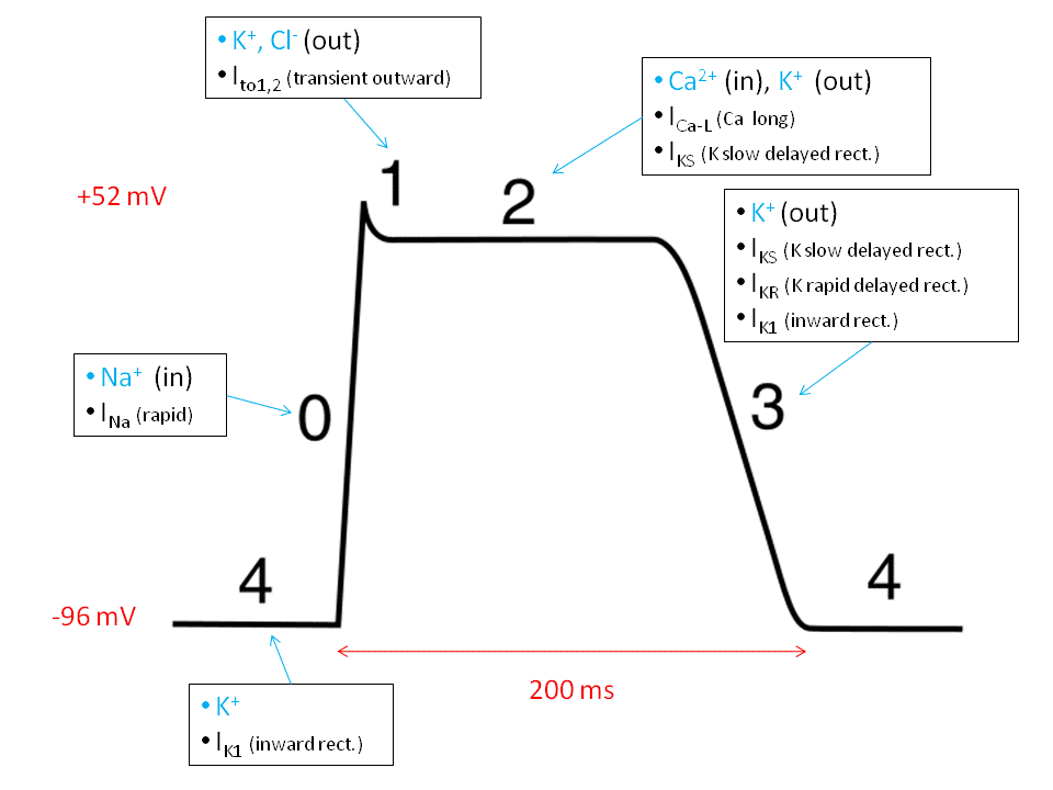
Of these ions, it is the Ca2+ influx that is responsible for signal transduction, for setting into motion pathways that tell the cell how to respond to external and internal stimuli. As such, calcium homeostasis is of critical importance for cell function. A rapid influx of supraphysiological levels of Ca2+, as was evidenced in the study above, would induce a hyper-contractible state, disturb the timing of the electrical signals, and eventually, completely overwhelm the capacity to clear the excess, invoking apoptotic and necrotic death pathways depending upon mitochondrial ATP availability. Each of these markers has been found in the COVID-infected heart. These same disturbances may be also found in a condition called wet beriberi – thiamine deficiency that affects the heart. Similarly, cardiomyopathy, dyspnea (breathlessness), and elevated troponin, other common findings attributed to the COVID heart, are also associated with cardiac beriberi. Given my research on thiamine deficiency/beriberi, I cannot help but wonder if what we are seeing in COVID heart patients is some manifestation of the same process.
Although electrochemical research on both COVID and thiamine deficient myocardial function is lacking, as much of the research appears to focus on gross morphological changes mediated by inflammatory infiltrates, and we do not know if the Ca2+ tsunami identified in the aforementioned study is common across both disease processes, it is logically possible. Inasmuch as mitochondria provide the energy substrate that permits contraction and relaxation of the cardiomyocyte as well as Ca2+ management and sequestration, mitochondrial function may sit at the nexus of virally mediated cardiac events. To the extent that thiamine is critical for each of the key pathways involved in ATP production, insufficiency would impair these actions. And finally, since mitochondrial fitness is unique to the individual, this might explain why, even when there is clearly visible cardiac damage, symptom expression is mild or absent altogether in some individuals but deadly in others. This may be what is happening with athletes who are COVID positive and display limited symptomology but develop myocarditis. This may have been the case with me as well. Again though, without proper testing, this is speculation on my part. Nevertheless, the Ca2+ tsunami aptly describes a potential underlying mechanism for what I experienced.
If this turns out to be correct, the questions then become 1) why does COVID preferentially affect the heart and not the lungs in some individuals, 2) are the mitochondria the determining factor of severity and survival, and if so, 3) are there things we can do to support the mitochondria and maximize survival? While I do not know the answer to question 1, I have argued from the beginning of this pandemic that the determining factor in COVID morbidity and mortality comes down to mitochondrial fitness. To the extent mitochondrial fitness is determined by nutrient availability, thiamine especially, the answer to question 3, is yes, we can do something to improve outcomes. In a subsequent post, I will explore these questions more closely.
We Need Your Help
More people than ever are reading Hormones Matter, a testament to the need for independent voices in health and medicine. We are not funded and accept limited advertising. Unlike many health sites, we don’t force you to purchase a subscription. We believe health information should be open to all. If you read Hormones Matter, and like it, please help support it. Contribute now.


Sounds like you had the gastric form of COVID-19, which I also had in March 2020. It took over a year for me to recover and no one thought I had it since regar testing came back negative. But the tests were not so great, either.
It is not a good idea to draw a conclusion of the incidence of heart disease in Japan based on iodine ingestion without considering their diet. It is low in calories and high in non caloric nutrients, whereas the very opposite is true of the average American diet. That is why I agree with the erudite discussion of Dr Marrs, that mitochondrial dysfunction from thiamine deficiency is the core issue of the degree of severity in Covid in America.
Iodine is protective of the heart. Japan has an extremely low covid death rate. They consume 12+ mg a day on average. They have the lowest heart disease mortality in the world.
I’ve been taking Nicotinamide for about a year now. I’m sure there’s COVID research being done in association with nicotinamide.