autoimmune disease

Hormones, Gut Bacteria, and Autoimmune Disease

6973 views

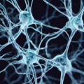
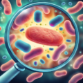
Women disproportionately suffer from autoimmune diseases at a rate of 3 to 1 compared to men. Most believe hormones influence the risk of autoimmune disease but determining the mechanisms has eluded researchers for some time. In animal research, when male rats are castrated, the incidence of autoimmune disease goes up, but when female rats are castrated – their ovaries are removed, the results are mixed. New research finds that the bacteria in the gut interact with hormones and provide the key to understanding the sex differences in the risks for and onset of autoimmune disease.

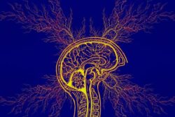
In a study published in the journal Immunity, Gender Bias in Autoimmunity is Influenced by Microbiota, researchers tested the potential signals between hormones and immune function, gut microbes and immune function, and hormones plus gut microbes and immune function in male and female mice. They wanted to delineate the relationship between hormones, specifically testosterone, gut bacteria and autoimmune factors. Is it solely the hormone signal that influences the types and distribution of gut bacteria that then initiates or regulates the immune response or do the gut microbes regulate the hormones which then influence the immune response, or is it some combination of the two? It appears to be the latter; hormones and gut microbiota interact synergistically to regulate the expression of various immune factors and each other.

In a series of experiments, researchers were able to show that the gut bacteria in post pubescent male mice, both quantitatively and qualitatively differed from those of the female mice and testosterone was the key. In male mice, there appeared to be a feedback loop in which testosterone increased gut bacteria and the gut bacteria in turn increased testosterone, to some degree. Once the minimum threshold was reached, higher concentrations of testosterone did not influence gut bacteria any longer. Because these bacteria in turn control the balance between inflammatory and anti-inflammatory factors researchers postulate that the relationship between gut microbiota and testosterone may be key in understanding the gender bias in autoimmune disease.

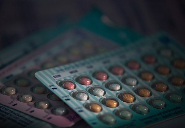
Questions remain. Testosterone is not the only androgen within the hormone pathway. There are other androgens that have noted and direct influence on immune factors. Moreover, while testosterone may be implicated that does not necessarily rule out a role for the estrogens, as testosterone is the precursor hormone for estrone, estradiol and the other estrogens. The downstream conversion from testosterone to estrone or estradiol may be an important consideration for future research. Nevertheless, this study highlights the importance of hormones in disease, and more specifically, the critical cross-talk between hormones, gut bacteria and inflammatory/anti-inflammatory factors.

We Need Your Help

More people than ever are reading Hormones Matter, a testament to the need for independent voices in health and medicine. We are not funded and accept limited advertising. Unlike many health sites, we don’t force you to purchase a subscription. We believe health information should be open to all. If you read Hormones Matter, like it, please help support it. Contribute now.

Yes, I would like to support Hormones Matter. 

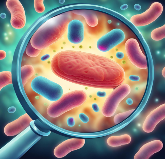
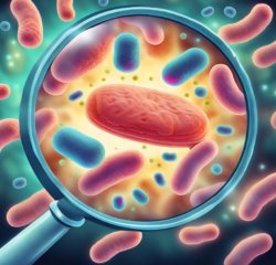
Image created using Canva AI.

This article was originally published on September 12, 2013. 

Digging Deeper into Mitochondrial Dysfunction

15125 views

When you have a hammer, everything becomes a nail, or so they say. I worry about this as I dig deeper into mitochondrial dysfunction. Could all of these disparate symptoms and conditions have their roots in the mitochondria? Could it be that simple? Perhaps. More and more, as I search for explanations for the devastating symptoms that so many of our readers report, the research I find points to mitochondrial dysfunction. Sure, changes in gut microbiota and function are apparent and often related and certainly immune dysregulation is a component of these illnesses, but the underlying connection among these disturbances seems inevitably and inextricably linked to dysfunctional mitochondria as the central hub of illness. Heal the mitochondria, heal the body is quickly becoming my new mantra.

Mitochondria as Danger Sensors

Some researchers argue that the mitochondria are the danger sensors for host organisms; having evolved over two billion years to identify and communicate signs of danger to the cells within which they reside. The signaling is simple and yet highly refined, involving a series of switches that control cellular energy, and thus, cellular life or death. When danger is present, energy resources are conserved and the immune system fighters are unleashed. When danger is resolved, normal functioning can resume.

If the danger is not resolved and the immune battles must rage on, the mitochondria begin the complicated process of reallocating resources until the battle is won or the decision is made to institute what can only be described as suicide – cell death. Cell death is a normal occurrence in the cell cycle of life. Cells are born and die for all manner of reasons. But when cell death occurs from mitochondrial injury, it is messy, and evokes even broader immune responses, setting a cascade in motion that is difficult to arrest.

And, if the on the battlefield, the host army is understaffed and under-resourced, no matter how hard the immune fighters battle, the fight will be lost, maybe not immediately, but eventually. All sorts of mechanisms will be employed to reallocate and reinforce needed battlements, but they will be for naught, further depleting already scarce host resources, until the decision is made, within the mitochondria, to begin pulling back, withdrawing, and ultimately casting the final orders of cell death.

It’s not Autoimmunity, but Impaired Immunity

I never much liked the war model of health and disease, but it seems to work well as metaphor for immune functioning, as it is far more illustrative and useful than the self-versus non-self-characterization. Really, what army with two billion years of experience, one that contains all of the memories and skills of battles past, would misidentify itself and begin broad scale fratricide  – kill itself and its brethren for no other reason but mistaken identity and do so for years on end?  Sure, there can be errors, over compensation and other weaknesses in the immune system, but not continued aggression towards itself in some maladaptive response. That makes no sense and contradicts the very notion and function of an immune system – to keep the host organism alive and well. Indeed, when we consider the trillions of microbes – clear non-self entities – that live inside and upon us, the idea that the immune system evolved simply to kill the non selves seems laughable. And so, I reject the concept of autoimmunity, not because the patients who suffer from continued immune system activation are not ill, they are, but because the concept of autoimmunity belies the very nature of immune function and severely limits possible approaches to recovery.

The Naming of Things

Many of you might be thinking ‘what the heck does what we name things have to do with understanding illness?’  Well, the language and the characterization of disease impacts therapeutic choices. In a system where autoimmunity dominates the discussion, survival is predicated on suppressing the invading immune army. Consequently, most therapeutic options for autoimmune disease are immunosuppressant, and mostly they fail. In contrast, if one characterizes immune function by its ability to protect and sustain life by fending off dangers or threats to survival, be they self or non-self, it does not matter, then we can be open to finding causes for those failed battles. We can ask questions like: what resources are missing that would allow the immune army to fend off the danger once and for all or what could heal the damaged cells, scavenge toxicants and oxidants or re-calibrate mitochondrial energy production? When we re-frame the discussion in this way, we open the door to a deeper understanding of health and disease. It is from this perspective, one that says chronic immune activation is not a disease itself but a symptom of an on-going and failing immune battle, that we can get to the mitochondria as the central hub for chronic ill-health.

Evolution and the Mighty Mitochondria

Mitochondria are interesting little buggers, having evolved from the very parasites our immune system sought to protect us against. Called symbionts, the mitochondria were microbial intruders swallowed by the host. In a brilliant move of survival, they somehow convinced the host organism not only not to kill them but to let the mitochondria, a parasitic intruder, run the host’s energy supply. The mitochondria proved their utility and developed a symbiotic relationship with the cells within which they resided. Over time, mitochondria developed a myriad of intricate communication and resource allocation mechanisms to ensure not only their survival but that of their host organism. And so, in many ways, the mitochondria evolved as part of a cooperative and collaborative ecosystem; one in which they sense and communicate danger to the rest of the organism, and if need be, initiate the final death programs; something, they should be loath to do, since their survival depends entirely on host survival.

Clearly though, and from the very beginning, the mitochondria positioned themselves as the brains of the operation. Mitochondria control energy. There is no other resource more important to the living organism than energy. Consider the most consistent sickness behaviors across all illness include, lethargy, fatigue, sleepiness, often followed by muscle and body aches, anorexia or the loss of interest in eating. The reduction in energy is purely a function of mitochondrial resources. The achy muscles are moderated by mitochondrial retractions of energy and the loss of appetite too, mitochondrial diminishments – recall the orexin/hypocretin system. By whatever pathway, declining mitochondrial energy production arises when danger signals, or more appropriately, cell damage signals are communicated. It is then that the immune armies are activated and inflammation sequences unleashed.

What Does Mitochondrial Dysfunction Look Like?

Everything and nothing at the same time. Mitochondrial dysfunction doesn’t lead to one, clear cut disease, even when there are clear genetic markers, but predisposes one to everything. Where mitochondrial damage is felt and the subsequent immune events present is complex and dependent upon the interactions among the host organism’s innate predispositions, environmental exposures and nutritional status, with the latter two significantly influencing each other. The microbial composition of the host, especially in the gut, but also on the skin and the various mucous membranes that interface with the outside world, can also moderate or trigger the danger signals that lead to mitochondrial dysfunction. More often than not though, mitochondrial damage is felt where oxygen demands are greatest, the brain, the heart, the gut, the muscles. Diseases that are currently identified discretely might all have common symptoms – the mitochondria.

Certainly, chronic fatigue should be considered mitochondrial in nature. I don’t think there is a more clear-cut example of mitochondrial dysfunction than severe fatigue, muscle pain and weakness. The question becomes, from where does the dysfunction originate and how can it be fixed or healed?

Migraine, seizures, ataxias and other neurological disorders are emerging as mitochondrial, particularly was more work is done on the hypocretin/orexin system.

Autonomic dysregulation, recognized under the umbrella as dysautonomias are mitochondrial in nature.

Thyroid dysfunction is likely mitochondrial in nature; the interaction between thyroid hormones and mitochondria is direct. Given the mitochondria’s role in steroidogenesis, other hormone systems are likely modulated by mitochondrial functioning.

Research is emerging suggesting that gastrointestinal disturbances, particularly those of dysmotility like IBS, gastroparesis, constipation and pseudo obstruction but also anorexia are mitochondrial in nature. Indeed, the GI system has its own nervous system, called the enteric nervous system. Only 10-15% of GI motility is controlled from brain’s autonomic system. The rest is controlled on site by the enteric system, mostly from cells called the interstitial of cells Cajal – the smooth muscle cells that propagate contractility and rhythm and form the gut barrier between the inside contents and the rest of the body.  The mitochondria control energy usage and production here. Mess with these cells, diminish oxygen usage, pull back energy production and all sorts of things go wrong. We can get ill-timed contractions or no contractions at all, making the movement of food stuffs through the GI impossible. Poor absorption and metabolism of nutrients, and increased permeability of the tight junctions allowing for the leaky gut scenario common in many chronic conditions, become prominent and are also symptoms of mitochondrial dysfunction. Even anorexia, the will to eat, can be disturbed significantly by mitochondrial damage.

And I suspect, although I have no evidence to support this claim, in women, mitochondrial damage can express itself in the reproductive organs, especially when oxygen demands are greatest, menstruation and pregnancy. Consider the increasingly painful muscle contractions for some women involved in shedding the uterine lining during menstruation, just as diminished oxygen and energy in other muscles begins the cycle of lactate production and buildup that initiates pain, so too might this happen in the uterus. As mitochondrial deficiencies persist, uterine dysfunction would grow and compensatory immune system mechanisms increase until the compensatory mechanisms take a life of their own. When we consider the mitochondrial influence on GI motility, their influence on uterine function is not difficult to imagine. I have an inkling that endometriosis, the excessive growth of endometrial cells first within the uterus and then in regions of the body where they ought not be, is a protective mechanism, albeit an aberrant and problem causing one, that indicates increased mitogenesis and cell growth as a compensatory reaction to some original mitochondrial inadequacy. How this might happen molecularly provides some intriguing possibilities.

Immune function and inflammatory reactions are directly controlled by mitochondrial signals of danger and so to the extent we see chronic inflammatory conditions, one can look towards mitochondrial resources and the ensuing danger signals for clues towards reducing these reactions. While much of the clinical research is nascent, more and more clinicians, often from disparate specialties and sometimes without recognizing the immune-mitochondrial connections, have made great inroads towards healing and restoring mitochondrial function through diet and nutritional supplementation paired with the reduction and removal of environmental and medical toxins and dietary inflammasomes.

Is Everything a Nail, When One has a Hammer?

Maybe, but I can’t help but thinking that this mitochondrial hammer might be the one to hit the nail on the head and finally make some inroads towards reducing the suffering and burden of chronic disease. Only time will tell. For the moment, however, this is a hammer that deserves more recognition and whether it turns out to be the final clue or not one thing is clear, mitochondrial health is critical for human health. Deny the mitochondria their due and chronic, complicated illness will persist.

We Need Your Help

More people than ever are reading Hormones Matter, a testament to the need for independent voices in health and medicine. We are not funded and accept limited advertising. Unlike many health sites, we don’t force you to purchase a subscription. We believe health information should be open to all. If you read Hormones Matter, like it, please help support it. Contribute now.

Yes, I would like to support Hormones Matter. 

Image credit: OpenStax, CC BY 4.0, via Wikimedia Commons

This article was published previously on Hormones Matter in June 2014.

Vitamin D’s Role in Preventing and Treating Multiple Sclerosis

7023 views

Modern lifestyles are countering nature’s intentions to keep us healthy. Since the advent of the Industrial Revolution in the late 18th century, we have migrated from farms to factories and office buildings. Nature intended for us to live and work outdoors in the sun—without sunscreen. Today most of us live and work indoors—often wearing sunscreen, or cosmetics containing sunscreen. By doing so, we have denied our bodies one of the most fundamental sources of health: the ultraviolet B (UVB) rays of the sun that initiate vitamin D protection in our skin.

Compelling scientific evidence over the past century indicates the significant role vitamin D plays in protecting us from developing a wide variety of medical conditions including autism, autoimmune disorders, cancer, cardiovascular disease, diabetes, and thyroid disorders. It is not a coincidence that the prevalence of these diseases has emerged during “modern” times. These medical conditions, many of which are serious, chronic, and life-threatening, frequently result in health, financial, and social burdens to the patients and their families.

What is Multiple Sclerosis?

Multiple sclerosis (MS) befits a disease of modern civilization. First identified by French neurologist Jean-Martin Charcot in 1868, MS is a chronic, neurological autoimmune disorder that damages the myelin sheath, the multiple layers of fatty tissue that surround and protect the nerves in the brain, spinal cord, and optic nerves. When the myelin sheath is intact, electrical impulses are carried through the nerves with accuracy and speed. When the myelin sheath is damaged (sclerosis is the scar tissue formed by damaged myelin), the nerves do not conduct electrical impulses normally. The impulses are distorted or interrupted, resulting in a range of symptoms including numbness, blindness, paralysis, and brain damage. MS also can result in death.

Who is at Risk of Developing MS?

Despite the identification of MS almost 150 years ago, MS has no cure. Over 2.5 million people around the world have been diagnosed with MS including about 400,000 Americans. Women are to two to three times more likely to develop MS than men. Although MS is usually diagnosed between the ages of 20 and 50, the disease can strike at any age. In addition, Caucasian women of Northern European descent are more frequently diagnosed with MS than African Americans, Hispanics, and Asians.

As part of the Environmental Risk Factors in MS Study (EnvIMS), researchers at the University of Bergen in Norway sought to understand better the association between MS and sun exposure measures by studying a total of 1,660 MS patients and 3,050 controls from Norway and Italy. The researchers’ findings included significant connections between infrequent summer outdoor activity and sunscreen use and an increased risk of MS. Published in the January 10, 2014 issue of Multiple Sclerosis, the study’s conclusion stated, “Converging evidence from different measures underlines the beneficial effect of sun exposure on MS risk.”

It is not surprising that incidences of MS in the equatorial region occur much less frequently than at the higher latitudes. Epidemiological studies over the past several decades however indicate that women who live at higher latitudes have an increased risk of developing MS. For example, University of Oxford researchers studied MS patterns in Scotland by examining hospital admissions throughout the country between 1997 and 2009. The research team discovered a “highly significant relationship between MS-patient-linked admissions and latitude” across Scotland. This study was published in a 2011 issue of the Public Library of Science (PLoS) One journal.

In addition, a seasonal risk factor also exists for MS. Researchers at Queen Mary University of London conducted a systematic review of data for 151,978 MS patients to ascertain the link between month and location of birth, and the risk of developing MS. They found that babies born in April had the highest risk of development of MS, and infants born in October enjoyed the lowest risk of MS. The researchers also noted a direct correlation between the latitudinal location of expectant mothers and MS risk. The study, published in a 2012 issue of the Journal of Neurology, Neurosurgery, and Psychiatry, suggests the importance of maternal vitamin D supplementation in particular during the winter season.

What Causes MS?

The definitive cause of MS remains unknown but medical research suggests genetic and environmental factors influence one’s risk of developing MS. Interestingly enough, science has demonstrated that vitamin D plays a role in influencing environmental and genetic factors that may affect how likely one is to develop MS.

A landmark study at the University of Oxford, published in a 2009 issue of Public Library of Science (PLoS) Genetics, examined how genes and the environment interact in MS. A gene variant called HLA-DRB*1501 is associated with an increased risk of developing MS. The research team discovered how vitamin D influences the HLA-DRB*1501 gene variant. As we know, the amount of vitamin D synthesized by UVB sunlight exposure fluctuates from season to season. Therefore, women who give birth during the spring, carry the HLA-DRB*1501 gene variant, and have low vitamin D levels are more likely to produce children with a higher risk of developing MS.

The study’s author Dr. Sreeram Ramagopalan suggested that adequate vitamin D3 supplementation during pregnancies may decrease the risk of children developing MS in later life. The combination of carrying the HLA-DRB*1501 gene variant and lacking adequate vitamin D levels may impair the ability of the thymus, an immune system organ, to delete rogue T cells, a type of white blood cells, that play an important role in maximizing the immune cells. The rogue cells would attack the body, causing demyelination of the central nervous system.

How Can Vitamin D Protect Against MS?

MS is a neurological autoimmune disorder. Scientific research over the past few decades solidifies the connection between vitamin D and autoimmunity. Vitamin D plays an integral role in the regulation of the adaptive immune system.

Adequate vitamin D in our bodies can protect us from autoimmunity because adaptive immune cells contain vitamin D receptors (VDRs). These receptors are attached to the surface of the adaptive immune system’s antibodies and sensitized lymphocytes. When the VDRs receive adequate amounts of vitamin D, they enable the adaptive immune system to function properly by attacking new and previous invaders.

When the VDRs attached to the adaptive immune system’s cells do not contain sufficient vitamin D to attack invaders, autoimmunity may kick in, causing the death of healthy immune cells. Thus, vitamin D deficiency can contribute to the development of autoimmune disorders such as MS.

How Can Vitamin D Treat MS?

The scientific community is delivering hope to MS patients by investigating vitamin D intake as a treatment for the disease. Research suggests that higher vitamin D levels are associated with reduced disease activity in MS sufferers.

Dr. Alberto Ascherio of Harvard University’s School of Public Health and colleagues recently concluded that vitamin D appears to be connected with MS disease activity and progression in patients who experienced an initial episode suggestive of MS and were treated with interferon β-1b. The researchers found that 20 ng/mL-increases of vitamin D levels within the first 12 months of experiencing an initial episode predicted a 57 percent lower rate of new active lesions as well as a lower risk of relapse. In addition, the results included a 25 percent decrease in annual T2 brain lesion volume and a 0.41 percent lower yearly loss in brain volume over four years. The Harvard study was electronically published on January 20, 2014 in JAMA Neurology.

According to a study published in a 2012 issue of the Annals of Neurology, a University of California, San Francisco research team examined 469 male and female MS patients over five years to ascertain how vitamin D affected disease progression. The researchers discovered that for each increase of 10 ng/mL in vitamin D levels, the MS patients benefited from a corresponding 15 percent decrease in new brain lesions as well as a 32 percent lower risk in inflammation of the myelin sheath.

A Finnish study, published in a 2012 issue of the Journal of Neurology, Neurosurgery, and Psychiatry, concluded that vitamin D3 supplementation significantly reduced the number of brain lesions in MS patients undergoing interferon β-1b treatment.

Paving a Way to Better Health and Quality of Life

Adequate vitamin D levels in our body may indeed protect us from developing MS. If you have experienced a possible initial episode or have been diagnosed with MS, please consider how vitamin D3 supplementation may decrease the severity of your symptoms.

We must take ownership of our health by understanding the importance of vitamin D as well as other micronutrients. Why wait years, or decades, to garner the results of further studies and clinical trials to define the exact relationship between vitamin D status and MS. We can be proactive by taking daily vitamin D3 supplements and enjoying moderate sunlight exposure to increase our vitamin D levels.

It is imperative to take enough vitamin D so this essential nutrient will be stored in your cells to help regulate your immune system. The greater your vitamin D level (easily obtained from a simple blood test called 25(OH) vitamin D), the more likely you will benefit from a stronger immune system that will protect your body’s cells from attacking one another.

No one wants to endure the health, financial, and social burdens of a chronic debilitating disease. By empowering yourself with adequate vitamin D, you may not only reap lots of health benefits but enjoy a better quality of life.

We Need Your Help

More people than ever are reading Hormones Matter, a testament to the need for independent voices in health and medicine. We are not funded and accept limited advertising. Unlike many health sites, we don’t force you to purchase a subscription. We believe health information should be open to all. If you read Hormones Matter, like it, please help support it. Contribute now.

Yes, I would like to support Hormones Matter. 

To Learn More


Image credit: Stephanie021299, CC BY-SA 4.0, via Wikimedia Commons
This post was published here originally on March 4, 2014. 

Copyright © 2014 by Susan Rex Ryan. All rights reserved.

What is Immunity?

3642 views

Arguably, my primary intellectual concerns around modern day infectious disease management and “prevention”, is an acute awareness of how little we know about our relationship to microbes in and around us, and about our immune system. We are just beginning to appreciate the role of the several trillion bacteria that inhabit our guts, dictate our immune responses, and synthesize nutrients. Add to this very steep learning curve, multiple layers of epigenetic expression and biochemical individuality and we have a recipe for disaster with a one-size-fits all vaccination schedule and rampant application of antibiotics.

Antibody-Response: Is that Immunity?

We have been led to believe that antibody-response to vaccine administration is in any way equivalent to protection from illness. This sadly rudimentary model of “immunization” is antiquated beyond acceptability, and in no way encompasses what we have learned about the relevance of the innate immune system, cytokines, and the role of nutrient sufficiency in vulnerability to infection.  Beyond the well-documented incidence of outbreaks of illness such as pertussis, mumps, measles, tetanus, polio, rotavirus, and chicken pox, in highly vaccinated populations, we have also learned that antibodies often play no role in the course of infectious diseases such as lethal vesicular stomatitis virus, discussed here. We also know that agammaglobulinemics (those born without limited capacity for immunoglobulin antibody production) contract and recover from measles in the usual fashion. So, it seems like we may have fundamentally misunderstood the role of antibodies in immunity.

This would be an excusable and understandable step in the evolution of biological sciences if we weren’t wielding the application of this misunderstanding in a lethal and morbid way. Room for primary vaccine failure based on fundamental misattribution of disease-protection to antibody production (which is always temporary) is one thing, but inducing chronic disease, atopy, neurodevelopmental delay, inflammation, autoimmunity, and death as a part of this effort, is quite another.

Auto-immunity and Evolving Theories of Immune Function

We are witnessing epidemic rates of autoimmunity in the American population and we are learning that vulnerability is more than genes + environment.  In fact, theories of immunity have evolved considerably since the 1950s when it consisted only of self vs non-self mechanisms.  The most all-encompassing theory is called the Danger theory, which posits that the immune system targets self-tissues when there is a “danger signal” or inflammation from the tissue itself.  Here is where the role of oxidative stress and inflammation play into immunity and autoimmunity in a significant way, and why the “terrain” is, in fact, everything and the germ is, in fact, nothing.

Evaluating the Safety and Efficacy of Vaccines and Medications

The fact that there is such an evolving conceptualization of immunity and one that only begins to account for the role of diet, environmental toxins, and gene expression variation should serve as a serious wake up call to those who believe that modern-day physicians and pharmaceutical companies are in any position to make recommendations, let alone mandates, about how we, as individuals, should manage our risks of infection. The truth is, once interventions such as vaccines and antibiotics have perturbed our natural mechanisms, there is very little that Western medicine can to do help. Chronic disease and autoimmunity are not the forte of the average doc, so gambling with that potential risk should certainly be done with thought and care.

To that end, there are so many tremendous resources out there, but the latest and greatest is Dissolving Illusions, which takes you on a meticulously documented tour of the role of hygiene and diet in the epidemiology of infectious disease and the misconceptions surrounding vaccinology and health.

For more practical tips, Saying No To Vaccines is an important guide for new parents to educate yourselves about each and every vaccine, because each and every one is a major medical intervention that should be scrutinized independently.

We need to remain humble about what we don’t know, measured in our assumptions about the safety and efficacy of our pharmaceutical interventions, and reliant on time-tested ways to support natural immunity through nutrient dense diet, minimized environmental chemical exposures, and stress reduction. We need to lose the fear we have been conditioned to bring to conversations about infectious disease.

After all, germs are all around and within us, we need them, and they need us.  We’ve spent quite a long time developing a sophisticated language with which to communicate, and we are only beginning to decode it.

About the author. Dr. Brogan is an M.I.T/Cornell/Bellevue-trained psychiatrist specialized in holistic women’s health. She is a mother of two and has a busy practice in Manhattan. A passion for understanding the intersection between health, nutrition, and the environment are the bedrock of her wellness approach with patients and at home. Visit her site at: Kelly Brogan, MD, Holistic Women’s Health Psychiatry.


Guts and Brains and Hormones, Oh My!

4887 views

When a woman experiences fatigue, brain clouding, flat mood, PMS, and constipation, we call it anxiety or stress and we stick her on an antidepressant that she will likely take for the rest of her life. Where in this protocol have we investigated why she is feeling that way?  How have we personalized the treatment to her unique biochemistry?  What is the plan for side effects including new and different psychiatric symptoms resulting from this prescription?  We haven’t.  We’ve applied a one-size-fits all treatment to mask symptoms without consideration for the cause.

The Immune System and Depression

Psychiatry has known about the role of the immune system in certain presentations of depression for the better part of the last century, and more recently, pioneering thinkers like Maes, Raison, and Miller have written about the role of altered immune set points and inflammation in models of depression. Our immune systems are largely housed in the gut and the interplay between the gut and the brain is a complex and profoundly important relationship to appreciate.

We all recognize that anxiety or nervousness can impact our guts – most of us have had butterflies before a date or even diarrhea with extreme performance anxiety?  We are just learning that this relationship is bidirectional; however, and that the gut can also communicate its state of calm or alarm to the nervous system.  We think that the vagus nerve is a primary conduit of information and that inflammatory markers are the vehicles traveling this highway. Scientists have studied the “protective effects” of severing this nerve when animals are exposed to gut-related toxins that normally cause depressive symptoms.  We are getting ahead of ourselves; however, because we need to better elucidate why inflammation matters, where it comes from, and why it is the universal driver of chronic illness.

How Does Inflammation Start?

When a woman feels foggy, run-down, easily overwhelmed, and flat, we know that  her hormones as messengers between her gut and brain are out of balance. From my perspective; however, hormone derailment is a downstream effect of cellular dysfunction from oxidative stress and inflammation. Inflammation stems from many sources, including, hallmarks of the modern American lifestyle:

  • Sugar. Sugar, particularly in the form of fructose and sucrose, spikes insulin and triggers release of inflammatory cytokines. It forms advanced glycation endproducts when it binds to proteins, and oxidizes lipids which form cell and mitochondrial membranes.
  • Chemicals. Pesticides, environmental pollution from industrial waste, hormonally-modulating plastics, fire retardants, and cosmetic additives all stimulate our immune systems to varying extents and disrupt optimal production of energy on a cellular level, particularly in vulnerable tissues like the thyroid.
  • Pathogens. The aforementioned culprits, and notably herbicides, gluten grains, and genetically modified foods, promote intestinal permeability, changes in our intestinal flora that facilitate growth of pathogenic bacteria, yeast, and fungus which keep our immune systems in a state of alarm,
  • Stress. This catch-all term, broadly defined, represents the ultimate link between hormones and inflammation, because stress, whether it’s psychological or physiologic, triggers the release of cortisol. Cortisol helps to mobilize blood sugar so that you can run effectively and efficiently from that tiger chasing you. It also acts as a systemic immune suppressant, lowering levels of secretory IgA, an important body guard of the gut mucosa.

Cortisol and insulin are like stress-response sisters, and high cortisol states will also contribute to insulin resistance, or high insulin and high sugar while the cells, themselves, are starving.  Insulin protects fat storage (inhibits lypolisis), and fat cells secrete their own inflammatory signals in addition to aromatizing testosterone to estradiol contributing to states of estrogen dominance, while also increasing DHEA and androgens to fuel that process (as well as acne, hair growth, and agitation).

Cortisol also inhibits the conversion of storage thyroid hormone to active hormone leading to states of hypothyroidism even with normal-looking labs.

What Does Inflammation Do?

Once inflammation is active, it is highly self-perpetuating. These inflammatory cytokines travel throughout the body causing oxidating stress to the fragile machinery of the tissues and mitochondria, specifically.  In the brain, inflammation serves to shunt the use of tryptophan toward production of anxiety-provoking chemicals like quinolinate, instead of toward serotonin and melatonin. They produce a replicable collection of symptoms called “sickness syndrome”, noted for it’s overlap with “depressive” symptoms: lethargy, sleep disturbance, decreased social activity, mobility, libido, learning, anorexia, and andhedonia. Psychiatric researchers have observed that patients with higher levels of inflammatory markers (like CRP) are less likely to respond to antidepressants, and more likely to respond to anti-inflammatories.

Where Do We Begin to Heal?

How is any of this good news? This approach to chronic illnesses like depression views it as a complex, non-specific symptom reflecting a state of bodily disharmony.  It isn’t that you were born with bad genes or low serotonin.  It is far more likely that you are experiencing an unhealthy inflammatory balance, driven by cortisol dysfunction, and stemming from a sick gut.  We can come at modifying your system from many angles, but here is a basic starter kit:

  • Exercise – Burst exercise is my primary recommendation.  It is the most bang for your buck in terms of cardiovascular benefit and specifically enhancing mitochondrial health because it puts a special kind of stress on the body when you move to your max for 30 seconds that then recover for 90.  I recommend 8 intervals 1-3x/week.
  • Meditation – The effects of stimulating the relaxation nervous system, even through listening to a 20 minute guided meditation, can be far-reaching.  Enhanced genomic expression of anti-inflammatory genes and suppression of inflammatory ones was demonstrated in this study.
  • Diet – I recommend a diet that controls for glycemic fluctuations through elimination of refines carbs and grains, and through high levels of natural fats to push the body to relearn how to use fats for fuel.  This is the brain’s preferred source.  I discuss some therapeutic foods here.
  • Strategic supplementation – Natural anti-inflammatories like polyunsaturated fats (evening primrose oil and fish oil), curcumin (the active component of turmeric), and probiotics to name a few, can help promote a synergy of beneficial effects from the above interventions.

In my practice, despite some suggestion that antidepressants may actually be having their effect through an anti-inflammatory mechanism, these medications have become obsolete.  An appreciation of the role of inflammation and immunity in driving hormonal imbalance which directly impacts mood, energy, and wellness, is at the core of personalizing the definition of “depression”. Don’t be lured into the simplicity of a one disease-one drug model.  There’s no room for you in that equation.

About the author. Dr. Brogan is an M.I.T/Cornell/Bellevue-trained psychiatrist specialized in holistic women’s health. She is a mother of two and has a busy practice in Manhattan. A passion for understanding the intersection between health, nutrition, and the environment are the bedrock of her wellness approach with patients and at home. Visit her site at: Kelly Brogan, MD, Holistic Women’s Health Psychiatry.

References

Inflammation and its discontents: the role of cytokines in the pathophysiology of major depression. Miller et al Biol Psychiatry. 2009 May 1; 65(9): 732–741.

Cytokines and cognition – The case for a head to toe inflammatory paradigm. Wilson et al. JAGS 50:2041–2056, 2002.

A randomized controlled trial of the tumor necrosis factor antagonist infliximab for treatment-resistant depression: the role of baseline inflammatory biomarkers. JAMA Psychiatry 70:31–41.


Vitamin D3 and Lupus

3408 views

The untimely death of 23-year-old Sasha McHale, daughter of professional basketball’s Hall of Famer Kevin McHale, recently shocked the world.

Sasha inherited her father’s athleticism, energy, enthusiasm for life, and love of the northern U.S. state of Minnesota. An insidious disease called lupus prematurely snatched Sasha from her family, friends, and life.

Lupus is a chronic autoimmune disease that attacks the body’s cells, tissues, and organs, and results in severe inflammation, fatigue, and, in some cases, death. The medical name for the most common form of the lupus is “systemic lupus erythematosus” (SLE). According to the Lupus Foundation of America, about 1.5 million Americans, and over five million people globally, suffer from a form of lupus. Ninety percent of persons diagnosed with the disease are women, many of whom are in their child-bearing years.

Mounting evidence suggests adequate vitamin D3 in the body may protect against the development of autoimmune diseases including lupus. Genetic and environmental factors including vitamin D3 deficiency have been linked to lupus. Sensitivity to sunlight, the primary source of vitamin D3, is common among SLE patients. Scientific research indicates a high prevalence of vitamin D3 deficiency among people suffering SLE:

Researchers at the University of Toronto Lupus Clinic studied 124 female SLE patients to understand, inter alia, their circulating vitamin D3 levels. Eighty-four percent of the women had vitamin D3 blood serum levels less than a sub-optimal reading of 32 ng/mL.

The Medical University of South Carolina conducted a study of vitamin D3 blood serum levels of 123 individuals who had been recently diagnosed with SLE. The findings suggested vitamin D3 deficiency as a possible risk factor for SLE.

Researchers studied 25 Canadian women diagnosed with SLE and found that over half of these patients had less than 20 ng/mL of vitamin D3 circulating in their blood. The research also suggested that hydroxychloroquine (HCQ), a drug used to treat SLE, may inhibit vitamin D3 production.

A study published in a 2012 edition of the journal Dermato-Endocrinology not only documented the prevalence of low vitamin D3 in SLE patients but recommended oral vitamin D3 supplementation for SLE patients. The researchers lauded the safety, low cost, and wide availability of vitamin D3 supplements as well as their potential effectiveness against SLE progression.

Maintaining adequate vitamin D3 levels may forestall the development of autoimmune diseases including lupus. In addition, vitamin D3’s capability to reduce inflammation may alleviate lupus symptoms. Further research is required to confirm the extent of vitamin D3’s connection with lupus.

Copyright ©2012 by Susan Rex Ryan
All rights reserved.

Autoimmune Olympiads

1912 views

Well, the Olympics are over. Thank God! Now I can get some sleep. I was so sleep deprived. I could have won a gold medal in crankiness.

I am in awe when I see the super-human type feats executed by these athletes. And then, when I hear that some of these competitors have autoimmune diseases like me, it is astounding.

Diseased Olympiads…what an oxymoron.

But it just goes to show you, no one is immune to the auto-immune atrocities.

These athletes endure their body fighting itself, yet they practice multiple hours in a day. When I am on my third or fourth dream all snugly in my bed, they are waking up before the sun comes up to begin their training. Just can’t wrap my mind around that…

Here are a few of those ailing athletes who competed at this year’s 2012 Olympics in London:

Tennis -Venus Williams–Sjogren’s syndrome (The body attacks moisture producing glands) Advanced to the third round of the London Olympics.

Kayaking –Carrie Johnson-Crohn’s disease (Inflammation of the digestive tract) Advanced to the semifinals.

Soccer (or women’s football)-Shannon Box-Lupus (the body attacks healthy tissues) Won a Gold medal.

Track and Field-Sanya Richards-Ross-Behcet’s Syndrome (Vasculitis leading to ulcers particularly around the mouth, genitals and pupils) Two-time gold medal winner in London.

Track and Field, pole vaulting-Jenn Suhr-Celiac’s disease (hypersensitivity to gluten) Won gold medal.

All of them deserve the gold medal as far as I am concerned.

This article was originally posted on http://www.crazythyroidlady.blogspot.com and was re-posted with permission.