lexapro

Antidepressant Induced Toxic Encephalopathy: A Case Update

9637 views

I was prescribed Lexapro due to some work-related stress and took it for years without any knowledge of the ill effects to my brain. As a consequence of the Lexapro, I developed a number of neurological symptoms that resulted in diminished neurocognitive and physical capacity. The doctors call this toxic encephalopathy – drug induced brain damage. My primary symptoms continue to be significant cognitive and short-term memory decline, headache, pain, weakness, and balance/coordination issues. Seven years after the last pill these same symptoms persist. I have written about my experience (with the help of the HM editor) here, and here.  Many have inquired on this blog or directly for an update on my case. My medical condition has not changed. However, since my last updates, it has been even further defined by additional medical experts who have weighed in on my case.

Experts Continue To Confirm Toxic Encephalopathy

Nuclear Medicine Specialist/Radiologist-2020

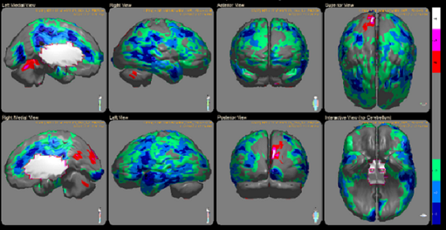
“This is a markedly abnormal brain SPECT indicating an advanced encephalopathy for which possible etiologies encompass a congruence of multiple possible factors including some or all of: vascular, neurotoxic (prolonged/extensive exposure to drugs, toxic substances, mold or iatrogenic effects) closed head injuries (multiple, cumulative), sleep problems, inflammation, anoxia, and neuro-vegetative dysfunctions.”

Neuropharmacologist-2021

“In conclusion, I can state with a significant degree of pharmacological certainty that the long term treatment with escitalopram/trazadone produced changes in Mr. CF’s brain responsible for reduction in the functions he has exhibited in the years following that exposure. This is based on the following:

  1. Symptoms are consistent with the side effects seen with these drugs.
  2. The removal of the drug with symptom reduction and its reinstatement in 2015 with a return of symptoms speaks strongly to a toxic encephalopathy.
  3. The literature is clear that although rare, toxic encephalopathy does occur and can be life-altering.
  4. The fact that Mr. CF is a slow metabolizer of the drug suggests that his chronic dosing was likely higher than normal consistent with a toxic encephalopathy, although this encephalopathy can occur with normal metabolism as well.”

Neurologist expert #1 -2021

Encephalopathy is a broad term that applies to brain dysfunction due to factors outside of the nervous system, such as systemic illness, trauma, metabolic disturbances, environmental exposures and drugs/medications. Symptoms include cognitive dysfunction, incoordination of movements, weakness, difficulty walking, problems with vision and speech. While symptoms can mimic stroke, neuroimaging can be useful in refuting cerebrovascular insult. Neurophysiological testing is often used to clarify and prognosticate cerebral dysfunction.

The claimant’s symptomatology and testing results corroborate encephalopathy as the leading clinical diagnosis. Specifically referring to drug-induced encephalopathy, any genetic polymorphism may influence the metabolism, excretion or action of the drug depending on single or multiple genes or by changes in gene expression. The claimant was proven to have slower metabolism of psychotropic medications (e.g., genetic pleomorphisms in CYP2C19, CYP2D6, and CYP3A4). The long-term effects of toxic levels of Lexapro and/or Trazadone predisposed him to encephalopathy.”

Neurologist expert #2 -2021

“CF’s case clearly demonstrates the importance of understanding medication half-life, CYP450 interactions in the development of medication toxicity, and an individual’s pharmacogenetics disposition. Genetic cytochrome P450 testing revealed that CF was found to have problems with his serotonin transporter gene SLC6A4 as well as reduced enzymatic activity of both CYP2D6 and CYP2C9 enzymes. Without question, his long-term exposure to long acting serotonergic medications has caused irreversible and permanent brain injury (toxic encephalopathy).”

QEEG PhD expert-2022 (after review of two QEEG’s performed in 2021)

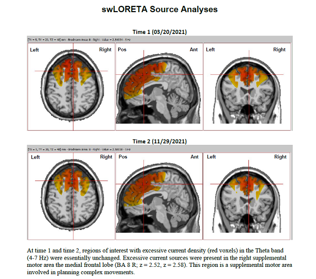
“SUMMARY: swLORETA source level analyses were significantly deviant from normal and findings between time 1 and time 2 were highly consistent. In particular, in both recordings there were elevated current sources in the Theta band were present in the same regions of the bi-lateral frontal lobe, especially in the medial and superior supplementary motor areas and the prefrontal cortex. The location of abnormality is given primary importance for linking regional brain dysregulation to neurological symptoms, but the frequency band provides an added layer of information.What is a QEEG

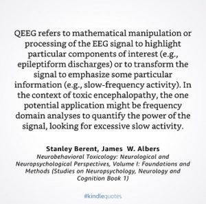
With regard to frequency interpretation, encephalographic patterns in CF’s brain activation were characterized by abnormal rhythms in the theta band and generalized slowing of the EEG. This is a common finding in patients with metabolic and toxic encephalopathy.

swLORETA connectivity analyses were deviant from normal, characterized by widespread hyperconnectivity within neurocognitive networks that play a major role in cognition, emotion, somatosensory, motor movement, vestibular, and autonomic functions. Hyperconnectivity is considered to be a primary compensatory response to neuronal injury with behavioral consequences from reduced processing speed and cognitive fatigue from added redundancy.

CF’s hyperconnectivity patterns were still present at time 2, including network connections to and from the cerebellum which play a vital role in finite motor sequencing, vestibular balance, and cognitive executive functions (e.g., attention). These connectivity results implicate white matter tracts which include the corpus callosum, cingulum, and superior longitudinal fasciculus.QEEG and encephalopathy

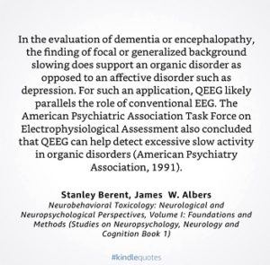
Mr. CF’s symptoms of executive dysfunction, chronic pain, headache, and fatigue may be explained by dysregulation involving these tract which connect 1) the frontal lobe (executive functioning, working memory, abstract thinking, motor control, expressive language, sequential planning, emotional regulation, and social skills), 2) the temporal lobes (auditory information processing, short-term memory, memory consolidation, and receptive language), 3) the parietal lobes (somatosensory processing, spatial information processing, proprioception, executive attention, receptive language, empathy control, and awareness of emotional expression) and 4) the occipital lobes (visual processing of color, form, movement, visual perception, and visual- spatial processing). The global extent of these abnormal electrical patterns found is consistent with Mr. CF’s severe limitations and impairments.

Together with abnormalities documented in his medical records, this qEEG record provides clear, objective, and overwhelming evidence of severe disability consistent with toxic encephalopathy.”

Psychiatrist, neuropharmacology and epidemiology expert- 2022

“In conclusion, it now appears that Mr. CF is suffering from a constellation of symptoms, severe enough to impair daily functioning. Regardless of the terminology which professionals may employ in diagnosing his condition – chronic toxic encephalopathy, Parkinsonism with dementia, or dementia due to multiple etiologies – it is with a high degree of certainty that antidepressant medications were the essential and causal agents of his disability.“

Neuropsychologist-2022

“In summary, the results of the neuropsychological re-testing (performed in late 2021) are consistent with Mr. CF’s symptoms, the diagnostic interview, interviews of witnesses, the medical records, including the claimant’s treating doctors and expert consultants, and my prior evaluations.

My diagnosis of the claimant remains the same as found in my prior reports of March 2, 2018 and May 14, 2020: Toxic Encephalopathy (ICD 10 G92) (a brain injury from prescribed drug); DSM-5 Substance/Medication Induced Major Neurocognitive Disorder (ICD F13.97).

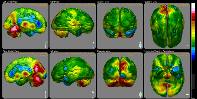
The medical record, including brain imaging and QEEG abnormalities reported previously and above, support the diagnosis of Toxic Encephalopathy and Substance/Medication Induced Major Neurocognitive Disorder. Note the concordance of the above-referenced specific qEEG abnormalities and the report of symptoms and the neuropsychological test results. SPECT scans also are interpreted as showing abnormalities in areas that support neuropsychological functions. These abnormalities support the opinion of an enduring brain injury resulting in long-term disability. This condition cannot be treated to remission by methods as standardly accepted in the practice of neuropsychology or medicine.”

In addition to all of this, I had a recent brain MRI showing white matter lesions in the same location as was in my 2017 brain MRI.

Final Thoughts

It seems like ages ago that doctors initially rejected my claims of the medication causing the decline in my health. My conditions/diagnoses described above are now being treated holistically with good care/support by a neurologist, a neuropsychiatrist and internal medicine specialist. However, it still irks me today that I had to go through all of this to prove myself. Doctors should remember their loyalty should be more aligned with their patients and not the pharmaceutical companies.

We Need Your Help

More people than ever are reading Hormones Matter, a testament to the need for independent voices in health and medicine. We are not funded and accept limited advertising. Unlike many health sites, we don’t force you to purchase a subscription. We believe health information should be open to all. If you read Hormones Matter, like it, please help support it. Contribute now.

Yes, I would like to support Hormones Matter. 

My Brain after Long Term Lexapro: Chemically Induced TBI

64127 views

In December 2015, I was given Lexapro. I had taken Lexapro previously for 12 years, but because of abnormal blood chemistry and building side effects, my doctor suggested that I cease taking the medication. After five months without the drug, I was improving slightly, but because of life events, my doctor and I decided to begin taking Lexapro again. During the reinstatement of the drug, I immediately began to experience serious neurological reactions that I have come to describe as a brain kindling of sorts. It felt like my head was on fire chemically and electrically. Some of the symptoms that developed included: fatigue, confusion, eye pain, leg weakness, headaches, visual processing disturbances, coordination difficulties, lack of concentration, and short-term memory problems.

Within a few weeks of taking the medication, these symptoms were worsening but my physician was not concerned. In fact, he suggested what I was experiencing was normal and that I continue taking the medication. I did, for a while, until the side effects were so bad that I decided to stop. I published my story on Hormones Matter a year ago. You can read it here: A Kindled Brain: Long Term Lexapro Use Reactions. A year and dozens of doctors later, I have become completely disabled. This is a follow up to my story, told with much help from the editor of Hormones Matter.

Two Years Post Lexapro Damage

I no longer take Lexapro or other medications but the damage was done. I still cannot work, watch TV, read, drive or walk more than a few feet. I have had what seems like a two year long headache and severe vision processing issues. It is going to be another great Christmas at my house.

Though I received diagnoses of Chronic Fatigue Syndrome (CFS) and Fibromyalgia they really were just symptoms of something bigger. Shortly after the  CFS diagnosis, an MRI revealed white matter lesions and demyelination. The neurologist dismissed a Lexapro connection. Research suggests otherwise. In the summer of 2017, I was finally given a provisional diagnosis of Lexapro induced neurotoxicity. Just recently, the extent of the brain damage was shown on both on a quantitative electroencephalograph (QEEG) and single-photon emission computed tomography (SPECT) brain scans. Both neurotoxicity and brain injuries were identified by both tests and later confirmed again by additional physicians who reviewed the results independently.

QEEG Results

The QEEG is an EEG with a visual mapping component. It measures the electrical activity or signal transduction between electrodes placed in specific locations on the brain. My test results showed that in certain areas of the brain there was very little organized electrical activity and other areas where there was unusual amount of high activity. What that means is that in some areas of my brain, the signals are very low and essentially not sufficiently strong for the activities that need to be performed but in other areas of my brain, there was way too much electrical activity. Specifically, the report said:

“The patient was found to have a significant amount of low frequency (8-9 Hz) in the right posterior temporal (T6) area. There is an extreme amount of low beta (12-15 Hz) predominantly in the prefrontal and frontal areas that may interfere with executive functioning, organizing, decision making and may also account for fatigue. There is a very unusual amount of high activity for all measured frequencies (1-50 Hz) in the right posterior (O2) area, specifically in the right inferior occipital gyrus (Broadman areas 17, 18, and 19). This may result in sub-optimal functioning in visual processing of color, form, movement, visual perception and spatial processing…

…The analysis of amplitude asymmetries were found to be abnormal for low frequencies (1-12 Hz) stemming from right posterior temporal (T6) area. Also, there are abnormalities for high beta (18-30 Hz) stemming from primarily the right posterior area (02). Both findings are substantiated by LORETA analysis and resembles TBI or other brain damage…

…The TBI index indicated that there was an 85% probability that the patient’s brain is functioning as if there is a TBI 0.95 on a scale of 0-10.”

This was the first confirmation of the cognitive dysfunction that I have been living with for the last few years. Doctor after doctor told me that my symptoms were not real and somehow made up (and some still do, as the QEEG is used mainly in research circles and not always accepted in conventional medicine). The QEEG clearly showed the abnormal electrical activity underlying my symptoms. Next up, the SPECT scan, a test I had to fight for, because again, no one seems to believe that a drug like Lexapro could cause such damage.

SPECT Scans

SPECT imaging shows brain activity (blood flow, which represents local brain metabolism and energy use). The technology uses radiolabeled isotopes that are then reconstructed with algorithms to display in color 3-D images of brain activity. The images below are some of my brain scans. Regions displayed in blue represent a state of very low activity called hypoperfusion and those in red represent hyperperfusion – excessive activity.

SPECT scale
Figure 1. SPECT color coding scale.


CF spect scans
Figure 2. Brain activity after long term Lexapro use.

From my images, you can see several areas of both abnormally low and abnormally high activity with very little normal brain function anywhere. From the report:

  • At rest, the overall cortical activity was reduced in a diffuse, decreased, patchy pattern.
  • Focal areas of abnormal cortical hypoperfusion were noted in the bilateral anterior frontal (L>R), left posterolateral frontal, left orbitofrontal, right dorsolateral prefrontal, bilateral anterior and medial temporal (L>R), bilateral superior parietal and bilateral occipital areas.
  • Focal areas of abnormal subcortical hypoperfusion were noted in the anterior aspect of the pontine portion of the brainstem, bilateral caudate and right lentiform areas.
  • Focal areas of abnormally increased cortical perfusion were not noted.
  • Focal areas of abnormally increased subcortical perfusion were noted in the bilateral thalamic and left lentiform areas.

The doctors conclude:

“The nature (diffuse, patchy), location (cortical and subcortical), and pattern (involving all lobes of the brain) of these abnormalities is primarily consistent with the scientific [literature] pertaining to a toxic/hypoxic/neuroinflammatory process and the patient’s clinical history, as obtained, of a medication reaction which was received after the blind review. These results agree in large part with outside EEG data showing hypofrontality and NM report showing left frontal abnormality, likely not to be artefactual.”

What This Means

My brain is a mess. According to the Hormones Matter editor, the areas of low activity in the various regions of the prefrontal cortex would explain my difficulties executive function, things like planning, decision-making and lack of concentration, while the reduced activity in the temporal and occipital lobes would explain my memory and vision difficulties, respectively. The reduced brainstem activity would connect to my walking and balance difficulties, but also, poor to autonomic regulation (the autonomic nervous system controls all automatic bodily functions including things like breathing, heart rate, temperature control, digestion, sleep/wake cycles, etc.). The areas of my brain on hyperdrive may reflect compensatory reactions, last ditch efforts to kick start some of the underactive regions. The thalamus (plural thalami), in particular, acts as a relay station between the brainstem and the rest of the brain for motor, sensory signals and ‘emotional’ signals via its connections to the limbic system. Mine seem to be screaming ‘wake up’ to the rest of the brain.

Please be careful using these medications, particularly psych meds and especially when prescribed for minor issues like a fear of flying or work-related stage fright. If I knew then what I know now, I would have never taken Lexapro.

*TBI: traumatic brain injury

Update: Neurocognitive Testing

In October 2017, I had a battery of neurocognitive assessments. The results have only recently become available (March 2018). According to the report, the decrements in cognitive ability were consistent with the QEEG and SPECT results indicating probable neurotoxicity with brain damage. Some of the findings included:

  1. An approximately 9 point drop in IQ from premorbid (pre-medication reaction) levels.
  2. Impairment of perceptual reasoning, working memory, processing speed, and full scale IQ (FSIQ), which is now at the 7th percentile.
  3. Significant declines memory function including auditory memory, visual memory, visual working memory and immediate memory (2nd percentile).
  4. Significant decline in manual dexterity with performance below the 1st percentile.
  5. Severe decline in executive functioning.

According to the doctor’s assessment:

“The neuropsychological testing indicates disability from gainful employment for the foreseeable future. The results of this work is a diagnosis of Toxic Encephalopathy (ICD 10 G92); DSM-5 Substance/Medication Induced Major Neurocognitive Disorder (ICD F13.97).”

He also indicates that

“…the abnormal neuropsychological findings consistent with neurotoxicity; personality testing consistent with neurotoxicity; abnormal advanced brain imaging and QEEG findings; and no other reasonable explanation for the claimant’s illness and resulting disability, in my opinion, Lexapro and associated drug interactions caused this neuropsychological decline.”

Again, I cannot stress strongly enough how dangerous these drugs are. Please consider my story before taking these medications.

Postscript: In 2020, I had an evaluation by an independent forensic medical examiner. She concluded:

Within a reasonable degree of medical certainty, it is my opinion that the decline in your health has been caused by the combination of DNA variants for several CYP450 genes, with reduced metabolic capacity together with prescription psychoactive medication which caused drug-gene, drug-drug and drug-drug-gene interactions leading to severe toxicity and consequently brain damage.

Share Your Story

If you have suffered from a medication reaction and would like to share your story, contact us.

We Need Your Help

More people than ever are reading Hormones Matter, a testament to the need for independent voices in health and medicine. We are not funded and accept limited advertising. Unlike many health sites, we don’t force you to purchase a subscription. We believe health information should be open to all. If you read Hormones Matter, like it, please help support it. Contribute now.

Yes, I would like to support Hormones Matter.