lupron mitochondria

Lupron, Brain Function, and the Keto Diet

8826 views

Reproductive senescence, the time in a woman’s life marked by the slowing and eventual cessation of reproductive function, frequently coincides with an increased risk of a host of neurodegenerative disorders from memory impairment to dementia and Alzheimer’s disease. Researchers have long postulated that the loss of ovarian hormones was responsible; estradiol, in particular, but likely others as well.

This begs the question, what happens to the brain when we abruptly and artificially derail ovarian hormone synthesis in young women using drugs such as Lupron (leuprolide) and the other GnRH agonists and antagonists or by removing the ovaries altogether as in surgical oophorectomy? Is it the same damage we see in aging, only expedited and perhaps magnified, or does it run a different course? Along those same lines, though perhaps a topic for another day, what happens when we chronically supplant endogenous ovarian hormone production with synthetic hormones such as those used in hormonal birth control or menopausal hormone replacement therapies? I suspect, and there is evidence to back up my suspicions, that in all cases brain function is altered, and not for the better.

Estrogens and the Brain

The mechanisms by which estradiol and other steroid hormones influence brain function are myriad and complicated. Beyond just reproduction, steroid hormones influence all aspects of neurological function, with estrogen, androgen, glucocorticoid (cortisol), and mineralocorticoid (electrolyte balance, blood pressure) receptors located throughout the brain. Steroid hormones produced in the body, because of their fat solubility, easily cross the blood-brain barrier where they bind to their receptors and regulate all sorts of processes. Perhaps even more remarkable, the brain has all of the machinery to synthesize its own steroid hormones and so when body concentrations fall, at least for a time, the brain can compensate. Eventually, however, brain synthesis declines and that is where we begin to have problems. Fortunately, natural reproductive senescence occurs later in life and the risk of neurodegenerative diseases is just that, a risk, not a foretold conclusion. This suggests that other variables are at play, ones that we may be able to modulate to improve health, offset and/or reduce the severity of the natural neurodegenerative processes. Again, however, we must ask, what happens when we induce reproductive senescence in young women? By all accounts, the effects are often devastating, leaving many to wonder if they will ever recover.

Estradiol and Mitochondrial Energy

Among the myriad of functions mitochondria control, perhaps the most important is energy production. That is, mitochondria take the nutrients supplied by diet and convert them into adenosine triphosphate (ATP), the energy currency that cells use to perform all of the functions that keep us alive. The loss or diminishment of ATP is deleterious to health and can ultimately be deadly, by invoking a series of complicated processes

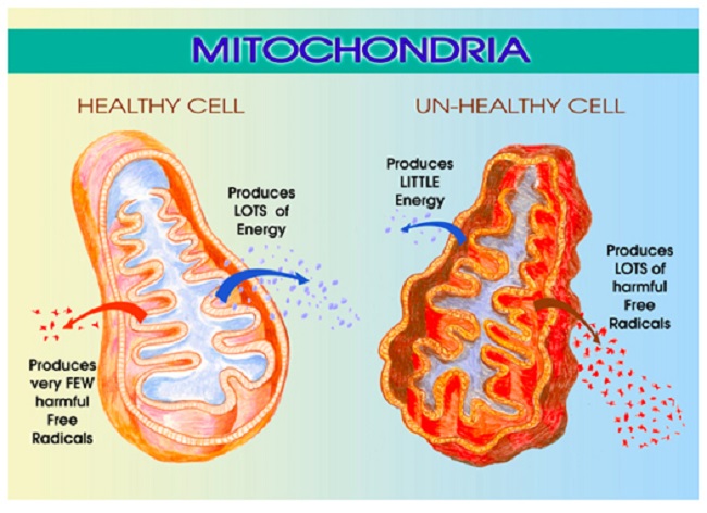
Estradiol is a critical component of that process and directly impacts mitochondrial energy production. That’s right, estradiol is part of the mitochondrial bioenergetic machinery such that when estradiol wanes, so too does energy production or ATP. As one might suspect, waning ATP is deleterious to brain health. In previous posts, I detailed the research showing how the loss of estradiol deforms mitochondrial morphology essentially disabling mitochondrial membrane potential while turning the mitochondria into misshapen donuts and blobs ripe for a slow, messy necrotic death; a process that evokes all sorts of deleterious reactions.

The Lupron Brain and Ketosis

Just recently, I stumbled upon research showing yet another mechanism of damage. In the absence of estradiol, brain glucose transport diminishes significantly. This effectively starves the brain for energy inducing severe bioenergetic deficiencies with all of the concordant neuronal damage one might expect. The reduction in glucose affects the mitochondria severely. Recall that glucose is one of the major fuel substrates of the brain, particularly where the Western diet predominates. The decline of glucose transport, therefore, is significant, and alone, without any other changes to the mitochondria, elicits a cascade of deleterious reactions. Oxidative phosphorylation and associated enzymes are downregulated, ATP production wanes, and ultimately may initiate the deformation of the very shape of the mitochondria, as observed in the research cited above. The ensuing reduction of ATP starves the brain of critical energy but also induces a state of hypoxia with the mitochondria incapable of utilizing molecular oxygen. With that hypoxia, inflammatory pathways are initiated further cementing mitochondrial death spirals and associated neuronal damage.

Interestingly, this reduction in aerobic activity coincides with the emergence of a ketogenic phenotype. That is, with the loss of one fuel substrate, ketones become the dominant source of fuel and the associated enzyme machinery is upregulated. Unfortunately, the Western diet is highly dependent upon carbohydrates and so a woman experiencing this loss of estradiol is not likely to consume sufficient fats and proteins to effectively weather this shift. Nevertheless, it does provide an opportunity for recovery. What if women who have lost the ability to produce sufficient estradiol either because of surgically (oophorectomy) or chemically (Lupron and other GnRH analogs) induced menopause adopt a ketogenic diet? Could we maximize the preferred energy source of the post-menopausal brain and reduce the neurological symptoms? I do not know the answer to that question, but given the severity of the suffering with surgical and chemical menopause, it seems worth the try.

We Need Your Help

More people than ever are reading Hormones Matter, a testament to the need for independent voices in health and medicine. We are not funded and accept limited advertising. Unlike many health sites, we don’t force you to purchase a subscription. We believe health information should be open to all. If you read Hormones Matter, and like it, please help support it. Contribute now.

Yes, I would like to support Hormones Matter.

Image by Angelo Esslinger from Pixabay.

This article was originally published on November 15, 2018.

A Rant about Lupron and Oophorectomy, Some Mechanisms and Clues to Recovery

10508 views

A Rant

Women who have been given Lupron (leuprolide) and the other GnRH agonists and antagonists and women who have had their ovaries removed are thrust violently into menopause. Overnight. There is no gradual decline of ovarian hormones that allows for molecular adaptations to the new state of chemical senescence. No, none of that. Just the chemical trauma of the loss of hormones.

The experience is akin to castration; something that should never be considered as a viable treatment option for any benign disease process, especially in young people. Yet millions of young women and children (Lupron for precocious puberty, gender dysphoria, and now autism) are prescribed these drugs and undergo these procedures annually without as much as the slightest recognition that there might be negative sequelae. Indeed, women are told routinely that what they experience with Lupron, its analogs, or upon oophorectomy is not real, that it does not exist, and that their symptoms are no more than some form of psychosomatic stress – the long vestige of Freudian acquiescence called hysteria. It is not. The symptoms these women experience is real and directly related to how the loss of estradiol and other ovarian hormones damage the mitochondria.

Some Mechanisms

Estradiol regulates mitochondrial energetics via multiple mechanisms both directly and indirectly. I have written about the mitochondrial damage provoked by the loss of estradiol previously, see here, here and here. In brief, the loss of estradiol fundamentally changes the shape and functionality of the mitochondria, effectively disabling not only their ability to produce ATP (cellular energy), but also, reducing their capacity to perform the myriad of vital functions for which the mitochondria are responsible. The reduction of ATP is alone sufficiently damaging and I would argue the root of all disease. Without sufficient ATP all sorts of vital functions grind to a halt, but when one considers that the mitochondria are responsible for managing immune and inflammatory signals, the production of steroids, the sequestration of Ca+ (cell excitability), the removal of toxicants, both endogenous and exogenous, and even cell life/death cycles, the logic behind using a form medically induced castration as a treatment for any disease becomes suspect.

For all the young women who suffer through the loss of estradiol, either via Lupron, its analogs or via oophorectomy, the damage is believed to be irreparable, mostly because there is neither recognition of the ill-effects nor any research into possible recovery options. Some of the damage may be permanent, unfortunately. It is impossible to tell at this point. However, and this is a big however, the human organism is remarkable in its ability to heal and sustain life despite our best efforts to the contrary. I believe strongly that the body can recover from just about anything, save except death. One just has to give it what it needs to heal. That is, if we provide the core substrates, the appropriate fuel sources and nutrients and if we remove the toxins, healing can occur. It will not be quick and it may not be complete, but it will happen. This is where basic research comes into play. Though not ideal, it can guide us. I will explain, but first, let us review some components of mitochondrial illness.

Understanding Mitochondrial Damage

Mitochondria are central to cell survival, and thus, our survival. Mitochondria take the foods we eat and through a series of enzymatic reactions convert the food into chemical energy called ATP. ATP fuels everything. Without ATP, cells struggle to function, become hypoxic, protein synthesis and repair processes falter producing aberrantly folded proteins, until the damage becomes too great, overwhelming their capacity to function. Cells die, tissues and organs die, and eventually, we die. Before we die, however, a whole host of seemingly random and complex illnesses and symptoms emerge as a direct result of the diminished ATP. That is, in an effort to keep us alive, the mitochondria and the cells in which they reside initiate survival cascades, key among them, inflammation and immune reactivity. These cascades, if left unchecked, evoke even more damage, illness, and eventually death. Yes, mitochondrial damage can precipitate death or a life so painful that death may seem preferable. The pain and suffering these women experience is real.

What Causes Mitochondrial Illness?

Mitochondrial illness can be initiated via a whole host of interacting variables. Genetics play a role, but so too do epigenetics – environmental stressors that activate or deactivate genes. Diet is a huge contributor. Too much sugar, processed food, alcohol and not enough nutrients are key variables determining mitochondrial illness or health. Nearly every, if not every, pharmaceutical damages mitochondria via one mechanism or another. All environmental and industrial chemicals damage the mitochondria. Hormones too influence mitochondria. Estradiol is top among them, but likely not the only hormone influencing mitochondria, only the most frequently studied.

Presentation of Mitochondrial Illness

Mitochondrial illness breaks all the rules of modern medicine. The symptoms are highly varied and individual. They do not fit into our discretely compartmentalized view of illness, and how could they? Mitochondria are the engines of every cell in the body, powering all life sustaining functions, from the brain and nervous system to the heart, the GI system to the musculature and everything in between. So when the mitochondria struggle, we have symptoms everywhere, in every compartment of the body, but exactly how those symptoms present is as individual as we are.

When the mitochondria are struggling, the systems that require the most energy are taxed most, but depending upon the individual’s genetic/epigenetic and nutritional liabilities, all sorts of weird symptoms may emerge due to the lack of ATP, the survival cascades initiated, and the other mitochondrial functions that now struggle. In that regard, even folks with the same constellation of genetic variables, express their symptoms differently. The wild variability in symptom expression makes mitochondrial disorders simultaneously the most difficult and the easiest to diagnose; most difficult if one subscribes to a compartmentalized, organ specific form of medicine and easiest if one looks to root causes. At the root of every disease process, whether cause or consequence, are struggling mitochondria. All disease begins and ends in the mitochondria. As such, unless and until the needs of the mitochondria are addressed, healing cannot occur. The flipside, of course, is if we support mitochondria, healing becomes possible.

Mitochondria, Estradiol and the Problem with Medically Induced Menopause

As mentioned previously, when the mitochondria are deprived of estradiol, the membranes surrounding the mitochondria become deformed. The increased permeability of these membrane causes all sorts problems, but top among them, the transfer of nutrients is less efficient. Mitochondria require at least 24 vitamins and minerals to efficiently convert the food we eat into ATP.  Though it is not clear what happens first, whether the enzyme machinery responsible for processing ATP diminishes heralding the increased permeability and deformation of the mitochondrial membranes or whether the loss of estradiol deforms the membranes first and that then results in a loss of ATP, it is clear that the loss of estradiol severely constricts mitochondrial functioning. In both cases, the loss of functionality initiates the survival cascades, and what many call the death spiral, begins.

Clues to Recovery

The easy answer would be to replace the lost estradiol. The problem with that, however, is that the ovaries synthesize a whole host of hormones, not just estradiol, and our ability to replicate those hormones in the appropriate concentrations is just not there yet. Nevertheless, working with a physician or pharmacist experienced in bioidentical hormone replacement may be helpful, at least in the transition period while the body is adapting to the new state of chemistry. The use of synthetics, however, are likely to do more damage than they are worth.

I believe a more prudent approach would be to tackle the health and efficiency of the mitochondria themselves; an approach that had it been undertaken prior to the use of Lupron or oophorectomy may have been able to reduce the impetus for these procedures in the first place. In fact, these recommendations are not specific to Lupron or oophorectomy, but carryover to any type of mitochondrial illness.

Vitamins, Minerals, and Diet

Considering the mitochondrion’s primary purpose is to convert the food we eat into life sustaining ATP, the simplest and most often ignored component of recovery, is to feed the mitochondria what they need to function and avoid the stuff they do not. In other words, eat well and avoid chemical toxicants. In practice, however, this seems to be exceedingly difficult for most of us. Decades of processed food marketing has skewed our preferences away from ‘real food’, and in many ways, disconnected us from the purpose of eating – to fuel our bodies.

Mitochondrial Nutrients to Rescue Mitochondria

There are 24 of vitamins and minerals required to power the mitochondrial machinery responsible for converting food into ATP. Ensuring an adequate supply of nutrients is critical to repairing mitochondrial damage, perhaps more important than any other aspect of recovery. Top among them are thiamine and magnesium. We have written about this extensively on Hormones Matter and in our book. Thiamine is the gatekeeper to the mitochondrial factory, involved in the initial enzymatic reactions required to convert consumed carbohydrates, fats and proteins into ATP and just about every enzyme reaction throughout the process. More so than any other nutrient, thiamine is critically important to mitochondrial functioning, but its deficiency is least likely to be recognized, even though it causes a host of serious disease processes including death. Magnesium is an essential cofactor for thiamine. Deficiencies in thiamine and magnesium may well account for the vast majority of modern illnesses.

The other B vitamins are also important, as are a variety of minerals. Which ones and in what doses are required for recovery is individual, but often, the dosages are far higher than what is proposed by the RDA and in combinations that are not typically found in standard, over-the-counter (OTC) supplements or even in marketed mitochondrial cocktails, though some of these products are considerably better than the OTCs. In the case of mitochondrial recovery, vitamins and minerals are used at pharmacological doses in order to kick start, and in many cases, compensate for the increased nutrient demands caused by the damage and/or by genetic or epigenetic variables. For individuals with severe deficiencies and/or poor absorption, repeated intravenous vitamins/minerals may be warranted. The details of dosing are covered in our book.

The Mitochondria Diet: Eat Real Food

The second component of healing mitochondria involves diet more broadly. That’s right, what we eat can harm or heal us. Processed foods must be eliminated, as should alcohol and other toxicants. Diets should be high in protein and good fats and low in carbohydrates, and to the extent feasible, organic. Why proteins and fats? Chemistry. Health demands that protein synthesis outpace protein breakdown. That requires a ready supply of protein from the diet and the appropriate nutrients listed above to process them. There is much debate about how much protein, but broadly, and depending upon level and type of illness, one must consider.08 – 1.5kg of protein per kg of weight per day. In research of critically ill patients, higher protein consumption (~1.5kg/kg/day) is associated with better outcomes. With our current predilection for excess carbohydrates, most folks are nowhere near even the lower end of these requirements and that creates a barrier to healing.

The integrity of mitochondrial and all cellular membranes require fatty acids. For women recovering from the loss of estradiol, and the subsequent deformation of mitochondrial membranes, a diet poor in essential fatty acids would be doubly debilitating. Indeed, for most people, an increase in fatty acids would improve health considerably. Fatty acids are also a key fuel substrate for mitochondrial ATP (which just so happens to be thiamine dependent as well).

In general, lower carbohydrate intake is warranted. High carbohydrate intake exacerbates thiamine deficiency and in fact, by itself, with no other risk variables, can induce thiamine deficiency. Researchers from USC have shown that immediately following the loss of estradiol, brain glucose transporters begin to downregulate forcing the brain into ketosis.  That is, at least in mice, the loss of estradiol forces a metabolic shift towards using ketones as the primary fuel source to produce ATP. This means that our traditionally carbohydrate dense diets may not meet the brain’s energetic demands when estradiol is absent. Granted, this research used mice and is preliminary, but it suggests an additional route to healing post Lupron and oophorectomy would be to increase dietary fat. Ketosis may be an option to repair mitochondrial damage.

Having said that, there are disorders of fatty acid metabolism which make lower carbohydrate diets very difficult and sometimes even dangerous. Full blown manifestations of these disorders usually occur in infancy or childhood, but more subtle manifestations brought on by single nucleotide polymorphisms (SNPs) in key genes may remain somewhat latent until triggered. Anecdotally, the inability to metabolize fats seems relatively common among the folks who interact with us, manifesting in what can only be described as energetic collapse post ingestion of fats and proteins. In the cases where SNP analyses are available, the difficulties are related to what have been traditionally considered ‘rare’ variances in key enzymes. Nevertheless, the parameters of these recommendations hold true for most people. Eat real, nutrient dense foods. The specifics of the diet, like the specifics of the nutrient replacements is all that varies.

Avoid Toxicants

Finally, with poorly functioning mitochondria, taxing these organelles further by ingesting the chemically laden foodstuffs produced by conventional agriculture and livestock practices makes healing that much more difficult. To the extent dietary toxicants can be avoided, they should. Similarly, since most, if not all medications damage mitochondria and deplete vital nutrients by one mechanism or another, medication use should be evaluated thoughtfully assessing potential benefits against likely risks.

Is it Really That Simple?

Yes and no. In many ways, it is very simple. Feed the mitochondria and they will do the rest, but in other ways, it is not so simple. Diet is absolutely critical, but mitochondrial recovery also includes using supplements at pharmacological doses, sometimes intravenously, to rescue damaged mitochondria and induce a more favorable mitochondrial replication process. This is difficult for some folks to access. Few physicians have any background in this and many simply do not believe that mitochondrial damage exists or evokes illness. Worse yet and despite evidence to the contrary, many physicians are steadfast in their belief that pharmaceuticals have no bearing on mitochondrial function, and thus cannot possibly be responsible for these illnesses. This means that to the extent pharmaceuticals do not treat and often make matters worse in these cases, fuel to the ‘it-must-be-hysteria-or-psychosomatic’ fire is added. In many ways, the biggest stumbling block is the notion that mitochondrially-mediated disease processes exist at all. A close second, is that nutrients can be used to recover mitochondria and in many cases, override even genetic defects.

Making matters even more complicated, there is no one-size-fits-all diet or supplement protocol. While it is true that thiamine is critical and must be addressed if there is any hope of recovery, everything else downstream must be considered on an individual basis. We each carry a host of unique genetic, epigenetic and environmental exposures that combine to make a complicated chemistry; one that no one wants to untangle and few have the skills to do so. As a result, it is much easier to dismiss the symptoms as psychosomatic and prescribe an antidepressant or other pharmaceutical. I would also mention, even among those of us who have a background in this stuff, we do not know everything and cannot know everything. Untangling these patterns takes time, effort from the patient, lots of research from both parties, and a fair degree of trial and error. Finally, recovery is neither a straight line nor an absolute. There are setbacks, sometimes serious ones, and health has to be actively managed from this point forward. Even with all of these complications, however, better health is possible if one tends to the mitochondria.

We Need Your Help

More people than ever are reading Hormones Matter, a testament to the need for independent voices in health and medicine. We are not funded and accept limited advertising. Unlike many health sites, we don’t force you to purchase a subscription. We believe health information should be open to all. If you read Hormones Matter, like it, please help support it. Contribute now.

Yes, I would like to support Hormones Matter. 

This article was published originally on August 6, 2018. 

Post Lupron Mitochondrial Collapse: A Case Story

5511 views

Dr. Marrs and I became aware of the symptoms and some of the laboratory results in a 38 year old woman. We wish to describe the case because it represents what should be an entirely new approach to medicine in general.

Before Lupron

The patient had been an active 38-year-old woman caring for her home and two children, ages 7 and 9 that were her focus, prior to Lupron. She had walked her dog daily, worked out at the gym twice a week and she had renovated her home in the summer of 2017. She had had her gallbladder removed in 2016, said to be because of a polyp. She had received allergy shots once a month for a couple of years. She took birth-control pills. Her history revealed that she had had a severe reaction to penicillin as a child, resulting in a rash and joint swelling causing inability to walk for a short time. She also had a history of frequent sinus infections and antibiotic treatment. In 2017, she began experiencing heavy menstrual bleeding and hysterectomy was recommended. She refused this and a second doctor recommended Lupron in an attempt to change her hormonal balance before hysterectomy.

After Lupron

She had received an injection of Lupron into her left hip and side effects, beginning within a few days, included:

  • Fatigue
  • Skin redness
  • Severe weakness in the legs, and tingling in the left side of the head and right leg.
  • A few days later she developed severe headaches and vomiting.

An initial estradiol patch caused no improvement, so another one at double the dose was prescribed with some improvement (presumably in menses).  By November 2017 the patches were discontinued. She began to experience

  • Joint pain
  • Pain throughout her body
  • Nervousness
  • Shaking
  • Panic attacks
  • Changes in personality.
  • In December she experienced cyclic vomiting and weight loss.

The estrogen patches were prescribed again. She saw an endocrinologist and some laboratory tests were abnormal, including what was described as a borderline high blood glucose.

These symptoms continued and in February 2018, three drugs were prescribed (Celexa, Neurontin, Ambien). They were discontinued two weeks later because there was no relief of symptoms.

In March, she experienced severe fatigue and had episodes of difficulty in walking which were intermittent and described as “almost like being paralyzed”.

In April, she saw a geneticist and some lab tests were performed that I will comment on shortly. A diagnosis of fibromyalgia and possible chronic fatigue syndrome were each entertained. The endocrinologist said that test results indicated that she was not producing estrogen or progesterone.

Based upon conversations with us, she began supplements of thiamine, fish oil, alpha lipoic acid, B complex, folinic acid, ferrous sulfate and methyl B12. Estradiol patches were resumed. We suggested the use of intravenous water-soluble vitamins, since Dr. Marrs and I agreed that giving the nutrients by mouth probably could not reach the necessary concentration of vitamin therapy needed. This was not followed through on by her current physician.

Discussion of Symptoms and Side Effects

The patient’s medical history indicated that she had experienced many different symptoms throughout her life. These included a severe reaction from penicillin and multiple sinus infections. The side effects from Lupron were fatigue, leg weakness, headaches, general body pain, panic attacks and cyclic vomiting. In other words, she had been a classic “problem patient” to her physicians. Since the symptoms could not be defined by usual and customary laboratory evidence the general conclusion was repeatedly that this was evidence of psychosomatic disease. Curiously, this common diagnosis in modern medical circles appears to be that the patient is thought of as inventing her symptoms neurotically without ever considering an underlying mechanism. Even worse, polysymptomatic disease of this nature is usually experienced by the brightest and the best. This is because high intelligence is developed within a brain which is more energy consuming than that of a less intelligent person. Such individuals are much more prone to unforeseen stress events, making them more susceptible to side effects from medication and inoculations. A car engine uses more energy to climb a hill. Stresses that we meet in life are like “hills to be climbed” and involve a commensurate supply of energy.

Laboratory Results: Low Amino Acids, Vitamin Deficiency and Defective Energy Metabolism

Many tests were performed on this patient. Two amino acid tests were performed, one measuring the amino acids found in blood plasma, the other measuring those excreted in urine. A word of explanation is necessary. Amino acids are the building blocks of proteins in the body and finding a given amino acid in very low or unusually high concentration can be used to define important aspects of body chemistry. Of 34 amino acids recorded in the plasma of this patient, aspartic acid, serine, ethanolamine, and tyrosine were severely decreased, while glutamine, histidine, alanine, ethanolamine and tyrosine were severely decreased in urine. All the others were in their expected normal concentration.

Amino acids are used in the body to create proteins, and this is an energy consuming mechanism. One of the deficient amino acids was aspartic acid whose metabolism is important in a mechanism known as transamination. The enzyme that carries out this function requires vitamin B6.

Two of them, ethanolamine and serine, play an important part in transmethylation, a mechanism that is dependent on folate and B12.

The fourth one was tyrosine and it is involved in the synthesis of thyroid hormone.

These low levels suggested that their respective vitamin dependent mechanisms were at fault. Since all the vitamins involved are water-soluble, it invited their administration by intravenous infusion. However, because they were energy dependent reactions, it is likely to construe the possibility that the underlying common fault was energy synthesis.

Was there any evidence from these laboratory results for defective energy metabolism? Yes.

Isocitric and citric acids were reported to be low in the urine and they are vital metabolites in the citric acid cycle, the “engine” of the cell. Also, there was a deficiency of pyruvic acid and this is the fuel that enables the citric acid cycle to function. This constituted strong evidence for energy deficiency with its major effect on the brain and nervous system.

Mitochondrial Energy Synthesis

Our bodies consist of 70 to 100 trillion cells that are being broken down and reconstructed throughout life. Relatively simple molecules are acted on by enzymes in a series of chemical reactions known by biochemists as “pathways”. Each enzyme requires a vitamin and/or essential mineral that assists the action of the enzyme and are known as cofactors to the enzyme. Several pathways reflect the synthesis of energy that is stored in the cell as ATP (adenosine triphosphate). ATP is a little like a battery that is being continuously charged and discharged and most of this occurs in the mitochondria. All the other pathways consume energy, either in enabling function or rebuilding cells. They might be compared loosely to the transmission in an automobile. In other words, the healthy body functions because energy synthesis meets energy demand. The abnormally low amino acids each could be used to suggest a defect in the energy consuming pathways and possibly a reflection of missing cofactors, making the “transmission” defective.

Vitamin Cofactors, Energy Deficiency, and Symptomology

The symptoms expressed by this unfortunate patient pointed strongly to cofactor deficiencies derived from diet that could easily be tested by their administration and clinical effect. The net effect is produced by a gap between energy synthesis and its utilization to meet the stresses of life in general. The administration of cofactors does not necessarily answer the underlying question because of the possibility of unknown genetically determined factors. However, it is safe, non-toxic, may have an epigenetic effect and is relatively cheap. It therefore should be the first approach. The greater the urgency or the severity of symptoms, the stronger the indication for intravenous administration of all the water-soluble vitamins. I have successfully treated many polysymptomatic patients this way, suggesting that mitochondrial function is as much an acquired disease as well as being genetically determined.

A Note About Oxidants and Antioxidants

Think of the body as a machine that consumes fuel by uniting it with oxygen to produce energy. This combination is called oxidation. Like a fire or any form of burning, it can be slow or fast and cellular oxidation seeks an intermediate level. If the oxidation is too slow, energy production is imperiled. If it is too fast or too vigorous, oxygen atoms are “thrown out” of the oxidation process like sparks are thrown out of a vigorously burning fire. These are referred to as “free oxygen radicals”. Like sparks from a fire, they can do damage. Some vitamins act to assist or accelerate oxidation, an example being B complex. They are known as oxidants. Others quench the free oxygen radicals (sparks) and are known as antioxidants. Vitamins C and E are examples.

Without going into the highly technical details, thiamine acts as an oxidant and an antioxidant, thus increasing its importance in metabolism. From this it is easy to see the essential importance of these substances that are obtained from naturally occurring foods and why their deficiency causes disease. Of course, we have known this for a long time, but current medical belief is fixed in the concept that “vitamin deficiency disease has been conquered and the resultant diseases are only of historical interest”. For example, this patient had “borderline high glucose”, something that would occur in the thiamine deficiency disease beriberi. She also had frequent “infections”, now known to be related to free oxygen radical production, indicating that her regulation of metabolism was extremely inefficient. The  amino acids that were extremely low in the plasma and urine could be used to interpret the possibility of missing cofactors, reflecting a chaotic state of metabolism. I must end this by saying that the use of vitamins and minerals in this manner is not (repeat not) simple vitamin replacement. We believe that the vitamin/mineral combination used in high-doses is resuscitating the activity of the corresponding enzyme and it is therefore acting as a drug. Identifying the underlying biochemical lesion is the essential nature of future diagnosis.

We Need Your Help

More people than ever are reading Hormones Matter, a testament to the need for independent voices in health and medicine. We are not funded and accept limited advertising. Unlike many health sites, we don’t force you to purchase a subscription. We believe health information should be open to all. If you read Hormones Matter, like it, please help support it. Contribute now.

Yes, I would like to support Hormones Matter. 

Share Your Story

If you have a Lupron story to share, send us a note and we’ll help you put it together. For more information on Lupron, click here.

Image Credit: Image by Steve Buissinne from Pixabay

This article was published originally on July 25, 2018. 

Lupron Side Effects Survey Results Part One: Scope and Severity

29712 views

Survey History

A few years ago, I embarked on a massive research project involving medication adverse reactions. I launched several online studies on the drugs that were popping up on our radar screen as having the most frequent and serious side effects; side effects that were largely ignored in the medical literature. At the time, this work was unfunded and, to a large degree, remains so today (feel free to contribute to our efforts). Despite the lack of funding, I thought it was important to investigate drug side effects from the patient’s perspective. Why was it that patients were reporting such a long list of devastating side effects while evidence in medical literature was largely absent? Were these patient experiences simply anomalies that we happened to be seeing, or was there something real going on? Without more quantitative data, these questions could not be answered.

To explore these questions, I designed several large studies, launched them online, and began collecting data, hoping that somehow the money would come in to fund the data analysis. It didn’t. And so these studies remained online, month after month, year after year, collecting data; data that needed to be analyzed and made public.

Earlier this year, I was fortunate to garner a grant for a new online project involving birth control and blood clots. The woman responsible for the grant understood the need to get the data from these legacy studies out to the public. The grant afforded me, not only the assistance of another researcher and a writer, but a much needed statistician. Per her wishes, the statistician could begin tackling the data analysis for these legacy studies, when not needed for the birth control project, e.g. during the periods of data collection. We are so very grateful that she recognized the importance of these projects.

A Note about the Data Sets

The surveys I designed were comprehensive, the data sets are massive and because they were conducted using survey software not designed for such large projects, the data sets are messy and require a tremendous amount of preparation to do even the most basic statistics. Notwithstanding the help of a part-time statistician, it will take us some months to ferret through these data sets. Nevertheless, we will get the data out. Again, if you’d like to help expedite this process, funding for a full-time statistician or even multiple statisticians would be most welcomed.

Beginning with Lupron

First in line, is one of my least favorite drugs used in women’s healthcare: Lupron. For those new to our blog, we’ve written a lot about Lupron over the years. Lupron or Leuprolide is a GnRH agonist prescribed for endometriosis, uterine fibroids, cysts, undiagnosed pelvic pain, precocious puberty, during infertility treatments, and to treat some cancers. I am not a big fan of this drug. If you have read the personal stories of the devastation caused by Lupron, or the research showing the mechanisms by which it induces damage, I doubt you would be either. Despite the decades of anecdotal evidence of serious side effects and the bevy of lawsuits filed and/or settled there is very little quantitative research delineating the scope and severity of these side effects. Given that Lupron chemically castrates its recipients, diminishing gonadal hormone production entirely, we might expect a little more research and certainly more caution in prescribing this drug. This doesn’t appear to be the case, as tens of thousands of women, men, and even children are prescribed this drug every year and have been for decades. Perhaps this is because Lupron is profitable, very profitable. In 2015, Lupron netted its manufacturers over $800 million in revenue.

What I find striking about Lupron, is not that are very few studies to support its safety and efficacy, or even that its manufacturers have been embroiled in lawsuits for decades all the while maintaining its safety and efficacy: that is common practice in the pharmaceutical industry. What is striking about Lupron is that it is a drug that effectively shuts down gonadal steroidogenesis, a key component of human health, and only a few in the medical industry think this is problematic. Here is a drug that could be used to induce chemical castration in pedophiles and rapists, if that were considered ethical or safe (and it’s not), but is used in tens of thousands of women, year in and year out, under the auspices of an effective treatment, and sometimes, even as a diagnostic for endometriosis. Worse yet, it is used on children in cases of precocious puberty, and to spur growth; two completely contraindicated uses.

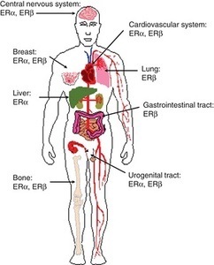
How is that a drug that blocks hormones so completely, hormones that have receptors and therefore regulatory roles in every tissue and organ of the body (brain, nervous system, heart, GI system, fat cells, immune cells, muscle, pancreas, gallbladderliver ) be considered safe? distribution of estrogen receptors Am I missing some great medical insight that suggests we really don’t need those pesky hormones after all; that all of those hormone receptors located all over the brain and body are there just because? How can a drug like this be used so cavalierly in young women? I don’t have answers for those questions beyond a collective insanity that has permeated medical science where women’s health is concerned. Absent answers, however, what I can begin to provide are data regarding the scope and severity of potential side effects associated with this drug; data gathered from the women themselves, unfiltered by industry bias or potential economic gains. Indeed, I suspect, once the full results of this study are published, industry will be none too happy with me or with our little project. Cue trolls.

All snark aside, results from this project, and from our others studies, are critical to an emerging discussion about medication safety. Patient experiences tell us a great deal about drug safety and efficacy, if we ask. I think it’s high time that we begin asking.

Study Design

The Lupron Side Effects Survey was designed to assess potential side effects across all organ systems. Hormones have receptors everywhere: it stands to reason then, if we deplete those hormones rapidly and continuously, there will be effects wherever those hormones play regulatory or modulatory roles. Of course, since estradiol, the primary hormone affected by Lupron, is critical to mitochondrial morphology, and thus mitochondrial energetics, anywhere there are high demands for energy, the nervous system, the heart, GI, musculature, we might anticipate a high degree of effects in those systems as well, especially with longer term use and as the damage accrues.

What we didn’t know, and won’t until we fully analyze the data (what is presented here is only the beginning), is in which systems the side effects are most severe (do they follow the path of mitochondrial energetics or some other yet to be identified pattern ) and when (do they present early or late, relative to use?); which women were more likely to have side-effects (are there health characteristics that make side effects more likely or more serious?); is there a dose-response curve for side effects (do higher doses mean greater side effects – sometimes with hormones, this isn’t the case); or how the side effects cluster together (did they cluster by organ or tissue system or by some other variable, like energy demands?). Most importantly, we didn’t know whether Lupron was clinically effective at reducing the symptoms for which it was prescribed. There is some evidence to suggest that while pain symptoms associated with endometriosis may show a statistically significant reduction, particularly while the woman is taking the drug, the reduction was neither clinically meaningful nor long lasting. That is, symptoms may diminish by a few points on a pain scale while on the drug, but not abate completely, and then inevitably return upon cessation. In light of the potential side effects induced by this medication, one would expect nothing less than a large clinical reduction, even remediation of the disease process itself, as the only fair trade off. It is not clear whether Lupron can provide those benefits. None of these questions have been answered in the medical literature, despite the use of this drug for many decades.

Lupron Side Effect Survey Basics

The study was launched in 2013. The goal was to get 500 respondents, evaluate, redesign and relaunch follow-up studies. We reached the 500 respondents within a few months, but absent funding, were not able to perform the analyses. So I left the study up to collect data passively (no longer advertising it), until recently.

When we closed the study, we had over 1400 partially completed surveys. For robustness, we analyzed only those surveys that were over 90% complete. That netted data from over 1000 respondents (the number of respondents for each question varies and is listed below with each item).  The survey was anonymous, voluntary, and included informed consent.

Survey respondents were asked to provide basic demographic information, answer questions about pre-existing conditions, reasons for Lupron prescription, Lupron dosage, and degree of efficacy pre, during, and post Lupron usage. To capture the range and severity of potential side effects, survey respondents were asked to indicate the presence/absence and severity of symptoms experienced relative to their Lupron usage with a 0-4 Likert-type scale (0=None, 1=Mild, 2=Moderate, 3=Severe and 4=Life threatening). And yes, we recognize that ‘life-threatening’ is not an appropriate indicator for some types of symptoms. For consistency, however, we used the same rating scale across symptoms. One hundred and eighty possible symptoms were assessed.

This post will review range and severity of Lupron side effects. Subsequent posts will address efficacy, side-effect clustering, patient characteristics predicting side effects, side-effect dose-response curves and other topics.

Demographics

Survey respondents (n=1064) were largely Caucasian – 86.6% (African American -4.7%, Hispanic -2.6%, Other – 2.3%, Asian – 2%, American Indian/Alaska Native 1.6%) and educated (30% – some college, 34% – BA/BS, 15% – MA/MS). The average age of the survey respondent was 35.36 (SD – 8.63), while the average age at which Lupron was prescribed was 29.9 (SD – 8.2). Among these respondents, Lupron was most commonly prescribed for endometriosis (88%), painful periods (33.5%), heavy bleeding (26%), ovarian cysts (18%), PCOS (4%), IVF (4%), anemia (3.9%), breast cancer (1%), ovarian cancer (.4%), precocious puberty (.2%), other (8%).  Respondents could select multiple answers.

For the discussion that follows, see the interactive graphic below.  We will be discussing symptom categories from left to right. The categories of side effects are grouped, to some extent, by system involved or by symptom characteristics. To view the side effects, click any of the boxes below. The side effects within that category will appear, along with the number of respondents who answered that question. Click again on a particular side effect and the severity of the side effect is displayed by percentage of women who experienced each severity level. Click in the white space within the graphs to move up a level (note, this is a little tricky in the categories with lots of symptoms). The size of the graphic does not display well on mobile phones and/or when using the internet browser Internet Explorer. For the best viewing, please use a computer screen.

Patient Reported Side Effects Associated With Lupron

General Side Effects and Allergic Reactions

Compared to the frequency and severity of other symptoms experienced in association with Lupron, side effects relative to the injection itself and those that would be characterized as allergic reactions, itching, swelling, etc., were uncommon in most of the respondents, except for injection site pain, which was experienced in varying degrees of severity by over 70% of the respondents.

Sex and Libido

As one might expect with medication that chemically castrates its users, reductions in libido and other symptoms whose net result diminishes sexual interest and ability were common. Some degree of a loss of interest in sex was experienced by all but 23% of the women, with 38% reporting a severe diminishment in sexual interest. Nearly 44% of respondents reported moderate to severe pain during sex which may explained to some degree by the almost equal percentage of women reporting moderate to severe vaginal dryness. Other symptoms reported included breast pain, swelling, and to a much lesser extent, discharge.

Muscle and Joint Pain

Up to 50% of the women reported moderate to severe muscle and/or joint pain. This is notable inasmuch as for the majority of the women prescribed Lupron, pelvic and abdominal pain associated with endometriosis is the driving factor for the use of this drug. It appears that we may be trading pain in one region of the body for another.

Gastrointestinal and Related Symptoms

Here again we see that a large percentage of women (from 15-50% depending upon the symptom) report moderate to severe gastrointestinal disturbances from nausea, vomiting and diarrhea through constipation and even bowel obstruction. Moderate to severe bladder pain was common (~31%) as was difficulty in urination (~19%) and bladder control (~18%). Since bladder pain and interstitial cystitis are co-morbid with endometriosis, it is difficult to determine if these symptoms were precipitated or exacerbated by the Lupron or simply associated with the endometriosis and thus, not remediated by the medication. We will attempt to disentangle those relationships with further data analysis and in subsequent studies.

Of note, gallbladder disease (~6.2% – all categories combined), gallstones (~3%), kidney disease (2%), kidney stones (4.5%) and renal failure affect a smaller but noticeable number of Lupron recipients. This consistent with adverse event reporting elsewhere. Similarly, non-alcoholic fatty liver was noted in ~6.6% of the survey respondents.

Bone, Skin, and Related Symptoms

Bone formation is particularly hard hit by the diminishment of estradiol. Bone related symptoms are some of the most commonly reported side effects ascribed to Lupron. Research suggests Lupron induces a 5-6% decline in bone mineral density over just 6-12 months of use. Read more on the mechanisms by which Lupron induces bone loss.

Almost 20% of the women who completed the survey reported some degree of osteoporosis, and 16% reported cracking or brittle bones, 42% reported toothaches (9% severe) and 26% had cracking teeth. Osteonecrosis was reported by 3% of the respondents. Skin and hair symptoms were common and affected a sizable percentage of the respondents as well. What we failed to ask about were fractures in the spine and pelvis or osteoporosis in the jaw; side effects that commonly appear in post Lupron discussion boards. We will do so in subsequent surveys.

Temperature Dysregulation

As expected by a drug that induces a rapid menopausal state, vasomotor symptoms with temperature dysregulation were prominent afflicting ~90% of the respondents. Severe hot flashes and night sweats were reported by over half of the study population.

Metabolic Symptoms

Estradiol affects insulin regulation and general metabolism, so it stands to reason that if concentrations are diminished significantly, metabolic disruption would ensue. Hypoglycemia was reported by about 15% of the women, while hyperglycemia was reported by about 6%. Similarly, increased hunger and thirst were prominent at least 50% of the population, along with rapid weight gain (mild 19.2%, severe 25.9%). In contrast, rapid weight loss was reported by 12% of the respondents. New onset diabetes, Type 1 and Type 2 was reported by ~1% and 2.8% respectively. As we perform more advanced analyses, we will try to more fully characterize the metabolic changes in different groups of women.

Cardiovascular and Respiratory Symptoms

We know that the estrogens and androgens affect heart function via multiple mechanisms, both at the receptor level and via more global changes to mitochondrial functioning. What we don’t know is what impact blocking those hormones so abruptly and completely and sometimes even chronically, has on heart function. Clinically, the results of this survey point to dysregulated blood pressure (BP – 12.5%) and heart rate or rhythm (24.7%) with a trend towards the elevated measures for blood pressure (24.4% – all, 13.3% moderate to life threatening) and heart rate (27.7% – all, 22% moderate to life-threatening. However, there was a noticeable percentage of women who experienced lower blood pressure (16.4%) and heart rate (6.1%). At least 10 women experienced a heart attack, 36 women developed mitral valve prolapse, 10 women developed blood clots in the leg and 8 women had pulmonary emboli. Difficulty breathing and sleep apnea were reported by 22.2% and 15.6% respectively. As we do further analyses we’ll be able to more fully characterize the pattern of cardiovascular symptoms.

Brain and Nervous System Symptoms

The next five sections of the graphic represent the scope and severity of symptoms associated with the nervous system. Though categorized distinctly for purposes of display, the distinctions are somewhat arbitrary as the symptoms within each category represent those related to the brain and nervous system. Arguably, many of the cardiovascular symptoms discussed previously may also represent nervous system symptoms, possibly suggesting some degree of autonomic system dysregulation.

Headache, Migraine, Dizziness, and Seizures. A large percentage of women experienced headache and migraine pain, frequently rated as severe or life-threatening (27.2% and 28.3% respectively). Dizziness was common (69.4%), as was vertigo (46.9%). Sleepiness (68%) and fatigue (87.3) were common, but interestingly, also insomnia (76.4). Seizures reported by 5.1% of respondents. Falling (17.1%) and difficulty walking (27.9%), perhaps indicating balance issues, were also reported. TIAs were reported by 11 women and full strokes by 3 women.  (Nervous System Symptoms).

Myoclonus and Neuropathy. Shaking, jerking, numbness, spasms and tingling were experienced to some degree by 15%-35% of the survey respondents. A sizable percentage of women reported moderate to severe symptoms. Muscle weakness was reported by 11-34% of the respondents whereas limb and/or facial paralysis was experienced 3-4% of the women.  (Neuromuscular, Sensory Perception and Motor Control).

Hearing and Vision Disturbances. Some degree of blurred vision was experienced by 46.5% of the women, with a little over 20% rating the symptom moderate to severe. Partial loss of vision was reported by almost 10% of the women, half of them indicating moderate to severe loss. Similarly, almost 20% reported some hearing impairment. Similarly, hypersensitivity to light or to sound was indicated by 35% and 37.5% respectively. (Sensory and Motor Symptoms).

Speech and Language Disturbances. Fully 20-50% of the respondents reported difficulty with basic communication, everything from difficulty speaking and finding words to difficulty understanding speech, reading and writing. (Sensory and Motor Symptoms).

Mood, Memory, Mental Health and Affective Behavior. The brain is a major target of and source for steroid hormones. Estrogen receptors are co-localized on neurons and affect neurotransmission, neurite outgrowth, synaptogenesis and myelin growth  and estradiol is generally considered neuroprotective. The prefrontal cortex, hippocampus, and amygdala, responsible for regulating directed behavior, memory, and emotion, have high densities of estrogen receptors. Depleting estradiol would be expected to have a significant impact on these functions, and it did. It is here that we see some of the most troubling and least well appreciated (by the medical profession) side effects associated with Lupron. A significant percentage of women reported severe psychological disturbances ranging from depression and anxiety (>50%) to suicidality (15% severe to life-threatening). Visual or auditory hallucinations were experienced by ~12%, with >6% reporting moderate severe issues. Moderate to severe frontal cortex issues like dulled or inappropriate emotions, lack of motivation, impulsiveness were reported in 25% to over 50% of respondents. Moderately to severely altered mental states (delirium, disorientation, confusion) were reported by 6%-25% of the women.  Moderate to severe diminishment in memory capacity was reported by at least a third of the women. This is in addition the difficulties with language reported above. (Mood, Memory and Mental Health).

General Impressions

Consistent with the case stories and patient comments on message boards related to Lupron side effects, the majority of women feel rotten while on this drug. This is to be expected given the global distribution of estrogen receptors. The brain and nervous system seem particularly hard hit. Again, this is understandable given the density of estrogen receptors in the brain and the modulatory role it, and other steroid hormones, play in neurotransmission. By depriving the estrogen receptors of their cognate ligand, estradiol, Lupron fundamentally alters brain chemistry, abruptly and thoroughly. Perhaps even more troubling, estradiol is required for mitochondrial functioning. By depleting estradiol, the mitochondria are impaired, and with that impairment comes a long line of compensatory mechanisms that will ultimately derail not only mitochondrial capacity but also the capacity of all of the cell functions that require healthy mitochondria. The fact that we see such severe side effects attributable to nervous system function would be expected with estradiol depletion.

We Need Your Help

This post was published originally on Hormones Matter on September 1, 2016. Since then, we have lost our funding to complete this and the other ongoing studies. We have enormous data sets like this one on medication adverse reactions waiting to be analyzed and published. Without funding, however, these data will never see the light of day. If these issues are important to you, please contribute. If you know of an organization or benefactor interested in understanding short and long-term medication and vaccine reactions, please refer them to us.

Yes, I’d like to support Hormones Matter.

Photo by Diana Polekhina on Unsplash.