epigenetics

Oral Contraceptives, Epigenetics, and Autism

10795 views

Oral Contraceptives and Autism

Over the last several months, I have published a series of articles exploring the potential connection between the use of oral contraceptives and the increased prevalence of Autism Spectrum Disorder (here, here, here, here, and here). It is my hypothesis that the synthetic hormones in oral contraceptives, which were created to imitate natural human hormones and disrupt endocrine function to prevent pregnancy, may be causing harmful neurodevelopmental effects in the offspring of women who use them [1].

The mechanisms by which oral contraceptives instigate neurodevelopmental changes is slowly emerging. It appears that in addition to preventing pregnancy, synthetic hormones like ethinylestradiol, used in most birth control formulations, initiate epigenetic alterations in the oocytes (eggs) causing persistent changes in expression of the estrogen receptor beta gene (ERβ). When those eggs become fertilized and conception ensues, the changes in the estrogen receptor gene impact the expression of autism and other neurodevelopmental disorders.

Ethinylestradiol is an Endocrine Disruptor

Here is how the ethinylestradiol used in oral contraceptives adversely modifies the condition of the oocyte. Bear with me, this is a bit complicated, but if you are woman who uses or is contemplating using oral contraceptives, this information is important to understand.

Ethinylestradiol is a known endocrine disruptor. Anything that disrupts endogenous hormones can be considered an endocrine disruptor. Evidence is emerging that ethinylestradiol may trigger what is called DNA methylation of the estrogen receptor gene. This then causes decreased messenger RNA resulting in impaired brain estrogen signaling in offspring [2]. Let’s think more deeply about this.

Methylation means that, by way of a chemical process, a gene is turned on (hypomethylation) or turned off (methylation) by an enzyme or protein. Researchers believe that methylation is one of a number of mechanisms by which environmental interactions influence genetic activity. In this case, ethinylestradiol silences or turns off some important processes that are associated with estrogen signaling, namely receptor activity.

Methylation and other epigenetic reactions influence health and disease processes across generations. This is called transgenerational transmission. So, I suspect that the deleterious effects of ethinylestradiol on the estrogen receptor gene are transgenerational. This is possible because the estrogen receptor gene may be an imprinted gene. Imprinting is a dynamic epigenetic phenomenon by which certain genes are expressed in a parent-of-origin manner. If the allele, an alternative form of the same gene, inherited from the father is imprinted, it is thereby silenced, and only the allele from the mother is expressed. If the allele from the mother is imprinted, then only the allele from the father is expressed.

If the estrogen receptor gene is an imprinted gene and silenced, then the oral contraceptive-induced methylation marks could be protected from global demethylation. Global demethylation is a protective process which is believed to occur throughout somatic cell differentiation and happen only twice during development, in primordial germ cells and in the pre-implantation embryo. If the methylation marks are protected from global demethylation, they will be preserved through fertilization and beyond to progeny generations.

To sum this up, durable changes to the function of the cells would be passed on by the aberrant methylation that piggybacks on the normal imprinting mechanism that protects epigenetic markings from reversal or demethylation. Ethinylestradiol, while successful at preventing pregnancy, may be damaging stored oocytes in such a manner that the offspring that emerge from those oocytes carry that same damage.

In addition, deleterious effects of exposure to oral contraception could perpetuate or even increase over generations as a result of both transgenerational transmission of the altered epigenetic programming and the continued exposure across generations. This has the potential to impart disease sensitivity at a later point in time [3,4,5]. While this concept, in the case of oral contraceptive use, is speculative, transgenerational imprinting was first studied in human beings in cases of nutritional factors [6,7,8]. In addition to nutritional factors, animal studies have shown that estrogens, androgens, progestagens, or similar receptor-level acting molecules, such as endocrine disruptors, can have harmful transgenerational effects [4,9,10].

How Impaired Estrogen Receptors and Estradiol Regulation Affects Brain Function

Estrogen receptors affect the regulation of endogenous estradiol concentrations. Estradiol is the primary estrogen our body synthesizes to regulate a variety of reproductive and non-reproductive functions. Estrogen receptors are located all over the body, in the heart, lungs, fat cells, and in the brain.

Maintaining appropriate estradiol concentrations in the brain is critical for mood, memory and a number of other cognitive functions. Estradiol is critically important because it directly influences brain function through the estrogen receptors located on neurons in many areas of the brain. Estradiol has direct protective effects on neurons and helps with the maintenance and survival of neurons. Endogenous estrogens, like estradiol, stimulate creation of nerve growth and viability, repair of impaired neurons, and influence dendritic branching. Estradiol also increases the concentration of neurotransmitters such as serotonin, dopamine, and norepinephrine and affects their release, reuptake, and enzymatic inactivation. In addition, estradiol increases the number of receptors for these neurotransmitters.

Synthetic estrogens, like ethinylestradiol used in many birth control formulations, may adversely affect the equilibrium of the endogenous estrogens like estradiol by disrupting sensitive hormonal pathways and impairing estrogen receptor expression. When the estrogen receptors become impaired, not only are hormone concentrations likely affected, but those functions that this hormone and receptor are responsible for regulating, are altered as well; functions like mood, memory and cognition.

Estrogen Receptors, Mood, and Cognition

Impaired estrogen receptor expression has been associated with altered emotional responses, depression, mood disorders, cognitive dysfunction, brain degeneration, and many other endocrine-related diseases [11-16]. In addition to confirmation that estrogen receptors are a factor in emotional responses [11], there is compelling evidence for estradiol’s involvement in the regulation of mood and cognitive functions [12,13,14]. Because the hippocampus, entorhinal cortex, and thalamus seem to be estrogen receptor beta (ERβ)-dominant areas, this suggests a function for ERβ in cognition, non-emotional memory, and motor functions [13,14]. Children with autism have notable difficulties in all of these areas.

Research also shows that estrogen is able to regulate the serotonin (5-HT) system, which has been associated with affective disorders [13,14]. Furthermore, recent studies using estrogen receptor knockout mice have assisted in defining the function of estrogen receptors in brain degeneration [15]. In vivo and in vitro studies also show that estrogen receptors are mechanistically involved in endocrine-related diseases [16]. Given that ERβ is the main estrogen receptor expressed in the cerebral cortex, hippocampus, and cerebellum [17], it is not difficult to imagine that epigenetic mechanisms cause persistent changes in gene expression of estrogen receptor beta (ERβ) that result in neurodevelopmental disorders like autism.

Interestingly, a recent study discovered a significant association of the lowered levels of the ERβ gene with scores on the Autism Spectrum Quotient and the Empathy Quotient in people with autism [18].

Evidence of Dysregulation of Estrogen Receptor Beta

Motivation for this epigenetic hypothesis comes from a recent study by Pillai et al., Dysregulation of Estrogen Receptor beta (ERbeta), Aromatase (CYP19A1) and ER Co-activators in the middle frontal gyrus of autism spectrum disorder subjects. This study examined the brain tissue of people that had ASD’s. The scientists found that the ASD brain tissue had far lower levels of a key estrogen receptor and other estrogen-related proteins [19]. The scientists measured the expression of proteins involved with estrogen signally pathways in brain tissue measuring levels of estrogen receptor beta and aromatase, an enzyme that changes testosterone to estradiol. Pillai et al. found 35 percent less ERβ. In addition, they discovered much less messenger RNA of estrogen co-regulators SRC1, CBP and P/CAF at 34 percent, 77 percent and 52 percent respectively [19]. Their results provide compelling evidence of the dysregulation of ERβ and co-regulators in the brain of subjects with ASD. Their data suggest that the synchronized regulation of ER signaling molecules has a significant function in ER signaling in the brain and that this coordinated network may be compromised in people with ASD.

Growing research supports the hypothesis that epigenetic mechanisms are causing persistent changes in gene expression of estrogen receptor beta that result in autism in offspring of mothers who use oral contraceptives. What is perhaps most troubling, is that it may be that the adverse effects of DNA methylation of the estrogen receptor gene are transgenerational.

Final Thoughts

We are just beginning to understand how endocrine disruptors can modify the development of specific tissues that lead to increased vulnerability to diseases and disorders. And, we are just beginning to appreciate the critical roles that hormones play in neurodevelopment, including neuroendocrine circuits that control physiology and sex-specific behavior that could result in behavioral and psychiatric conditions. As women, we have a crucial decision to make about which kind of birth control we use. Because there are inherent risks in all medications that we take, it is important that we fully understand all of the risks of the drugs we choose to use. Although this research is in its early stages, there is a growing body of evidence that ethinylestradiol initiates epigenetic mechanisms that cause persistent changes in gene expression of the estrogen receptors that contribute to the risk of autism in offspring.

References

  1. Strifert, K (2015) An epigenetic basis for autism spectrum disorder risk and oral contraceptive use. Med Hypotheses. 2015 Sep 6. pii: S0306-9877(15)00323-0. doi: 10.1016/j.mehy.2015.09.001
  2. Strifert, K (2014) The link between oral contraceptive use and prevalence in autism spectrum disorder. Medical Hypotheses December 2014 Volume 83, Issue 6, Pages 718–725
  3. Skinner M (2008) Epigenetic programming of the germ line: effects of endocrine disruptors on the development of transgenerational disease. Reproductive BioMedicine Online Vol 16 No 1. 23-25.
  4. Skinner M (2014) Endocrine disruptor induction of epigenetic transgenerational inheritance of disease. Molecular and Cellular Endocrinology Jul 31. pii: S0303-7207(14)00223-8. doi: 10.1016/j.mce.2014.07.019.
  5. Vaiserman A (2014) Early-life Exposure to Endocrine Disrupting Chemicals and Later-life Health Outcomes: An Epigenetic Bridge? Aging and Disease Jan 28;5(6):419-29. doi: 10.14336/AD.2014.0500419.
  6. Kaati G, Bygren LO, Edvinsson S (2002) Cardiovascular and diabetes mortality determined by nutrition during parents’ and grandparents’ slow growth period. Eur J Hum Genet. 2002 Nov;10(11):682-8.
  7. Pembrey ME (2002) Time to take epigenetic inheritance seriously. Eur J Hum Genet. 2002 Nov;10(11):669-71.
  8. Pembrey ME, Bygren LO, Kaati G, Edvinsson S, Northstone K, et.al (2006) Sex-specific, male-line transgenerational responses in humans. Eur J Hum Genet. 2006 Feb;14(2):159-66.
  9. Csoka, A B, Szyf, M (2009) Epigenetic side-effects of common pharmaceuticals: A potential new field in medicine and pharmacology (Article). Medical Hypotheses Vol. 73, Issue 5, 2009, 770-780.
  10. Csaba G (2011)The biological basis and clinical significance of hormonal imprinting, an epigenetic process. Clinical Epigenetics August 2011, Volume 2, Issue 2, pp 187-196.
  11. Amin Z, Canli T, Epperson CN (2005) Effect of estrogen–serotonin interactions on mood and cognition. Behav Cogn Neurosci Rev 2005, 4:43-58.
  12. Berman KF, Schmidt PJ, Rubinow DR, Danaceau MA, Van Horn JD, et. al (1997) Modulation of cognition-specific cortical activity by gonadal steroids: a positron-emission tomography study in women. Proc Natl Acad Sci USA 1997, 94:8836-8841.
  13. Ostlund H, Keller E, Hurd YL (2003) Estrogen receptor gene expression in relation to neuropsychiatric disorders. Ann NY Acad Sci 2003 Dec;1007:54-63.
  14. Osterlund MK, Hurd YL (2001) Estrogen receptors in the human forebrain and the relation to neuropsychiatric disorders. Prog Neurobiol 2001 Jun;64(3):251-67.
  15. Mueller SO, Korach KS (2001) Estrogen receptors and endocrine diseases: lessons from estrogen receptor knockout mice. Curr Opin Pharmacol 2001 Dec;1(6):613-9.
  16. Candelaria NR, Liu K, Lin CY. (2013) Estrogen receptor alpha: molecular mechanisms and emerging insights. J Cell Biochem. Oct;114(10):2203-8. doi: 10.1002/jcb.24584.
  17. Bodo C, Rissman EF (2006) New roles for estrogen receptor beta in behavior and neuroendocrinology. Front Neuroendocrinol 2006, 27(2):217-232.
  18. Chakrabarti B, Dudbridge F, Kent L, Wheelwright S, Hill-Cawthorne G, et.al (2009) Genes related to sex steroids, neural growth, and social-emotional behavior are associated with autistic traits, empathy, and Asperger syndrome. Autism
  19. Crider A, Thakkar R, Ahmed A, Pillai A (2014) Dysregulation of Estrogen Receptor beta (ERbeta), Aromatase (CYP19A1) and ER Co-activators in the middle frontal gyrus of autism spectrum disorder subjects. Molecular Autism 2014, 5: 46. DOI: 10.1186/2040-2392-5-46.

We Need Your Help

More people than ever are reading Hormones Matter, a testament to the need for independent voices in health and medicine. We are not funded and accept limited advertising. Unlike many health sites, we don’t force you to purchase a subscription. We believe health information should be open to all. If you read Hormones Matter, like it, please help support it. Contribute now.

Yes, I would like to support Hormones Matter.

Image by jcomp on Freepik

This article was first published on October 15, 2015. 

Thiamine, Epigenetics, and the Tale of the Traveling Enzymes

6192 views

For some time now, we have been covering all the ways in which thiamine deficiency influences disease. The primary mechanisms are through the down regulation of mitochondrial enzymes critical for ATP production. The lack of thiamine impairs mitochondrial functioning significantly leading to complex, debilitating, chronic, and sometimes, deadly illnesses. With mitochondrial energy a requisite for cell functioning and survival it is easy to see how the diminishment of mitochondrial functioning would negatively impact health and how high-energy physiological systems like the nervous and cardiovascular systems might be particularly hard hit. More than just just derailing cell function, as if that wasn’t problematic enough, when mitochondrial energy production slows, the adaptive cascades that ensue include epigenetic modification, not only at the level of mitochondrial DNA (mtDNA), but also, at the level of gene expression from the cell nucleus or nDNA. This is a huge discovery with broad implications about health and disease. It means that all sorts of things considered innocuous, are directly influencing gene activation and deactivation by way of the mitochondria.

Epigenetics: How the Cells Adapt to the Environment

Epigenetic modification refers to the activation or deactivation of chromosomal gene expression absent mutation. These changes can be heritable and often are. Strictly speaking, epigenetics involves changes in methylation, histone modification and/or alterations in non-coding RNA that affect transcription. Epigenetics is the way our genome adapts to environmental circumstances and prepares our offspring to do the same. The majority of epigenetic work focuses on genomic changes. That is, those variables that affect gene expression from the cell nucleus or nDNA. There is a growing body of evidence, however, that mitochondrial DNA (mtDNA) are affected by epigenetic factors in much the same way as nDNA. In fact, given the mitochondria’s role in cell survival, one might suspect that mitochondria are more susceptible to environmental epigenetics, perhaps even the first responders and/or the initiators of chromosomal genetic changes.

Considering that nDNA accounts for over 90% of the proteins involved in mitochondrial functioning, how could damaged mitochondria, even functionally inefficient mitochondria, not affect gene expression in the cell’s nucleus? Though we tend to think of mitochondria as self-contained and discrete entities, black boxes of sorts, where stuff goes in and ATP magically comes out, this is not only not the case, it seems biologically illogical to think that way. So much of mitochondrial functioning is related to its environmental milieu. In fact, increasingly researchers are finding that the mitochondria are the center of the organismal universe, sensing and signaling danger, and effectively, regulating all adaptive responses, including epigenetic modification of the proteins encoded by the cell nucleus.

Beyond the strict definition of epigenetics, it seems to me that any factor that altered mitochondrial function, would eventually alter gene expression by any number of mechanisms, not just the direct ones. That is, because the mitochondria produce the energy required for cell survival, anything that derails their capacity to produce ATP, pharmaceutical and environmental chemicals, for example, should be considered, ipso facto, epigenetic modulators. Energy is a fundamental requirement for life. How could energy depletion not affect gene expression? It does, and now we know how.

Starve the Mitochondria, Alter the Genome

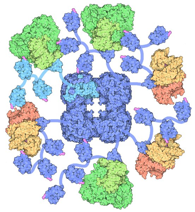
It turns out, when the mitochondria are starving and/or damaged the key enzyme complex that sits atop the entire energy production pathway and is critical for the production of the substrates involved directly (yes, I said directly) with gene expression, migrates from the mitochondria across the cell and into the cell’s nucleus where it then sets up shop and begins its work there. Sit with that for a minute. The entire enzyme complex decides that things are not working where it is, so it hitches a ride with some transporter proteins, several of them along the way, to find a more suitable home. The enzyme complex ‘knows’ that its functions are critical for survival and so it must move or die. What an incredible bit of adaptive capacity. A symbiosis, if you will.

To make matters even more interesting, the traveling enzyme complex just so happens to be the pyruvate dehydrogenase complex (PDC) and you guessed it, the PDC is highly, and I mean highly, thiamine dependent. But wait, there’s more. Several other enzymes involved in mitochondrial bioenergetics are also thiamine dependent. These include: alpha-ketoglutarate dehydrogenase (α- KGDH) in the tricarboxylic acid (TCA) or citric acid cycle, transketolase (TKT) within the pentose phosphate pathway (PPP), the branched chain alpha-keto acid dehydrogenase complex (BCKDC) involved in amino acid catabolism and, more recently, thiamine has be identified as a co-factor in fatty acid metabolism via an enzyme called 2-hydroxyacyl-CoA lyase (HACL1) in the peroxisomes (organelles that break down fatty acids before transporting them to the mitochondria). So thiamine deficiency is problematic. It not only causes dysfunction in the mitochondria, reducing bioenergetic capacity, but if severe enough and/or chronic, it alters the genome. And those changes are likely heritable. That is, your thiamine deficiency likely will affect your children’s ability to process thiamine.

Thiamine Deficiency and Gene Expression

Without sufficient thiamine, the PDC enzyme complex does not function well and because of its geographic position at the entry point into the citric acid cycle, when it is not working at capacity, everything below it eventually grinds to a halt resulting in severe neurological and neuromuscular deficits. Absent congenital PDC disorders, however, when it simply is inefficient or starved for its cofactors, metabolic disorders ensue because we cannot convert carbohydrates into ATP and the sugars that normally would be converted into energy, remain unmetabolized, floating around outside the cells and causing the whole cascade of effects that mark type 2 diabetes. When the PDC is inefficient, ATP levels wane and fatigue ensues. Systems that are highly energy dependent are hit the hardest. Think brain, heart, muscles, GI tract. When mitochondria are inefficient or damaged, reactive oxygen species (ROS) increase. Anti-oxidant capacity decreases and further damage to the PDC ensues.  Inflammation increases, immune function decreases. Cell level hypoxia grows. Alternative energy pathways are activated, those that are endemic of cancer. Yes, cancer can be considered a metabolic disorder.

When the PDC is inefficient, mtDNA heteroplasmy, the balance between mutated mtDNA and healthy mtDNA grows with each mitogenic cycle. This, of course, further derails mitochondrial capacity (and increases the need for thiamine). Absent available resources, mitochondrial death cascades are initiated. And now, we know at some point in this disease and death spiral, when ATP diminishes and the litany of adaptive measures fail to maintain sufficient energy availability, the PDC up and leaves its mitochondrion and sets up shop in the cell nucleus, in what is presumably a last ditch effort to save its cell and the organism as a whole. What a remarkable bit of adaptive capacity.

The researchers, who discovered this, ruled out the possibility that the PDC enzyme complex was already in the cell nucleus. It wasn’t. It traveled by way of several transporter proteins. They also found that once in the nucleus, it began producing acetyl-coenzyme A (CoA). Acetyl-CoA is requisite for the acetylation and deacetylation of histones, post translational changes in DNA, but also lysine acetylation, which further affects metabolism – mitochondrial energy homeostasis. With insufficient acetyl-CoA and insufficient acetylation, DNA replication is aberrant. Moreover, without sufficient acetyl-CoA, acetylcholine synthesis, an incredibly important transmitter for nervous system functioning, diminishes.

A Constitutively Active Enzyme: What Could Possibly Go Wrong?

Even more interesting, the PDC in the cell nucleus appears to be constitutively active. Unlike in healthy mitochondria where checks and balances prevail, when the PDC travels to the nucleus it has no feedback control to temper its activity. The enzymes that normally shut down PDC production (pyruvate dehyrodgenase kinase) in the mitochondria, were not present in the cell nucleus, and thus, once the PDC was turned on, it stayed on. That means when the PDC translocates to the cell nucleus it operates independently much like a Warburg effect in cancer metabolism; one that fully supplants healthier metabolic pathways. Can you say tumorogenesis?

This research has enormous implications for everything from cancer to Alzheimer’s disease and everything in between that involves metabolic disturbances like diabetes and heart disease. Metabolism begins and ends with the mitochondria, at the PDC, and the PDC is highly dependent on thiamine. Every pharmaceutical and environmental chemical damages the mitochondria in some manner or another and that damage inevitably reduces ATP capacity. Some chemicals also deplete thiamine directly, thus downregulating the activity of the PDC and the other thiamine dependent enzymes. Even when these chemicals don’t deplete thiamine directly, the western diet is more often than not thiamine deficient, sometimes only marginally, others time quite significantly so with symptoms already expressed, but very rarely recognized. We have known that thiamine was critical for health and survival for some time. Now we have one more reason to tread carefully with lifestyles and exposures that deplete thiamine.

We Need Your Help

More people than ever are reading Hormones Matter, a testament to the need for independent voices in health and medicine. We are not funded and accept limited advertising. Unlike many health sites, we don’t force you to purchase a subscription. We believe health information should be open to all. If you read Hormones Matter, like it, please help support it. Contribute now.

Yes, I would like to support Hormones Matter. 

David Goodsell, CC BY 3.0, via Wikimedia Commons

This article was first published on April 25, 2017. 

Silent Inheritance: Are You Predisposed to Depression?

2782 views

I am excited to announce the publication of my new book, Silent Inheritance: Are You Predisposed to Depression. As a long time contributor to Hormones Matter and other publications on the benefits of Vitamin D, this book is quite a departure for me. It is personal, representing my journey through depression. As a researcher, I could not help but investigate the causes of my illness and the book is result of that work.

Depression Across the Globe

The silence of depression is deafening. Over 300 million people of all ages across the globe succumb to depression, according to the World Health Organization. Yet few truly understand this condition. And millions suffer without proper treatment.

For decades, until I suffered from this condition, I viewed it with haughty judgment as a character weakness. Was I wrong! My own experience firmly put me in my place. This illness felt like a nasty fall down the abyss of mental despair and hopelessness.

Subsequently, I connected the research dots: Depression commonly runs in families. Realizing that a few family members had suffered, I delved into genetics—mine as well as understanding the basic science. Based on my methylation profile, I found that I am predisposed to this condition.

I also discovered what triggers this illness. Simply inheriting a genetic profile does not mean that a disease process will develop. Environmental stressors, however, can turn on disease processes by expressing the related genes. Called, epigenetics, we now know that environment plays a significant role in disease expression.

Furthermore, I learned that depression may be overcome with nutritional and conventional treatments that target particular neurotransmitter deficiencies. Nutritional treatments may work when taken according to one’s methylation profile. And, with all their perceived baggage, other treatments can indeed be effective.

My New Book

Silent Inheritance is part personal journey and part guide book. My goal was to guide the reader through the genetic components of depression and provide tools to overcome genetic predispositions through nutritional and conventional means. Throughout the book, I explain the genetic and environmental factors influencing depression including many that are silently inherited from biological parents. The book provides a fresh perspective on behavior and tools to manage that behavior.

Life is too precious to ignore depression. Please remember that you are not alone.

We Need Your Help

Hormones Matter needs funding now. Our research funding was cut recently and because of our commitment to independent health research and journalism unbiased by commercial interests we allow minimal advertising on the site. That means all funding must come from you, our readers. Don’t let Hormones Matter die.

Yes, I’d like to support Hormones Matter.

Navigating Health – A Video Talk about Endocrine Disruptors, Epigenetics and Energetics

3112 views

Last summer I was privileged to give a talk at a midwifery conference in Wisconsin, hosted by the wonderful people at Southwest Technical College and organized by my good friend Cynthia Caillagh.

The title of the talk was Endocrine Disruptors, Epigenetics and Energetics: Navigating Health in a Toxic World. It was weighty and depressing topic to be sure, but one that merits far more consideration than is recognized. The sheer number of environmental insults facing modern humans makes navigating health difficult at best and impossible for many.

The current generation is plagued with more chronic and complex health issues than any other in history. Why is that? The reasons for chronic illness are many, but the common pathway begins with pervasive toxicant exposures, poor nutrition, and limited exercise and ends with mitochondrial damage; damage that compounds generationally.

At some point, we have to break the pattern, for ourselves, our children and our grandchildren. Those in the birthing community are on the front lines of health and disease. Their influence and guidance can affect change for generations, and so, even though this topic is not one that would be typically presented at a childbirth conference, I thought it was important to provide a new framework through which to view the influence of midwifery.

Below is the video. It’s a little rough the first 8-10 minutes when I am pinned to podium mic, unable to see the computer and unable to move around, but after that, it’s pretty good. Enjoy.

We Need Your Help

More people than ever are reading Hormones Matter, a testament to the need for independent voices in health and medicine. We are not funded and accept limited advertising. Unlike many health sites, we don’t force you to purchase a subscription. We believe health information should be open to all. If you read Hormones Matter, like it, please help support it. Contribute now.

Yes, I would like to support Hormones Matter.

Assisted Reproductive Technologies, Birth Defects and Epigenetics

2675 views

Assisted reproductive technologies (ART) have grown in popularity and success over the recent decade. According to the CDC, in 2011 there were 61,610 babies born via ART, representing 1% of the US newborn population, nearly doubling ART use in just one decade. ART can be a blessing for the nearly 6% of US couples struggling with fertility issues. In the 30 years since ART began, there have been over 3.5 million children conceived using ART, many of whom are now adults of reproductive age. One wonders, what long-term, transgenerational effects might exist from ART; will those conceived via an assisted reproductive technology, also require reproductive assistance? Are the rates of cancer, especially reproductive cancers and hormone dependent cancers known to be epigenetic in nature, increased? Each of these questions remains to be addressed fully, but here is what is known so far.

The Basics – What is ART?

Assisted reproductive technologies refer to the techniques used to bring sperm and egg together in order to achieve pregnancy. The methods of assisted reproductive technologies include: in vitro fertilization – embryo transfer (IVF-ET), gamete intrafallopian transfer (GIFT), zygote intra-fallopian transfer (ZIFT), and frozen embryo transfer (FET). By far the most common is IVF- ET with fully 99% of couples using this method of assisted reproductive technology. IVF begins with intense hormone treatment to stimulate maternal oocyte production. Those eggs are removed and fertilized with the donor or partner’s sperm.  In most cases, eggs and sperm are placed in a petri dish and allowed to mix freely. In some cases, additional manipulation is required and the sperm is injected into the egg. This is called intracytoplasmic sperm injection or ICSI. IVF plus ICSI appears to account for a large subset of the birth defects associated with IVF.

Early Indicators of Birth Defects with Assisted Reproductive Technologies

A 2007 study of California couples found that children conceived using ART, especially those conceived with ICSI, had a 35% increase in risk for birth defects compared to those conceived naturally. Most common among them were eye abnormalities, heart defects and malformations of the genitourinary tract. Other studies have linked ART to an increased risk major structural malformations of the heart, cleft lip and palate, esophogeal atresia (the esophogus dead-ends in a pouch rather than into the stomach where it should be) and anorecto atresia – (a malformed anal opening).

Among the few studies addressing birth defects and developmental anomalies post infancy, a Chinese study found an increase in observed birth defects in ART males as time progressed, compared to females and compared to those observed at birth. In fact the rate of observed birth defects doubled over the course of the 3 years. Similarly, a study looking at one year olds conceived via ART found a doubling of the rate of multiple major birth defects including chromosomal and musculoskeletal defects.

Long Term Consequences of ART

Studies looking at longer-term difficulties, whether health or developmentally related are few and have had mixed results, always ending with the caveat that it is unclear whether the assisted reproductive technology or the original infertility itself was to blame for the defect. There does seem to be a near doubling of the risk of some rare cancers children conceived via ART, but again the data sets are small and the risk of theses cancers in general is low.

A more recent study compared cardiac function between children and young adults conceived naturally versus those conceived with ART. What they found was striking. The apparently healthy individuals with no visible malformations who were conceived by ART had significant decreases in cardiac and pulmonary functioning by a number of parameters. There was marked vascular dysfunction of the systemic and pulmonary circulation, to which the authors of the study suggest may lead to premature cardiac morbidity at a rate similar to rates seen in type 1 diabetes.

ART and Imprinting Errors

A number of ART epigenetic studies published have assessed the risk or rate of what are called imprinting errors. Imprinting errors occur when genes are incorrectly silenced. A individual normally gets one active imprinted gene, either from mom or dad. When errors occur, they may get two active or two inactive copies. Children born from assisted reproductive technologies have an increased risk of imprinting errors compared to the rest of the population. The common conditions that arise include:

As with the some of the other birth defects observed with ART, those using ICSI – the forcible injection of the sperm into the egg, seem to proffer higher risks and seem to affect males more than females (or perhaps, as is the case with most research, it is the male offspring that are studied more frequently). Of note, the combination of ICSI and environmental endocrine disrupting chemical exposures is linked to trends in demasculization and potential sterility.

Epigenetics and Assisted Reproductive Technologies

Thus far the notion of epigenetic changes in children conceived via assisted reproductive technologies has been limited to research on the aforementioned imprinting errors, also called epimutations. Research on the broader consequences ART, particularly in general health and reproductive health is lacking. The exposure to hyper hormonal states common in many assisted reproductive technologies has the possibility of disrupting critical hormone pathways across the lifespan of the offspring and may impact his/her reproductive health in subtle, and not so subtle, ways. Some effects may not appear until much later in life and certainly there is the possibility of transgenerational changes as those observed with DES, dexamethasone and other hormone exposures during embryonic and fetal development. Additionally, as evidenced by the study on cardiac-pulmonary function, it is conceivable that many of the epigenetic effects will be functional in nature versus more obvious structural malformations. However, because ART bypasses the natural buffers in human reproduction that might have otherwise selected out for specific traits, it is difficult to disentangle native ‘deficits’ – those of the mom and dad – versus those directly linked to the procedure itself. Only time will tell what the effects of ART are on the health and functioning of subsequent generations.

Way Too Much Biochemistry but Worth the Effort: Methylation Mutations

4356 views

Methylation and MTHFR Mutations

Louise’s Story Forty Years of Pain and Still No Diagnosis generated a lot discussion on Linkedin from physicians worldwide. Highlighted was the role of a common, but often untested genetic mutation on the MTHFR gene. The MTHFR gene affects how our enzymes process nutrients and regulate hormones, neurotransmitters and other chemical messengers in the body.  The following video is long, complicated and very technical, but well worth it if you or a family member suffers from one of the many complications, syndromes or diseases associated with these particular mutations.


Lucine Medical Disclaimer: All material on this website is provided for your information only and may not be construed as, nor should it be a substitute for, professional medical advice. To read more about our health policy, see Terms of Use.