oxidative stress

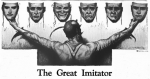
Psychosomatic Disease: We Have It All Wrong

5015 views

Psychosomatic disease (psyche: the mind, soma: the body). Illness is defined as “of, or relating to, the interaction of mind and body”. It is generally believed to be caused or aggravated by a mental factor such as internal conflict or stress. Synonyms are “psychological, irrational, stress-related, stress-induced, subjective, subconscious, unconscious”. There are two words in the synonyms that I disagree with because they give rise to false impressions. The first one is “psychological”, because this is often interpreted as “being invented by the patient”. The second word is “irrational”, because “it is thought by most physicians that there is no reason for the development of symptoms”. There is, however, one word that I do agree with and that is “subconscious”.

Understanding Stress and Stressors

The word “stress” is commonly used improperly and must be defined in order to understand what we mean. In the dictionary it is referred to as “a force demanding energy”. That means that everything, including human beings, is under stress from the surrounding environment. The ultimate long-term effect is rust, (metal oxide) and decay for animals like us. Essential to life, oxygen kills us in the end by causing the equivalent of rust.

The stress imposed on human beings is from changes in the environment to which we must adapt continuously throughout life. Why in fact do we sweat when it’s hot and shiver when it’s cold? Why do we get angry when we are insulted? The first two examples are responses to physical changes in the environment to which we have to adapt. Sweating produces fluid on the skin and its evaporation leads to cooling. Shivering is caused by muscular action and results in heat formation to compensate for its loss in a cold environment. We become angry from an insult because it produces an electrochemical reaction programmed in the brain to react that way. The insulting words signal the hearing mechanism that automatically sends a signal to the brain. Note that we do not usually smile and say thank you when we are insulted, although we can. It is therefore necessary to look at the construction of the human brain to try to clarify these issues.

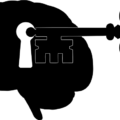
Basic Construction of the Brain

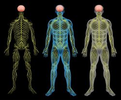
All the higher members of the animal kingdom have exactly the same basic brain construction that has evolved from more primitive to less primitive by adding layers of greater sophistication. It can be thought of as existing in two basic sections, the lower primitive brain and the upper cognitive or thinking brain. In humans, Freud called the lower brain the id and the upper brain the ego. The lower brain does not think. It simply reacts automatically to incoming stimuli. This part of the brain is sometimes referred to as “reptilian”, because that is the kind of brain possessed by reptiles. The executive actions can be modified, or they can be suppressed, by the ego. The id is a bit like a computer, while the ego may be compared with the person operating the computer. It was this idea that gave rise to the philosophy expounded by Descartes. Now we have to consider how these two brains cooperate with each other to produce what might be called normal, healthy human function.

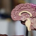
The Brain is a Machine

To suggest that the brain is a machine is very offensive to many people. Conventionally, they say that “there must be something more than whirling atoms and electrons to humans who have a soul as well as a body”. Unfortunately, whether we like it or not, the physical attributes of survival must dominate our thinking, in spite of our colossal ignorance. After all, we all know that we cannot survive without water and food; we die from disease and injury. This presentation therefore deals with the “now”, not the hereafter. When an infant is born, the only operative brain is the lower one, the id. This enables the infant to suck milk from the maternal breast, enables him to cry and control the urinary and bowel system, all automatic functions of survival. An automatic nervous system known as autonomic is controlled by this part of the brain, enabling signals to pass to the body and back from the body to the brain. As he grows, the upper brain gradually becomes “hardwired” as he proceeds from infancy to childhood, to adolescence and adult status. As the two parts of the brain become connected, what we call “free will” emerges. That is why the ego can be used voluntarily to suppress the reflex actions of the id. It is why we can voluntarily suppress bowel and bladder function in youth and fail to control it well in age. Oxidative decay affects brain cells in preference to most body cells, so mental changes usually occur first, as we age.

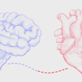
Chemistry and Electricity

All of this requires energy and it is derived entirely from the consumption of food. It is how this food is converted into energy that should dominate our attention in regards to the maintenance of health. The health of all body and brain cells is maintained by the efficient consumption of fuel in a process known as oxidation. The id that never sleeps is particularly vulnerable to loss of this efficiency and its actions become distorted in what might be an unexpected manner. Because the loss of oxidative efficiency represents a dangerous state, it  activates the well-known fight-or-flight reflex, producing a state of anxiety or fear, palpitations of the heart, unusual sweating and acceleration of the rate of breathing. Remember, this is a primitive reflex that is hardwired into the brain, preparing us to fight an enemy or flee from danger. The body is being prepared for action. Because the ego does not perceive any environmental threat, or perhaps the nature of the threat, this reflex is activated for no observable reason. It is obviously disturbing and causes the sufferer to seek medical advice.

The patient describes the symptoms to the physician, who carries out a physical examination and does a series of laboratory tests, all of which are perfectly normal. The patient is told that these are “panic attacks” and he may be given a “tranquilizer” to calm his nerves. The possibility of oxidative dysfunction is rarely if ever considered. This is a typical example of psychosomatic disease, but the distortions in the mechanism may give rise to an assortment of unpredictable physical or mental symptoms other than panic attacks. It is precisely because of the multiple disconnected symptoms, without laboratory evidence of pathology, that suggests the term psychosomatic. Even more importantly, the emphasis may be placed on the word “fight”, so let us see how this might have practical application. Readers on this website may be aware of a number of posts describing the loss of oxidative efficiency in brain, caused by an excess of dietary sugar inducing vitamin B1 deficiency, and the variable symptoms associated with it. The example below illustrates how the brain, and subsequently mood and behavior, are affected by the loss of oxidative efficiency.

A Path to Violence

A fictitious 12 year old boy indulges in the common American habit of consuming empty calories in the form of sweets, chocolate, candy, assorted carbonated, and even caffeinated, beverages and perhaps alcohol. He is hyperactive, inattentive in class, often rude but is otherwise considered to be in normal health. One day he is punished by his teacher in a perfectly appropriate manner. Because he does not have any conscious awareness of the distortions in his id, he believes the punishment to be unjust. He sees himself being put upon by the establishment and nurses his grievance in a state of chronic anger. Perhaps it grows and begins to dominate his hatred against the world, finally exploding in a form of violence that satisfies his hatred, his only motive. Note that he does not consciously understand why he hates the world, but to destroy some of it is indeed the motive.

If oxidative inefficiency activates the fight-or-flight reflex, it may be beyond the power of the ego to suppress it. Under these circumstances this boy may be perceived as being “more primitive” in his actions. The constant reiteration by the news media that “a motive for violent action is being sought” in conventional (voluntary) terms may be a useless exercise. To test this hypothesis, it would be so simple to make inquiries concerning the nutritional habits of the perpetrator.  If and when such an individual survives the inevitable shoot out, it might be possible to assess whether his sick mind is related to such an easily definable cause. If this was shown to be true, it would be an example of an extremely dangerous form of psychosomatic disease.

Conclusion

In the minds of many physicians, psychosomatic disease is almost an accusation of malingering. It is sometimes referred to as “functional”, demanding little thought on behalf of the patient as to its underlying cause. They see it as a response to stress and imagine that if the patient can only “pull himself together”, the symptoms will disappear. This gave rise to the use of drugs called “tranquilizers”, prescribed almost exclusively for this extraordinarily common condition. The irony is that it is indeed related to “stress”, but it is the abnormal chemistry of the brain that produces the symptoms and the solution is simple. There is no doubt that physicians should be taking a good hard look at the dietary indiscretion that is causing “functional” illness in so many people in the United States. What may be even worse is that if these symptoms are not recognized for what they represent and dietary indiscretion persists, permanent damage may follow in later years to become “chronic brain disease”, with variable pseudonyms.

We Need Your Help

More people than ever are reading Hormones Matter, a testament to the need for independent voices in health and medicine. We are not funded and accept limited advertising. Unlike many health sites, we don’t force you to purchase a subscription. We believe health information should be open to all. If you read Hormones Matter, like it, please help support it. Contribute now.

Yes, I would like to support Hormones Matter. 

Photo by Jose A.Thompson on Unsplash

This article was published originally on September 26, 2016. 

Beriberi: The Great Imitator

33212 views

Because of some unusual clinical experiences as a pediatrician, I have published a number of articles in the medical press on thiamine, also known as vitamin B1. Deficiency of this vitamin is the primary cause of the disease called beriberi. It took many years before the simple explanation for this incredibly complex disease became known. A group of scientists from Japan called the “Vitamin B research committee of Japan” wrote and published the Review of Japanese Literature on Beriberi and Thiamine, in 1965. It was translated into English subsequently to pass the information about beriberi to people in the West who were considered to be ignorant of this disease. A book published in 1965 on a medical subject that few recall may be regarded in the modern world as being out of date and of historical interest only, however, it has been said that “Those who do not learn history are doomed to repeat it”. And repeat it, we are.

Beriberi is one of the nutritional diseases that is regarded as being conquered. It is rarely considered as a cause of disease in any well-developed country, including America. In what follows, are extractions from this book that are pertinent to many of today’s chronic health issues. It appears that thiamine deficiency is making a comeback but it is rarely considered as a possibility.

The History of Beriberi and Thiamine Deficiency

Beriberi has existed in Japan from antiquity and records can be found in documents as early as 808. Between 1603 and 1867, city inhabitants began to eat white rice (polished by a mill). The act of taking the rice to a mill reflected an improved affluence since white rice looked better on the table and people were demonstrating that they could afford the mill. Now we know that thiamine and the other B vitamins are found in the cusp around the rice grain. The grain consists of starch that is metabolized as glucose and the vitamins essential to the process are in the cusp. The number of cases of beriberi in Japan reached its peak in the 1920s, after which the declining incidence was remarkable. This is when the true cause of the disease was found. Epidemics of the disease broke out in the summer months, an important point to be noted later in this article.

Early Thiamine Research

Before I go on, I want to mention an extremely important experiment that was carried out in 1936. Sir Rudolf Peters showed that there was no difference in the metabolic responses of thiamine deficient pigeon brain cells, compared with cells that were thiamine sufficient, until glucose (sugar) was added. Peters called the failure of the thiamine deficient cells to respond to the input of glucose the catatorulin effect. The reason I mention this historical experiment is because we now know that the clinical effects of thiamine deficiency can be precipitated by ingesting sugar, although these effects are insidious, usually relatively minor in character and can remain on and off for months. The symptoms, as recorded in experimental thiamine deficiency in human subjects, are often diagnosed as psychosomatic. Treated purely symptomatically and the underlying dietary cause neglected, the clinical course gives rise to much more serious symptoms that are then diagnosed as various types of chronic brain disease.

  • Thiamine Deficiency Related Mortality. The mortality in beriberi is extremely low. In Japan the total number of deaths decreased from 26,797 in 1923 to only 447 in 1959 after the discovery of its true cause.
  • Thiamine Deficiency Related Morbidity. This is another story. It describes the number of people living and suffering with the disease. In spite of the newly acquired knowledge concerning its cause, during August and September 1951, of 375 patients attending a clinic in Tokyo, 29% had at least two of the major beriberi signs. The importance of the summer months will be mentioned later.

Are the Clinical Effects Relevant Today?

The book records a thiamine deficiency experiment in four healthy male adults. Note that this was an experiment, not a natural occurrence of beriberi. The two are different in detail. Deficiency of the other B vitamins is involved in beriberi but thiamine deficiency dominates the picture. In the second week of the experiment, the subjects described general malaise, and a “heavy feeling” in the legs. In the third week of the experiment they complained of palpitations of the heart. Examination revealed either a slow or fast heart rate, a high systolic and low diastolic blood pressure, and an increase in some of the white blood cells. In the fourth week there was a decrease in appetite, nausea, vomiting and weight loss. Symptoms were rapidly abolished with restoration of thiamine. These are common symptoms that confront the modern physician. It is most probable that they would be diagnosed as a simple infection such as a virus and of course, they could be.

Subjective Symptoms of Naturally Occurring Beriberi

The early symptoms include general malaise, loss of strength in knee joints, “pins and needles” in arms and legs, palpitation of the heart, a sense of tightness in the chest and a “full” feeling in the upper abdomen. These are complaints heard by doctors today and are often referred to as psychosomatic, particularly when the laboratory tests are normal. Nausea and vomiting are invariably ascribed to other causes.

General Objective Symptoms of Beriberi

The mental state is not affected in the early stages of beriberi. The patient may look relatively well. The disease in Japan was more likely in a robust manual laborer. Some edema or swelling of the tissues is present also in the early stages but may be only slight and found only on the shin. Tenderness in the calf muscles may be elicited by gripping the calf muscle, but such a test is probably unlikely in a modern clinic.

In later stages, fluid is found in the pleural cavity, surrounding the heart in the pericardium and in the abdomen. Fluid in body cavities is usually ascribed to other “more modern” causes and beriberi is not likely to be considered. There may be low grade fever, usually giving rise to a search for an infection. We are all aware that such symptoms come from other causes, but a diet history might suggest that beriberi is a possibility in the differential diagnosis.

Beriberi and the Cardiovascular System

In the early stages of beriberi the patient will have palpitations of the heart on physical or mental exertion. In later stages, palpitations and breathlessness will occur even at rest. X-ray examination shows the heart to be enlarged and changes in the electrocardiogram are those seen with other heart diseases. Findings like this in the modern world would almost certainly be diagnosed as “viral myocardiopathy”.

Beriberi and the Nervous System

Polyneuritis and paralysis of nerves to the arms and legs occur in the early stages of beriberi and there are major changes in sensation including touch, pain and temperature perception. Loss of sensation in the index finger and thumb dominates the sensory loss and may easily be mistaken for carpal tunnel syndrome. “Pins and needles”, numbness or a burning sensation in the legs and toes may be experienced.

In the modern world, this would be studied by a test known as electromyography and probably attributed to other causes. A 39 year old woman is described in the book. She had lassitude (severe fatigue) and had difficulty in walking because of dizziness and shaking, common symptoms seen today by neurologists.

Beriberi and the Autonomic Nervous System

We have two nervous systems. One is called voluntary and is directed by the thinking brain that enables willpower. The autonomic system is controlled by the non-thinking lower part of the brain and is automatic. This part of the brain is peculiarly sensitive to thiamine deficiency, so dysautonomia (dys meaning abnormal and autonomia referring to the autonomic system) is the major presentation of beriberi in its early stages, interfering with our ability for continuous adaptation to the environment. Since it is automatic, body functions are normally carried out without our having to think about them.

There are two branches to the system: one is called sympathetic and the other one is called parasympathetic. The sympathetic branch is triggered by any form of physical or mental stress and prepares us for action to manage response to the stress. Sensing danger, this system activates the fight-or-flight reflex. The parasympathetic branch organizes the functions of the body at rest. As one branch is activated, the other is withdrawn, representing the Yin and Yang (extreme opposites) of adaptation.

Beriberi is characterized in its early stages by dysautonomia, appearing as postural orthostatic tachycardia syndrome (POTS). This well documented modern disease cannot be distinguished from beriberi except by appropriate laboratory testing for thiamine deficiency. Blood thiamine levels are usually normal in the mild to moderate deficiency state.

Examples of Dysfunction in Beriberi

The calf muscle often cramps with physical exercise. There is loss of the deep tendon reflexes in the legs. There is diminished visual acuity. Part of the eye is known as the papilla and pallor occurs in its lateral half. If this is detected by an eye doctor and the patient has neurological symptoms, a diagnosis of multiple sclerosis would certainly be entertained.

Optic neuritis is common in beriberi. Loss of sensation is greater on the front of the body, follows no specific nerve distribution and is indistinct, suggestive of “neurosis” in the modern world.

Foot and wrist drop, loss of sensation to vibration (commonly tested with a tuning fork) and stumbling on walking are all examples of symptoms that would be most likely ascribed to other causes.

Breathlessness with or without exertion would probably be ascribed to congestive heart failure of unknown cause or perhaps associated with high blood pressure, even though they might have a common cause that goes unrecognized.

The symptoms of this disease can be precipitated for the first time when some form of stress is applied to the body. This can be a simple infection such as a cold, a mild head injury, exposure to sunlight or even an inoculation, important points to consider when unexpected complications arise after a mild incident of this nature. Note the reference to sunlight and the outbreaks of beriberi in the summer months. We now know that ultraviolet light is stressful to the human body. Exposure to sunlight, even though it provides us with vitamin D as part of its beneficence, is for the fit individual. Tanning of the skin is a natural defense mechanism that exhibits the state of health.

Is Thiamine Deficiency Common in America?

My direct answer to this question is that it is indeed extremely common. There is good reason for it because sugar ingestion is so extreme and ubiquitous within the population as a whole. It is the reason that I mentioned the experiment of Rudolph Peters. Ingestion of sugar is causing widespread beriberi, masking as psychosomatic disease and dysautonomia. The symptoms and physical findings vary according to the stage of the disease. For example, a low or a high acid in the stomach can occur at different times as the effects of the disease advance. Both are associated with gastroesophageal reflux and heartburn, suggesting that the acid content is only part of the picture.
A low blood sugar can cause the symptoms of hypoglycemia, a relatively common condition. A high blood sugar can be mistaken for diabetes, both seen in varying stages of the disease.

It is extremely easy to detect thiamine deficiency by doing a test on red blood cells. Unfortunately this test is either incomplete or not performed at all by any laboratory known to me.

The lower part of the human brain that controls the autonomic nervous system is exquisitely sensitive to thiamine deficiency. It produces the same effect as a mild deprivation of oxygen. Because this is dangerous and life-threatening, the control mechanisms become much more reactive, often firing the fight-or-flight reflex that in the modern world is diagnosed as panic attacks. Oxidative stress (a deficiency or an excess of oxygen affecting cells, particularly those of the lower brain) is occurring in children and adults. It is responsible for many common conditions, including jaundice in the newborn, sudden infancy death, recurrent ear infections, tonsillitis, sinusitis, asthma, attention deficit disorder (ADD), hyperactivity, and even autism. Each of these conditions has been reported in the medical literature as related to oxidative stress. So many different diseases occurring from the same common cause is offensive to the present medical model. This model regards each of these phenomena as a separate disease entity with a specific cause for each.

Without the correct balance of glucose, oxygen and thiamine, the mitochondria (the engines of the cell) that are responsible for producing the energy of cellular function, cannot realize their potential. Because the lower brain computes our adaptation, it can be said that people with this kind of dysautonomia are maladapted to the environment. For example they cannot adjust to outside temperature, shivering and going blue when it is hot and sweating when it is cold.

So, yes, beriberi and thiamine deficiency have re-emerged. And yes, we have forgotten history and appear doomed to repeat it. When supplemental thiamine and magnesium can be so therapeutic, it is high time that the situation should be addressed more clearly by the medical profession.

We Need Your Help

More people than ever are reading Hormones Matter, a testament to the need for independent voices in health and medicine. We are not funded and accept limited advertising. Unlike many health sites, we don’t force you to purchase a subscription. We believe health information should be open to all. If you read Hormones Matter, like it, please help support it. Contribute now.

Yes, I would like to support Hormones Matter.

This article was published originally on November 4, 2015. 

The Fluoroquinolone Time Bomb – Answers in the Mitochondria

16547 views

Two of the more perplexing features of Fluoroquinolone Toxicity (an adverse reaction to a fluoroquinolone antibiotic – Cipro/Ciprofloxacin, Levaquin/Levofloxacin, Avelox/Moxifloxacin or Floxin/Ofloxacin) are delayed reactions and tolerance thresholds. Both of these features of Fluoroquinolone Toxicity can be explained by noting that fluoroquinolones have been shown to damage mitochondria and cause oxidative stress, and that delayed onset of a disease state, as well as tolerance thresholds, are features of illnesses brought on by pharmaceutical induced mitochondrial damage and oxidative stress.

Delayed Reactions and Tolerance Thresholds with Fluoroquinolone Reactions

By “delayed reactions” I mean that adverse reactions to fluoroquinolones can occur weeks, months or even years after administration of the fluoroquinolone has stopped.  For the lawsuit filed by Public Citizen on behalf of patients who tore or ruptured tendons after taking a fluoroquinolone, (a suit that prompted the addition of the black box warning on all orally and IV administered fluoroquinolones) notes that “Fluoroquinolones, including CIPRO®, are associated with an increased risk of tendinitis and tendon rupture in all ages. This risk is further increased in older patients usually over 60 years of age, in patients taking corticosteroid drugs, and in patients with kidney, heart or lung transplants”. Tendon tears and ruptures that occurred within one year of the patient taking the fluoroquinolone were accepted as being related to the patient’s fluoroquinolone use. Patient reports have noted that new adverse symptoms of fluoroquinolone toxicity have occurred years after administration of the fluoroquinolone has ceased.

Many patients also experience a tolerance threshold for fluoroquinolone use.  A patient can tolerate fluoroquinolones well, experiencing few or no side-effects, until his or her threshold is reached.  After the patient’s tolerance threshold is reached, multisymptom systemic illness ensues. This patient’s story, found on the Fluoroquinolone Wall of Pain, illustrates the issue of tolerance thresholds:

On April 15, 2013 I was prescribed Avelox. I had been on this drug many times for chronic sinus infections. This time was different. Within 10 minutes of the first dose I went into anaphylaxis. I stopped breathing, had numerous convulsions and two grand Mal seizures. Since that day I have suffered with seizures, convulsions, tremors, debilitating fatigue, muscle weakness, vision loss, severe neuropathic pain, vomiting, nausea, lack of appetite, tendon, and vein problems.

This patient tolerated Avelox (moxifloxacin – a fluoroquinolone) well until her tolerance threshold was reached. Once her tolerance threshold was reached, she experienced multi-symptom systemic illness.

I personally experienced both a delayed reaction to Cipro/Ciprofloxacin (also a fluoroquinolone) and a tolerance threshold for it. I took 7 500-milligram pills of Cipro in 2009 without notable incident. I was even able to hike the entire 500-mile Colorado Trail in 2010 (no peripheral neuropathy or weakness were present at that time). When I took 7 more 500-milligram pills in 2011 I experienced a severe adverse reaction that began two full weeks after I was done taking the pills. I experienced multiple musculoskeletal (I couldn’t walk more than a block) and nervous system symptoms (I lost my memory and reading comprehension), and I would describe the reaction as feeling like a bomb had gone off in my body.

Fluoroquinolone Time Bomb: It’s All About the Mitochondria

My experience of a delayed onset of systemic health issues after having previously tolerated Cipro/Ciprofloxacin well, is typical of diseases that are brought on by a pharmaceutical causing mitochondrial dysfunction. (Multiple journal articles have noted that fluoroquinolones cause mitochondrial damage and oxidative stress.)

In “Mechanisms of Pathogenesis in Drug Hepatotoxicity Putting the Stress on Mitochondria” it is noted that:

…damage to mitochondria often reflects successive chemical insults, such that no immediate cause for functional changes or pathological alterations can be established. There is indeed experimental evidence that prolonged injury to mitochondria, such as that which typifies oxidative injury to mitochondrial DNA or to components of the electron transport chain (ETC), has to cross a certain threshold (or a number of thresholds) before cell damage or cell death becomes manifest.

Each time mitochondria is injured, the patient gets closer to his or her personal tolerance threshold for mitochondrial damage. Once the threshold is crossed, cell damage and apoptosis occur – which manifest themselves in various states of illness.

It is further explained in “Mechanisms of Pathogenesis” that:

…approximately 60% of mitochondrial DNA must be deleted from the mouse genome before complex IV activity is compromised and serum levels of lactate are elevated. This non-linear response can be explained upon consideration that the molecules that subserve mitochondrial function (e.g., mitochondrial DNA, mRNA, and ETC proteins) are present in excess of amounts required for normal cell function. This reserve (or buffering) capacity acts as a protective mechanism; however, at a certain stage of damage, the supply of biomolecules needed to support wild-type mitochondrial function becomes compromised.

The lay person’s summary of the above excerpts is that we have excess mitochondrial DNA and that excess mitochondrial DNA keeps each of us from developing a systemic multi-symptom illness whenever mitochondrial DNA is adversely affected (many pharmaceuticals and environmental toxins adversely affect mitochondrial DNA). However, when mitochondrial DNA is depleted sufficiently, cellular dysfunction, oxidative stress and cell death, ensue.

Multiple studies have noted that fluoroquinolones deplete mitochondrial DNA (here, here and here).  When enough mitochondrial DNA are depleted, adverse reactions that are systemic and include multiple symptoms simultaneously, occur.

Multi-Symptom Reaction: Look to Mitochondrial Damage

It is often difficult for the patient who is experiencing a systemic multi-symptom illness to connect his or her illness to the mitochondria damaging drug or toxin that hurt him or her because of the time delay between the cause (mitochondria damaging chemical) and the effect (bomb going off in body and mind). Though the delayed onset of fluoroquinolone toxicity and mitochondrial dysfunction symptoms are noted in many articles (here, here), the reason for the delayed onset of symptoms is not known.  In “Mechanisms of Pathogenesis” it is hypothesized that “an initial adaptive response was followed by a toxic response” when cells are exposed to a mitochondria damaging chemical. Perhaps the delay in adverse reaction onset is due to a toxic response taking time to develop.

Many pharmaceuticals damage mitochondria. Bactericidal antibiotics (including fluoroquinolones), Statins, acetaminophen, some chemotherapy drugs, vaccines, and many others, cause mitochondrial dysfunction, oxidative stress and cell death. Mitochondrial dysfunction and oxidative stress are connected to a variety of ailments, from chronic fatigue syndrome to Alzheimer’s disease and obesity. However, the FDA and other drug regulatory agencies have systematically ignored damage to mitochondria caused by pharmaceuticals and “mitochondrial toxicity testing is not required by the US FDA for drug approval.”

The recognition of delayed adverse reactions and tolerance thresholds for mitochondrial damaging drugs and vaccines will go far in helping both doctors and patients to recognize mitochondrial damage related adverse drug reactions (and adverse vaccine reactions). Once the reactions are recognized, perhaps some pressure can be put on the FDA and/or the pharmaceutical companies to test how drugs affect mitochondria before they are released onto the market. After all, mitochondrial damage and oxidative stress are causally related to almost every chronic illness.  It would be nice if doctors, those in the pharmaceutical industry, the FDA regulators, and others, recognized the harm that drugs do to mitochondria, and the symptoms of iatrogenic mitochondrial dysfunction.

Information about Fluoroquinolone Toxicity

Information about the author, and adverse reactions to fluoroquinolone antibiotics (Cipro/ciprofloxacin, Levaquin/levofloxacin, Avelox/moxifloxacin and Floxin/ofloxacin) can be found on Lisa Bloomquist’s site, www.floxiehope.com.

Share Your Story

If you have suffered from a fluoroquinolone or any other medication reaction, please consider sharing it on Hormones Matter.

We Need Your Help

More people than ever are reading Hormones Matter, a testament to the need for independent voices in health and medicine. We are not funded and accept limited advertising. Unlike many health sites, we don’t force you to purchase a subscription. We believe health information should be open to all. If you read Hormones Matter, like it, please help support it. Contribute now.

Yes, I would like to support Hormones Matter.

Image was created using Canva AI.

This story was published originally on Hormones Matter in March 2014.


What is Immunity?

3637 views

Arguably, my primary intellectual concerns around modern day infectious disease management and “prevention”, is an acute awareness of how little we know about our relationship to microbes in and around us, and about our immune system. We are just beginning to appreciate the role of the several trillion bacteria that inhabit our guts, dictate our immune responses, and synthesize nutrients. Add to this very steep learning curve, multiple layers of epigenetic expression and biochemical individuality and we have a recipe for disaster with a one-size-fits all vaccination schedule and rampant application of antibiotics.

Antibody-Response: Is that Immunity?

We have been led to believe that antibody-response to vaccine administration is in any way equivalent to protection from illness. This sadly rudimentary model of “immunization” is antiquated beyond acceptability, and in no way encompasses what we have learned about the relevance of the innate immune system, cytokines, and the role of nutrient sufficiency in vulnerability to infection.  Beyond the well-documented incidence of outbreaks of illness such as pertussis, mumps, measles, tetanus, polio, rotavirus, and chicken pox, in highly vaccinated populations, we have also learned that antibodies often play no role in the course of infectious diseases such as lethal vesicular stomatitis virus, discussed here. We also know that agammaglobulinemics (those born without limited capacity for immunoglobulin antibody production) contract and recover from measles in the usual fashion. So, it seems like we may have fundamentally misunderstood the role of antibodies in immunity.

This would be an excusable and understandable step in the evolution of biological sciences if we weren’t wielding the application of this misunderstanding in a lethal and morbid way. Room for primary vaccine failure based on fundamental misattribution of disease-protection to antibody production (which is always temporary) is one thing, but inducing chronic disease, atopy, neurodevelopmental delay, inflammation, autoimmunity, and death as a part of this effort, is quite another.

Auto-immunity and Evolving Theories of Immune Function

We are witnessing epidemic rates of autoimmunity in the American population and we are learning that vulnerability is more than genes + environment.  In fact, theories of immunity have evolved considerably since the 1950s when it consisted only of self vs non-self mechanisms.  The most all-encompassing theory is called the Danger theory, which posits that the immune system targets self-tissues when there is a “danger signal” or inflammation from the tissue itself.  Here is where the role of oxidative stress and inflammation play into immunity and autoimmunity in a significant way, and why the “terrain” is, in fact, everything and the germ is, in fact, nothing.

Evaluating the Safety and Efficacy of Vaccines and Medications

The fact that there is such an evolving conceptualization of immunity and one that only begins to account for the role of diet, environmental toxins, and gene expression variation should serve as a serious wake up call to those who believe that modern-day physicians and pharmaceutical companies are in any position to make recommendations, let alone mandates, about how we, as individuals, should manage our risks of infection. The truth is, once interventions such as vaccines and antibiotics have perturbed our natural mechanisms, there is very little that Western medicine can to do help. Chronic disease and autoimmunity are not the forte of the average doc, so gambling with that potential risk should certainly be done with thought and care.

To that end, there are so many tremendous resources out there, but the latest and greatest is Dissolving Illusions, which takes you on a meticulously documented tour of the role of hygiene and diet in the epidemiology of infectious disease and the misconceptions surrounding vaccinology and health.

For more practical tips, Saying No To Vaccines is an important guide for new parents to educate yourselves about each and every vaccine, because each and every one is a major medical intervention that should be scrutinized independently.

We need to remain humble about what we don’t know, measured in our assumptions about the safety and efficacy of our pharmaceutical interventions, and reliant on time-tested ways to support natural immunity through nutrient dense diet, minimized environmental chemical exposures, and stress reduction. We need to lose the fear we have been conditioned to bring to conversations about infectious disease.

After all, germs are all around and within us, we need them, and they need us.  We’ve spent quite a long time developing a sophisticated language with which to communicate, and we are only beginning to decode it.

About the author. Dr. Brogan is an M.I.T/Cornell/Bellevue-trained psychiatrist specialized in holistic women’s health. She is a mother of two and has a busy practice in Manhattan. A passion for understanding the intersection between health, nutrition, and the environment are the bedrock of her wellness approach with patients and at home. Visit her site at: Kelly Brogan, MD, Holistic Women’s Health Psychiatry.


Fluoroquinolone Antibiotics Damage Mitochondria – FDA Does Little

9447 views

Most people assume that antibiotics kill bacteria, but leave human (and other eukaryotic) cells unscathed. Unfortunately, this assumption isn’t correct. Bactericidal antibiotics have been found to damage the mitochondria of human cells.

Mitochondria are present in almost every cell in the body (except for red blood cells and sperm). They are responsible for cellular energy production, cell signaling, apoptosis (the process through which the body kills unhealthy cells), and aging. Mitochondria are vitally important organelles and malfunctioning mitochondria have been linked to many diseases including fibromyalgia, chronic fatigue syndrome / M.E., autism, Gulf War Syndrome, Alzheimer’s Disease, Parkinson’s, diabetes, cancer, and others.

Focusing on Fluoroquinolones

Though several kinds of bactericidal antibiotics have been shown to damage mitochondria, I’m going to focus this article on fluoroquinolone antibiotics because, a) there are multiple journal articles that note that fluoroquinolone antibiotics damage mitochondria, b) fluoroquinolone antibiotics have been shown to not just damage mitochondria, but to deplete mitochondrial DNA as well, and c) adverse reactions to fluoroquinolones involve multi-symptom, chronic illness that typifies mitochondrial injury.

Fluoroquinolone antibiotics – cipro/ciprofloxacin, levaquin/levofloxacin, avelox/moxifloxacin and floxin/ofloxacin – are damaging human mitochondria. This has been shown repeatedly, and even the FDA (never first to the party) acknowledges that the mechanism for damage done by fluoroquinolones is mitochondrial damage and the ensuing oxidative stress that occurs when mitochondria are damaged.

FDA Report Notes that Fluoroquinolones Damage Mitochondria

In their April 27, 2013 Pharmacovigilance Review, “Disabling Peripheral Neuropathy Associated with Systemic Fluoroquinolone Exposure,” the FDA notes that the mechanism for action through which fluoroquinolones induce peripheral neuropathy is mitochondrial toxicity. The report says:

Ciprofloxacin has been found to affect mammalian topoisomerase II, especially in mitochondria. In vitro studies in drug-treated mammalian cells found that nalidixic acid and ciprofloxacin cause a loss of motichondrial DNA (mtDNA), resulting in a decrease of mitochondrial respiration and an arrest in cell growth. Further analysis found protein-linked double-stranded DNA breaks in the mtDNA from ciprofloxacin-treated cells, suggesting that ciprofloxacin was targeting topoisomerase II activity in the mitochondria.

The FDA Pharmacovigilance Report also notes that mitochondrial damage (and the ensuing oxidative stress that occurs when mitochondria are damaged) is related to multi-symptom, chronic diseases like optic neuropathy, neuropathic pain, hearing loss, muscle weakness, cardiomyopathy, lactic acidosis, Parkinson’s, Alzheimer’s and amyotrophic lateral sclerosis (ALS).

Bactericidal Antibiotics Damage Mitochondria

A study entitled “Bactericidal Antibiotics Induce Mitochondrial Dysfunction and Oxidative Damage in Mammalian Cells” that was published in Science Translational Medicine in 2013 notes that:

Clinically relevant doses of bactericidal antibiotics – quinolones (fluoroquinolones), aminoglycosides, and Beta-lactams – cause mitochondrial dysfunction and ROS overproduction in mammalian cells. We demonstrated that these bactericidal antibiotic-induced effects lead to oxidative damage to DNA, proteins and membrane lipids. Mice treated with bactericidal antibiotics exhibited elevated oxidative stress markers in the blood, oxidative tissue damage, and up-regulated expression of key genes involved in antioxidant defense mechanisms, which points to the potential physiological relevance of these antibiotic effects.

Fluoroquinolones Deplete Antioxidants

Fluoroquinolone antibiotics damage the cellular mechanisms that are needed to repair mitochondrial damage – namely intracellular antioxidant production. The FDA report notes that, “Under normal circumstances, there is a mechanism to remove or prevent the generation of ROS to avoid cellular damage such as lipid peroxidation, mtDNA mutations, and DNA strand breaks; if this does not happen, it can then lead to even more oxidative damage.”

A 2011 study of Indian patients who took various fluoroquinolones to treat urinary tract infections found that, “There was substantial depletion in both SOD and glutathione levels particularly with ciprofloxacin.” SODs are superoxide dismutases – enzymes that are necessary for converting superoxide – a powerful oxidant – into oxygen and hydrogen peroxide. Without adequate SOD, superoxide wreaks havoc on every cell in the body. Glutathione is often called the “master antioxidant” and without proper levels of it, oxidants damage cells. (Additionally, “Glutathione (GSH) has a crucial role in cellular signaling and antioxidant defenses either by reacting directly with reactive oxygen or nitrogen species or by acting as an essential cofactor for GSH S-transferases and glutathione peroxidases. GSH acting in concert with its dependent enzymes, known as the glutathione system, is responsible for the detoxification of reactive oxygen and nitrogen species (ROS/RNS) and electrophiles produced by xenobiotics. Adequate levels of GSH are essential for the optimal functioning of the immune system in general and T cell activation and differentiation in particular.” source)

As the FDA noted, when damaged mitochondria produce too much ROS, and the mechanisms to limit the damage (cellular production of antioxidants) aren’t working properly, an increasing amount of oxidative damage is done. This is often referred to as the “vicious cycle” of mitochondrial damage – damaged mitochondria produce excess ROS (reactive oxygen species – often called oxidative stress), those ROS further damage the mitochondria, the further damaged mitochondria produce more ROS, and so on, and so on. As noted above, mitochondrial damage, oxidative stress, and the vicious cycle between them has been linked to every chronic disease that plagues people – diabetes, Alzheimer’s, autism, etc.

Fluoroquinolone antibiotics damage mitochondria and cause an increase in ROS/oxidative stress. They also deplete cells of antioxidants such as SOD and glutathione, leaving the cells unable to neutralize ROS / oxidative stress. Excess cellular ROS / oxidative stress is related to every chronic disease there is.

See Evil, Hear Evil, but do Nothing About It

Rather than utilizing the FDA’s vast databases and the hundreds of Ph.D. scientists and statisticians on their payroll to determine whether or not fluoroquinolones – which have been shown to cause mitochondrial damage and oxidative stress, are related to the many chronic diseases that plague Americans – diseases that have been shown to be caused by mitochondrial damage and oxidative stress; the FDA simply added one more thing to the 43 PAGE warning label for fluoroquinolones – PERMANENT PERIPHERAL NEUROPATHY. They could have, and probably should have, added warnings that noted that fluoroquinolone use increases the risk of every disease that is linked to mitochondrial damage and oxidative stress – Alzheimer’s, Parkinson’s, ALS, diabetes, autism, depression, etc.

But they didn’t. They just added another warning that few physicians will read let alone understand and left these drugs on the market.

Fluoroquinolone antibiotics – cipro/ciprofloxacin, levaquin/levofloxacin, avelox/moxifloxacin and floxin/ofloxacin – are damaging human mitochondria and inducing large amounts of oxidative stress that wreaks havoc on many areas of health. The FDA knows about this, and they are doing nothing about it. Actually, worse than doing nothing, they are increasing the length of the warning label, so that when victims go to sue the companies (Bayer makes Cipro and Avelox and Johnson & Johnson makes Levaquin) that damaged their cells and gave them a multi-symptom, chronic disease, they are told that their symptoms are on the warning label and thus they gave informed consent to be crippled by an antibiotic.

It is absurd. Fluoroquinolones do severe damage to human cells, and the FDA is looking the other way. They’re not protecting our mitochondria, and they’re not protecting us.

Sources:

  1. Department of Health and Human Services Public Health Service Food and Drug Administration Center for Drug Evaluation and Research Office of Surveillance and Epidemiology, Pharmacovigilance Review, April 17, 2013, “Disabling Peripheral Neuropathy Associated with Systemic Fluoroquinolone Exposure
  2. Science Translational Medicine, “Bactericidal Antibiotics Induce Mitochondrial Dysfunction and Oxidative Damage in Mammalian Cells”
  3. Journal of Young Pharmacists, “Oxidative Stress Induced by Fluoroquinolones on Treatment for Complicated Urinary Tract Infections in Indian Patients
  4. Molecular Pharmacology, “Delayed Cytotocicity and Cleavage of Mitochondrial DNA in Ciprofloxacin Treated Mammalian Cells
  5. Molecular Neurobiology, “The Glutathione System: A New Drug Target in Neuroimmune Disorders

Information about Fluoroquinolone Toxicity

Information about the author, and adverse reactions to fluoroquinolone antibiotics (Cipro/ciprofloxacin, Levaquin/levofloxacin, Avelox/moxifloxacin and Floxin/ofloxacin) can be found on Lisa Bloomquist’s site, www.floxiehope.com.

Write for Us

Patient stories are important. Help spread awareness. Write for us.

Participate in Research

Hormones MatterTM is conducting research on the side effects and adverse events associated with the fluoroquinolone antibiotics, Cipro, Levaquin, Avelox and others: The Fluoroquinolone Antibiotics Side Effects Study. The study is anonymous, takes 20-30 minutes to complete and is open to anyone who has used a fluoroquinolone antibiotic. Please complete the study and help us understand the scope of fluoroquinolone reactions.

Hormones MatterTM conducts other crowdsourced surveys on medication reactions. To take one of our other surveys, click here.

To sign up for our newsletter and receive weekly updates on the latest research news, click here.

What Else Can I Do To Help?

Hormones MatterTM is completely unfunded at this juncture and we rely entirely on crowdsourcing and volunteers to conduct the research and produce quality health education materials for the public. If you’d like help us improve healthcare with better data, get involved. Become an advocate, spread the word about our site, our research and our mission. Suggest a study. Share a study. Join our team. Write for us. Partner with us. Help us grow. For more information contact us at: info@hormonesmatter.com.

To support Hormones Matter and our research projects – Crowdfund Us – Buy an Unsubscription.