progesterone

How Hormones Rise and Fall Throughout the Menstrual Cycle

1629 views

Fertility Awareness Method For Contraception

Back in 2012, I was really sick and while we were trying to figure out what was going on, my doctor recommended I discontinue hormonal birth control for a while. For about 6 months, I used conductivity monitoring to avoid pregnancy. Each morning, I’d record the conductivity of my salivary and vaginal secretions looking for a change to indicate I was approaching ovulation and another change to indicate ovulation had occurred.

Back then, it felt confusing to me and a little black box”ish”, so when I was cleared to go back on hormonal birth control, I went back on it and didn’t give another thought to Fertility Awareness Methods (FAMs), until I decided to ditch hormonal birth control again.

This time, I did a deep dive and discovered new methods alongside familiar methods of FAM, and I went head-over-heels into the science of it.

In the decade since I relied on FAMs last, at-home urinary monitors are now available, and being a data driven girl, this is the method I opted for. Qualitative devices such as the ClearBlue Fertility Monitor (CBFM) didn’t quite offer the numbers I craved, so I went with the Mira Fertility Monitor even though, to date, no FAM endorses the use of this monitor for contraception (though Marquette University is actively testing the Mira against the CBFM with its protocols).

This ability to monitor your hormones at home also revolutionizes maintaining healthy hormonal balance and body literacy. Indeed, body literacy and the natural rise and fall of hormones throughout a healthy cycle is the topic of this post.

Hormones of the Menstrual Cycle

In this article, we will discuss:

  • follicular phase and ovulation
    • follicular development, how follicles are recruited and begin maturing throughout a woman’s reproductive life span
    • how testosterone and estradiol are produced in the developing follicles
    • the role of the hypothalamus and pituitary glands in follicular development and ovulation
    • the role of progesterone in ovulation
  • luteal phase
    • key changes in hormone production during the luteal phase (second half of the cycle)
  • finally, the entire menstrual cycle will be summarized in a single graph showing the rise and fall of hormones throughout the cycle

Why does all of this matter? When you understand how the menstrual cycle works, it becomes much easier to determine hormonal imbalances and much easier to navigate fertility. Women are only fertile for around a maximum of 5 days during any given menstrual cycle and when you have a condition like PCOS (polycystic ovarian syndrome) or experience delayed ovulation (or anovulation) for any reason during a cycle, menstrual cycle literacy makes it possible to pinpoint your fertile days when trying to conceive and naturally improve your chances of conception in each cycle.

For women who are not trying to conceive, cycle awareness is profoundly beneficial to overall health because you are better able to determine which part of your cycle is unhealthy and better able to address the underlying imbalance simply by knowing how your cycle works. Maintaining a healthy cycle throughout your reproductive years is of utmost importance even when your intention is to avoid pregnancy because the reproductive hormones impact every system within your body and are critical for everything from maintaining a healthy weight to a healthy heart.

This particular article (while containing lots of information) is an overview of the topics bulleted above. You will find a more in-depth discussion of these topics in this post.

An Overview of Follicular Development

Non-cyclical follicular development: Early follicular development of pre-antral follicles (follicles that don’t respond to follicular stimulating hormone) happens in a way that is not well understood by modern science and this part of follicular development is not governed by the menstrual cycle but instead occurs throughout a woman’s reproductive years beginning at the onset of puberty and ending with menopause.

Cyclical follicular development: A follicle is a structure within the ovary and it contains an ovum (immature egg). Each ovary houses several hundred thousand follicles at birth and throughout a woman’s reproductive life, these follicles mature and are responsible for releasing the reproductive hormones, estradiol and progesterone, which control release of these hormones:

  • GnRH (gonadotropin releasing hormone) released by the hypothalamus in a pulsed pattern
  • FSH (follicular stimulating hormone) released by the pituitary gland
  • LH (luteinizing hormone) released by the pituitary gland

The brain’s role in follicular development and ovulation: The tempo at which GnRH releases from the hypothalamus controls the secretions of FSH and LH by the pituitary, and these two hormones influence ovarian hormone patterns and those ovarian hormones affect the tempo of GnRH pulses by the hypothalamus. This feedback loop is what the term, hypothalamic-pituitary-ovary (HPO) axis refers to. It is important to know about the brain’s involvement in follicular development and ovulation because when there is a problem with the menstrual cycle, practitioners generally look at where in this axis the misfire is occurring. Conditions like hypothalamic amenorrhea (HA) arise due to an issue with the release of GnRH from the hypothalamus and we will revisit this condition along with others caused by a dysregulation of hormonal release in the brain rather than the ovaries in future articles.

Selection of one follicle for ovulation: Once follicles have matured into antral follicles, further development is governed by FSH and the follicles need FSH to not only continue growing but also to prevent atresia (follicular death). More than one follicle matures during each menstrual cycle and because of the well-designed negative feedback between estradiol concentrations and FSH, the fastest growing follicle generally outcompetes all other follicles by releasing more estradiol, which then suppresses FSH production and starves out the remaining developing follicles. The dominant follicle survives this period of FSH famine because it has more FSH receptors. The additional FSH receptors make it better able to sequester the small amounts of FSH released at this time. It is also larger and has more energy reserves than smaller and slower growing follicles. This is why women typically release only one egg (mature ovum) at ovulation.

Testosterone and estradiol in follicular development: During follicular development, follicles produce both testosterone (and several other androgens [male hormones]) and estradiol (plus small amounts of estrone). The androgens are produced in the theca cell layers. The theca cell layers are not able to convert these androgens into estradiol or estrone because they lack the necessary enzymes. Instead, through diffusion, these androgens enter the granulosa cell layer of the follicle where the necessary enzymes are found (aromatase) to convert testosterone to estradiol and androstenedione to estrone. A separate enzyme converts the estrone into estradiol within the granulosa cells. In conditions like polycystic ovarian syndrome (PCOS), there is an imbalance in the androgen and estradiol ratio with higher levels of androgens suggesting a problem with conversion of these hormones in that condition. We will revisit this in future articles on PCOS.

Ovulation

Progesterone prompts ovulation. Historically, it was thought that the LH surge caused the follicle to release the mature ovum (egg) in a reversal of the negative feedback loop between estradiol and the pulse of GnRH which suppresses release of both FSH and LH from the pituitary. New research suggests that the adrenals release a small surge of progesterone that stimulates ovulation and prompts a rise in LH. This pathway explains why women who are under stress experience delayed ovulation.

Based on my own at-home hormone monitoring of urinary metabolites of estradiol and progesterone plus LH and FSH, I can confirm this pre-ovulatory temporal rise in progesterone. In fact, if this new theory proves correct, it may help explain the sudden shift in the electrolyte composition of vaginal secretions at ovulation.

Progesterone concentrations just prior to ovulation are much lower than concentrations mid-luteal phase, and so it is likely that the adrenal cortex, rather than the developing follicles, are producing the progesterone necessary to prompt the surge in luteinizing hormone (LH). It is also of note that high concentrations of progesterone (like those produced during the luteal phase and during pregnancy) inhibit ovulation. In in-vitro fertilization, when progesterone is given at doses to simulate the blood concentration seen during the luteal phase, this prompts the “vanishing follicle” phenomenon suggesting that a low progesterone concentration is vitally important to successful ovulation.

This theory may also explain why women under stress do not ovulate. It is common for women who develop a cold or illness during the peri-ovulatory phase to have either delayed ovulation or an anovulatory cycle. Other forms of stress (mental, over-exercise, disturbances to the circadian rhythm) are also known to delay ovulation. Considering that pregnenolone is the precursor to both cortisol and progesterone, this progesterone rise theory as the key event leading to ovulation evolutionarily fits the concept of conserving eggs or preventing reproduction when conditions aren’t favorable to pregnancy. Elevated demands for cortisol during times of high stress would deplete the body’s ability to create progesterone.

Role of LH: LH (luteinizing hormone) transforms the follicle into the corpus luteum. While the follicle primarily generated the hormones testosterone and estradiol throughout follicular development and leading up to ovulation, the corpus luteum releases progesterone and estradiol to maintain the uterine lining after ovulation.

Key Takeaways From the Luteal Phase and Menstruation

Progesterone released by the corpus luteum throughout the luteal phase is vitally important for pregnancy because it sustains the uterine lining providing nourishment to the developing embryo until the placenta fully forms around 12 weeks gestational age. It is especially important that concentrations of progesterone be maintained until implantation of the fertilized egg occurs. Luteal phase deficiencies, which we will talk about more in future posts, is one of the common causes of implantation failure.

In the absence of pregnancy, the corpus luteum atrophies between 10 and 16 days after ovulation. As the corpus luteum atrophies, levels of progesterone and estradiol both fall, resulting in atrophy of the uterine lining resulting in onset of menses.

An Overview of a Healthy Menstrual Cycle

In summary, a slowdown in the rate of release of GnRH from the hypothalamus prompts an increase in FSH secretion from the pituitary and this awakens further development in antral follicles within the ovaries. As these follicles mature, both testosterone and estradiol are made by the developing follicles increasing the amount of both these hormones within the body. Estradiol quickens the release rate of GnRH by the hypothalamus which reduces FSH secretions by the pituitary gland.

Historically, it was believed that once estradiol achieved a critical threshold, this negative feedback loop reverses, and FSH spikes along with an LH surge to cause ovulation. New research shows a transient rise in progesterone ahead of the LH surge. This rise in progesterone is about one-tenth the maximum rise in progesterone seen during the luteal phase of the cycle and is presumably produced by the adrenal cortex. If this theory (that a transient concentration-dependent rise in progesterone) prompts ovulation, then this better connects the dots between why stress and undereating cause anovulatory cycles.

Luteinizing hormone, which spikes around the time of ovulation, elicits key changes within the follicle allowing for rupture of the mature egg from the follicle and conversion of the follicle into the corpus luteum. The corpus luteum produces both progesterone and estradiol and in the absence of pregnancy naturally atrophies resulting in falling levels of progesterone and estradiol. As circulating blood concentrations of these two hormones, which are necessary for maintaining the uterine lining fall when the corpus luteum atrophies, the uterine lining itself also atrophies and sloughs off the walls of the uterus leading to the onset of menses between 10 and 18 days after ovulation in a healthy cycle.

hormones across menstrual cycle
Figure 1. Hormone concentration throughout the menstrual cycle.

In Summary

This very quick overview of the menstrual cycle (aka ovulation cycle) forms the basis of every single fertility awareness method (FAM) today. Whether the method involves monitoring changes in cervical mucus, cervical position, basal body temperature, electrolyte composition of salivary/vaginal secretions, and/or at-home urinary hormone monitoring, these methods are highly reliable for predicting ovulation and are so reliable that their efficacy for avoiding unplanned pregnancy vies that of hormonal birth control.

These methods are also invaluable for shining light on a woman’s reproductive health and elucidating where hormonal imbalance lies within her cycle when things are a bit off. FAMs also provide real time data for women who are tracking their cycles so that you are able to adjust diet and lifestyle to support hormonal balance.

I will refer back to this article often in future posts on FAMs and hormonal health.

We Need Your Help

More people than ever are reading Hormones Matter, a testament to the need for independent voices in health and medicine. We are not funded and accept limited advertising. Unlike many health sites, we don’t force you to purchase a subscription. We believe health information should be open to all. If you read Hormones Matter, and like it, please help support it. Contribute now.

Yes, I would like to support Hormones Matter.   

Photo by Towfiqu barbhuiya on Unsplash.

Hormones, Birth Control, and Insulin Resistance

1948 views

Little known fact. Your reproductive hormones influence how your body responds to insulin. The artificial hormones in hormonal birth control also play a huge role in how your body responds to insulin. And, your body’s response to insulin determines how well you are able to use glucose to supply your daily energy needs.

In this article, we will discuss the basics of how your body creates energy. In this first section, we will unpack:

  • How your body creates energy from glucose
  • Glucose vs. fatty acids as an energy source
  • How insulin resistance impacts the shift between glucose burning and fat burning
  • How glucose enters your cells to become fuel for energy
  • How insulin resistance interferes with the transfer of glucose into your cells

Then, we will tie in how your natural reproductive hormones, estradiol and progesterone, impact your body’s use of glucose as a fuel source and discuss how hormonal birth control disrupts this natural balance.

How the Body Creates Energy From Glucose

Many of your cell types are designed to run on glucose, a metabolic product of carbohydrates, as their main source of energy, and in fact, certain cells that don’t contain mitochondria (or contain very few mitochondria) like red blood cells and cells of certain parts of your eye (lens, retina, and cornea) rely either exclusively (as is the case for red blood cells) or primarily on glucose as an energy source.

The reason for this is that mitochondria are responsible for aerobic (oxygen required) energy creation processes within your body, and cells with no or very few mitochondria rely mostly on anaerobic (no oxygen required) energy creation by glycolysis in the cytoplasm of the cell. As we will discuss in more detail later, when your body uses fatty acids as a fuel source, this pathway is purely aerobic, so it is not possible for fatty acids to be used in anaerobic energy creation processes within your cells.

When you eat a meal containing sugar (sucrose) or carbohydrates, enzymatic processes begin breaking the sugar and carbs down into their basic structures within your digestive tract. The structure of both sugar and carbs contain glucose.

Glucose fuels the creation of ATP in a process known as glycolysis, which happens within the cell, and through oxidative phosphorylation (OXPHOS), which happens within the mitochondria (substructures within the cell). When ATP is broken down within your cells, it releases energy, which is harnessed to power your mitochondria and other important cellular functions. The by-products of that ATP creation (pyruvate and ATP) fuel additional energy production cascades within the cell.

How the Body Switches From Glucose to Fatty Acids for Energy

Even when particular cell types prefer carbs (glucose) as their energy source rather than fatty acids, most cell types are capable of using either of these macronutrients (and also, when necessary, amino acids) as a fuel in order to survive periods of fasting (including overnight fasting).

Insulin plays a key role in regulating whether your body uses glucose (glycolysis in the cell’s cytoplasm and OXPHOS in the mitochondria) or fatty acids (lipolysis in the cell’s cytoplasm and fatty acid oxidation in the mitochondria) as its preferred fuel source. This is because insulin impacts the ratio of two key enzymes (malonyl Coenzyme A and acetyl CoenzymeA) that determine which of these energy pathways is preferred (here and here). The ratio of these enzymes is dynamic, changing throughout the day in response to when and what you eat, and in response to this fluctuating ratio, your body preferentially uses carbs (glucose) or fatty acids as its fuel source.

In an insulin resistant state, your body does not easily shift between glycolysis/OXPHOS (glucose as fuel) and lipolysis/fatty acid oxidation (fatty acids as fuel) and instead remains in a state of using fatty acids as fuel. We will talk about why this is the case in the next section.

How Glucose Gets Inside Cells

The glucose released in your digestive tract from the food you eat is absorbed into your bloodstream, and when your blood glucose levels start to rise following a meal (or any drink containing carbs or sugar), it signals your pancreas to release insulin.

Insulin is the messenger that lets your cells (specifically, your skeletal muscle, fat, kidney, and liver cells) know there is glucose available in your bloodstream.  Insulin does this by binding to the cellular membrane, and this activates glucose transporters on the cellular membrane.

Once blood glucose levels start to drop, a healthy body clears insulin fairly quickly so that it can maintain adequate blood sugar levels. Insulin must be cleared so that blood sugar doesn’t drop too low.

What Is Insulin Resistance?

A number of factors influence how your cells respond to insulin. External influences (like stress, diet, and lack of sleep) along with internal factors (hormonal fluctuations) play a role in how the cells respond to insulin. And, different types of cells respond differently to insulin. Skeletal muscle cells are the most sensitive to insulin. Fat cells and liver cells are also sensitive to insulin, and so these cell types (skeletal muscle, fat, and liver) are the quickest to take up extra glucose from the bloodstream.

When your body becomes more insulin resistant, the cells are not as able to respond to insulin. My favorite analogy for this is to imagine that you are at a rock concert. You cannot easily hear the person next to you because the volume in the venue is so loud that your ears are overloaded by the background noise. In order to carry on a conversation, you must move to a quieter place. In this scenario, insulin is the background noise or the decibel level. When you are insulin resistant, your pancreas releases extra insulin to try to get your body’s cells to respond. This would be the same as somebody yelling at you in a concert hall so that you are able to hear them speak.

When you restore insulin sensitivity, it is like taking your body out of that loud concert hall and placing it somewhere quiet. Now, you are able to hear and carry on a conversation without any problems. When you restore insulin sensitivity, the cells are capable of responding to a much lower amount of insulin much more quickly and take the action of absorbing glucose from the bloodstream.

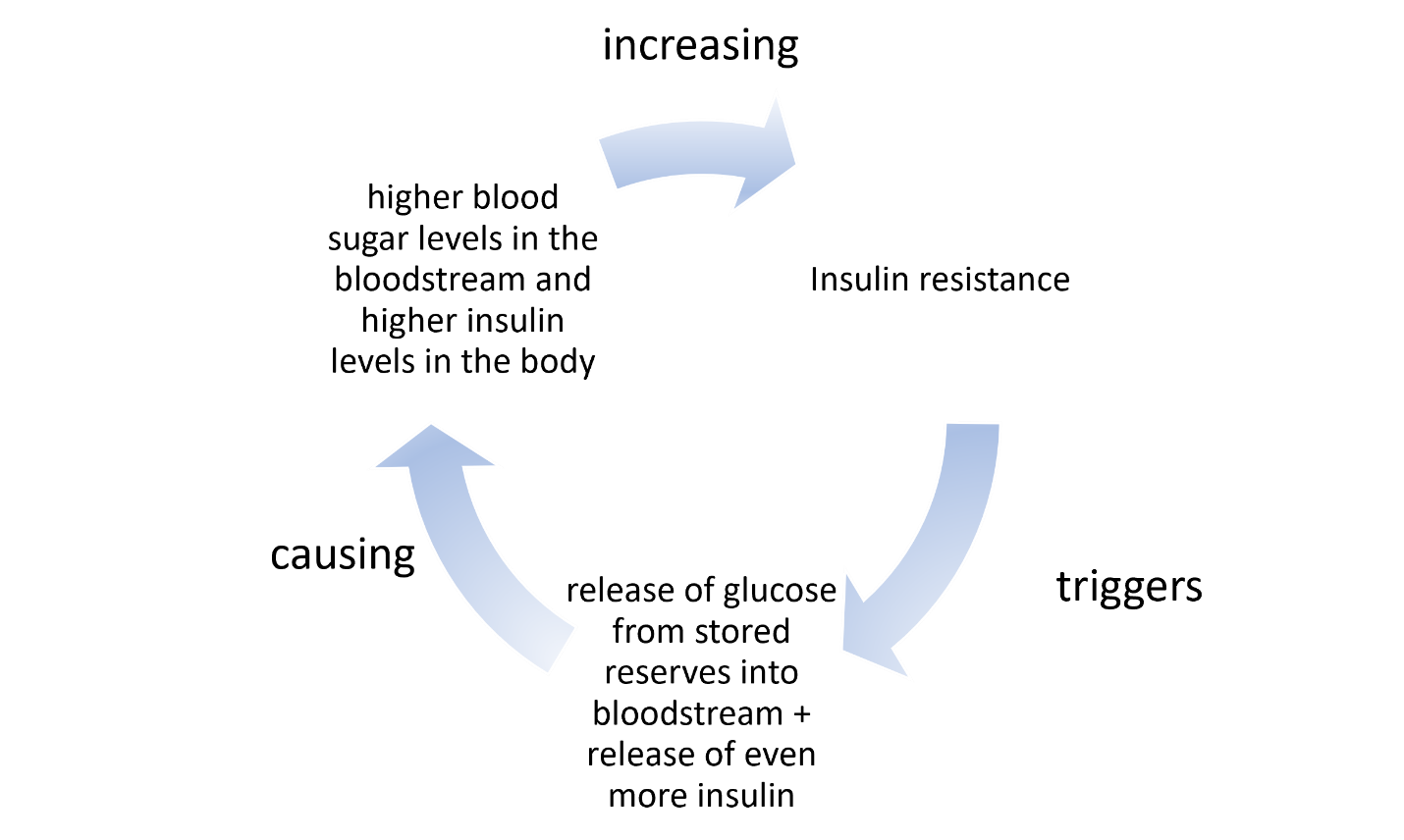
Insulin Resistance Begets Insulin Resistance

With insulin resistance, the cells are used to the high insulin environment (partially deaf to insulin), so they stop responding to insulin’s call. This prompts the pancreas to release more insulin in order to get your cells to hear the message to soak up the extra glucose circulating in the bloodstream. When insulin is unable to be heard because of the high background noise (because there is so much circulating insulin the cells are deaf to it), then glucose isn’t taken up by the cells. This then creates the false message from your cells to key organs to start releasing stored glucose (in a process called gluconeogenesis) to supply the body’s energy needs.

When we are talking about diabetes, this feedback loop often, but not in everyone with diabetes, results in a perfect storm of upward spiraling blood sugar levels.


insulin resistance cycle common in diabetes showing increased insulin resistance triggering gluconeogenesis resulting in higher blood sugar levels which increases insulin resistance
Figure 1. Insulin resistance begets more insulin resistance.

Even in conditions besides diabetes where blood sugar levels are dysregulated, you might have one condition (for example, insulin resistance), without the other (increased release of glucose from your body’s reserves).

With all of that in mind, let us take a look at how reproductive hormones impact insulin resistance and gluconeogenesis, the process of releasing glucose from stored reserves.

Estradiol, Synthetic Estrogens, and Insulin Resistance

Reproductive hormones play a key role in insulin resistance. Most scientific studies agree that estradiol (the endogenous estrogen produced primarily in the ovaries throughout the reproductive years) boosts the release of insulin from the pancreas. While at first glance, this looks like estradiol might contribute to insulin resistance because it prompts release of extra insulin, the opposite is actually true.

Estradiol is widely accepted as a potent compound to restore insulin sensitivity. Whether this is because of upregulation of insulin from the pancreas or whether it is also because of the influence estrogen has on the cells when it binds to estrogen receptors or a combination of both of these is not clear. What is clear, is that estradiol encourages cellular uptake of glucose and more rapid reduction of blood glucose levels after a meal. Estradiol also reduces gluconeogenesis in the liver suppressing the release of free glucose into the bloodstream from the body’s reserves, and this supports healthy blood sugar levels (here and here).

Estrogen Concentrations and Insulin Resistance

How estradiol affects insulin resistance is concentration dependent. Estradiol concentrations in the bloodstream within the normal circulating range (not more than 1 nanomolar abbreviated 1 nM) are associated with healthy insulin sensitivity and healthy blood sugar levels while concentrations higher than 1 nM are associated with insulin resistance. This may be why gestational diabetes is a common condition during pregnancy with up to 10% of pregnant women in America developing gestational diabetes. Progesterone also plays a key role in gestational diabetes as we will discuss in more detail below.

Non-bioidentical Estrogen and Insulin resistance

Ethinyl estradiol, the most common synthetic estrogen used in hormonal contraceptives here in America, also impacts insulin resistance, but like endogenous estradiol, the relationship is not straightforward. Ethinyl estradiol has been shown to impact insulin sensitivity and gluconeogenesis differently depending on:

  • its concentration in the hormonal birth control
  • what progestin (synthetic progesterone) it is paired with

Just as high concentrations of endogenous estradiol increase the chances of dysregulated blood glucose control, the synthetic estrogen, ethinyl estradiol, also increases chances of dysregulated blood glucose control. Chemical diabetes caused by hormonal birth control is also well documented in the literature. This is one of the reasons why, since the 1960s, the concentration of artificial estrogens in combined oral contraceptives has been dramatically reduced from upwards of 60 micrograms per pill to as low as 10 micrograms. Currently, most birth control options contain from 20 to 35 micrograms of ethinyl estradiol per pill.

Estrogen Binds to Insulin Receptors Affecting Insulin Resistance

Estrogens, whether synthetic or endogenous, affect blood sugar regulation differently at different concentrations because of their ability to bind to insulin receptors. This concentration-dependent effect of both endogenous estradiol and synthetic estrogens is often overlooked in the conversation regarding the impact of hormonal contraceptives on blood sugar control. Inasmuch as estrogens play a role in insulin sensitivity, insulin secretion, and in gluconeogenesis, and because estrogens are combined in hormonal contraceptives with a wide range of synthetic progestins, the effects on blood sugar regulation are quickly compounded and convoluted.

Progesterone, Progestins, and Insulin Resistance

As with estradiol, the concentration of progesterone also impacts whether progesterone improves or diminishes insulin sensitivity. It is generally accepted that higher concentrations of progesterone during pregnancy are a major contributor to gestational diabetes. Similarly, high concentrations of progesterone, even after menopause, are linked to an increased risk of developing type 2 diabetes.

The actions of progesterone on glucose metabolism is very much related to carrying a pregnancy to term, promoting glucose storage (rather than consumption of glucose for fuel) and promoting ketogenesis (fat burning) within the body. Even when not pregnant, progesterone is the dominant hormone during the luteal phase (second half of your cycle), and this effects how your body uses glucose and its sensitivity to insulin. This ties into common experiences during the second half of your cycle including carb cravings, potentially diminished appetite (if you are like me), and also weight gain.

Unlike artificial estrogens, of which there is only one used in the combined hormonal contraceptives available in the United States, for progestins, the synthetic forms of progesterone, there are four generations of progestins, with each generation containing progestins of different molecular structures. The class of molecules used in synthetic progestins are similar in structure to the endogenous progesterone molecule, but they are not the same. In other words, they are non-bioidentical.

Progestins bind differently to the progesterone receptors within the body (and also bind to a variety of other receptors), than the endogenous progesterone and their specific structure contributes to how much and whether insulin resistance increases. The molecular structure also affects how the body conserves glucose (increases glucose storage) or uses glucose (in the process of gluconeogenesis). It is generally believed that the androgenic nature of progestins determine their role in reducing insulin sensitivity (here and here).

Hormones and Body Composition

An interesting note, whether we are talking about natural reproductive hormones, estradiol and progesterone, or artificial hormones, ethinyl estradiol and the various progestins, these are all fat-soluble hormones. That means, these hormones may be stored in, and thus, impact the behavior of fat cells. One study evaluated the response of fat cells (adipocytes) in the presence or absence of treatment with artificial hormones and found that in the presence of artificial hormones, the adipocytes were more insulin resistant. This suggests that fat cells may serve as a reservoir for artificial hormones and endogenous hormones alike. They essentially soak up circulating hormones from the bloodstream, and these absorbed hormones in turn impact how the fat cells behave.

This finding means that body composition affects how you respond to hormones, whether endogenous or synthetic, and vice versa. It also suggests that, among other things, we ought to consider dosing hormonal contraceptives relative to body composition. Women with higher body fat may store more of the hormones than those with lower body fat and this may initiate or exacerbate insulin resistance.

Summary

In summary, reproductive hormones are intricately intertwined with metabolism, both with how the body creates energy and how it stores fats and carbs to meet energy demands between meals. Hormonal birth control impacts this finely choreographed dance between reproductive hormones and insulin sensitivity, and this seemingly small influence has a dramatic ripple effect. Insulin sensitivity dictates things like weight gain, oxidative stress, and even, as we will discuss in the next article, susceptibility to UTIs and UTI like symptoms.

Before you go, is there someone you know who needs to hear this message? Share this post with them.

We Need Your Help

More people than ever are reading Hormones Matter, a testament to the need for independent voices in health and medicine. We are not funded and accept limited advertising. Unlike many health sites, we don’t force you to purchase a subscription. We believe health information should be open to all. If you read Hormones Matter, like it, please help support it. Contribute now.

Yes, I would like to support Hormones Matter. 

Photo by isens usa on Unsplash.

Progesterone for Peripheral Neuropathy

76850 views

Some 20 years ago, during my very first neuro class taught by an accomplished neurologist from a prominent research university, I had a conversation about hormones and the brain. It was a brief conversation during which he admitted not only knowing nothing about how hormones affected the brain or nervous system functioning, but also, how he and others had no interest in considering the question. He believed hormones were too complicated to consider relevant. One didn’t ‘mess with hormones’ as he put it.

Lucky for us, some intrepid neurologists have moved the science of neuroendocrinology past the foibles of ‘don’t mess with hormones’ to hormones might be important therapeutic options. Nowhere is this more evident than in the areas of traumatic brain injury and diseases of demyelination. Here we see advances in hormones used as viable and important treatments where once there were none. Although the research is yet in its infancy and suffers from the typical one-size-fits-all approach, it marks a huge step forward in clinical neuroendocrinology.

What is Neuropathic Pain?

Neuropathic pain, the often chronic and difficult to treat pain that comes from nerve injury and demyelination affects approximately 3% of the population. The number of individuals suffering from neuropathy is likely much higher when one considers diseases such as endometriosis and the ill-understood, under-recognized neuropathy emerging post medication or vaccine adverse reactions. The experience of neuropathic pain in hands, feet, arms and legs is described as burning, freezing, electrical, tingling, prickling and more often than not, severe and unrelenting. As the nerve injury progresses and the pain continues, the rawness and intensity of the pain becomes indescribable to someone who has not experienced nerve pain firsthand.

Hormones and the Nervous System

Since the late eighties, researchers have known that steroid hormones, such as progesterone were not limited to reproductive functions and that many steroids were active in the nervous system. Not only were those steroids synthesized peripherally in ovaries, adrenals or adipose tissue able to cross the blood brain barrier, but all the core substrates for steroid synthesis were available in the brain too, meaning the brain could make its own steroids, de novo, from scratch. Researchers initially deemed steroids made or active in the brain as neurosteroids. Eventually, that nomenclature fell by the wayside as researchers realized there was tremendous crosstalk between peripheral and central hormones, no matter where the hormones were synthesized.

It should be noted, that hormones exert influence all over the body and brain via receptor binding. (A discussion on hormones and receptors can be found here: Promiscuous Hormones and Other Fun Facts.) In addition to steroid hormone receptors on (cell membrane) and in (nuclear) hormone-specific cells, like those in ovary, testes, adrenals, uterus, endometrium, hormone receptors are co-located on neurons, glial cells, oligodendrocytes and Schwann cells (myelin producing cells), immune cells, cardiomyocytes (heart), hepatocytes (liver), adipocytes – essentially every cell, organ or tissue in our body is modulated in some way by a hormone. Hormone influence is particularly important in the in the nervous system, where everything from neurotransmitter release and uptake to synaptic connections are modulated.

Traumatic Brain Injury, Peripheral Neuropathy and Hormones

When we talk about injuries to the nervous system, be it the brain and spinal cord, which is called the central nervous system (CNS), or all of the nerves that control movement and organ function in the body, the peripheral nervous system (PNS), there are two categories of injuries, those that develop acutely, post trauma, or those that develop chronically because of some metabolic dysfunction. In the case of the former, traumatic brain injuries (TBI) or  traumatic nerve injuries, the research points to progesterone for repair and regrowth. In the case of the later, where injuries develop as a result of internal and often chronic dysfunction, such as diabetic neuropathy, multiple sclerosis and other diseases affecting nerve fibers and myelin, less is known about progesterone and the thyroid hormone triiodothyronine (T3) is implicated, more strongly.

What is Myelin and How Does it Impact Neuropathy?

Myelin is the insulation that protects the axons of the neuron (in the brain) or nerve (in the body) to allow rapid conduction or messaging across the brain or through the body. Recall the axon is the part of the neuron/nerve that sends messages to other neurons/nerves, to other tissues, like muscle, or to organs like the heart and the liver. The dendrites receive messages and the nucleus processes messages. Myelin is like the plastic coating around the electrical wiring in your house. If the coating is too thick, conduction is blocked. If the coating is frayed or too thin, electrical sparks fly everywhere. Frayed myelin around axons is one of the mechanisms of neuropathic pain. Myelinated axons in the brain look white and therefore are called white matter. Whereas the grey matter, is where the nuclei of the brain reside. White matter in the brain consists of the oligodendrocytes – the type of cell that forms the myelin sheathing around axons. Myelin in the body, around the peripheral nerves, is made from cells called Schwann cells.

Progesterone, Myelination and the Nervous System

In the 1990s, Etienne-Emil Baulieu and colleagues recognized a role for progesterone (and other hormones) in central nervous system myelination. Over the next two decades, researchers uncovered the possible mechanisms and delineated more clearly for whom and in what types of injury progesterone seems most helpful. From studies of neurons (CNS) nerve cells (PNS), we now know that progesterone is key for myelination and neuron/nerve regrowth, at least in the acute stages. Progesterone stimulates myelination both directly by acting on oligodendrocytes and indirectly via actions on the neurons and the astrocytes that then message the oligodendrocytes to produce more myelin. Similarly in the PNS, progesterone aids in the remyelination and re-growth of nerve fibers, via the Schwann cells and via progesterone receptors located in what are called the dorsal root ganglia (DRG), the sensory neurons that carry information from the periphery to the brain. Whether in the CNS or the PNS, timing and length of progesterone administration are critical.

Animal Research – Progesterone, Nerve Injury and Neuropathy

The animal research has been mixed, but taken together, the results seem dependent upon the type of injury, the timing of the treatment and the methods of assessment. When treatment is begun early enough and extended long enough (this varies) and when the measure is neuropathic pain versus other potential outcomes (such as morphological changes to the nerve), there seems to be a favorable response. In rodents, single dose treatment does not seem to work, neither does treatment that is initiated too late after the injury or ended prematurely, though these criteria vary from study to study.

For example, using an induced model of diabetic neuropathy, researchers from Italy found that diabetes markedly reduced progesterone concentrations in male rodents within three months (females were not tested). This was the only study I could find that measured progesterone concentrations relative to treatment and outcomes. Chronic treatment (one month) with progesterone or one of its derivatives restored nerve function, increased key components of myelin production and reduced pain. Similarly, an induced model of trigeminal pain in male rodents found when progesterone was initiated early and at a high enough dosage, it tempered the experience of pain while increasing myelin producing proteins. Lower dosages did not work.

From Animals to Humans: Traumatic Brain Injury and Neuropathy

The research with animals, male rodents specifically, shows that progesterone treatment works best if given early enough, for long enough, and at high enough dosages. With acute or induced injuries under experimental conditions, early treatment is much easier than in real life where neuropathic pain develops much more gradually and often goes undiagnosed and untreated for some time. Would progesterone work in humans and would it work for chronic, well established neuropathy? The answers to those questions are not clear because the human research on progesterone and myelin focuses on acute injury, like the traumatic brain injuries. The human research also suffers from short duration dosing, includes mostly males, and without exception fails to address endogenous progesterone concentrations either pre or post treatment. Nevertheless, there are some indications that progesterone therapy may work.

Progesterone and TBI – Human Studies

In a smaller, single center open trial and two larger, double-blind, placebo-controlled, human trials, progesterone therapy was administered to individuals with severe traumatic brain injuries (Glasgow coma scale <8). In each case, the progesterone group did better, showed reduced morbidity rates than the placebo groups.

In the first study, 26 cases were treated with progesterone and 20 controls with placebo. At both 10 days and three months post injury and treatment, the progesterone treated group improved significantly more than the control group (abstract only).

In a second study, 159 patients, arriving to the treatment facility just eight hours post traumatic brain injury were randomized to receive either intramuscular injections of progesterone (82) or placebo at 1.0 mg/kg via intramuscular injection and then once per 12 hours for 5 consecutive days. Both intake neurological functioning and post treatment functioning were assessed and compared using a number of measures. Follow up assessment was conducted at 3 and 6 months post injury/treatment. The results were positive, albeit small. The progesterone treated group improved significantly across all measures showing consistently larger improvements compared to the placebo group. It should be noted that only 44 of the total subject population was female, 24 in the placebo group and 20 in the progesterone group. No analysis by sex was conducted and so it is not clear whether progesterone therapy works equally well in males and females.

In the third study, called ProTECT, a similar double-blind, placebo controlled, randomized methodology was used. Here, however, the randomization was 4:1 and favored progesterone treatment, whereas in the study cited above, the progesterone and placebo randomization was 1:1. Progesterone was given via IV for three days. The ProTECT study researchers found that patients in the progesterone had a lower 30-day mortality rate than controls (rate ratio 0.43; 95% confidence interval 0.18 to 0.99). While those who suffered more severe injuries had relatively poor outcomes at the follow up tests 30 days post injury, despite the treatment, and those who suffered only moderate traumatic brain injury and received progesterone were more likely to have a moderate to good outcome than those randomized to placebo (abstract only).

Two additional trials are on-going, hoping to test progesterone on thousands of patients: the ProTECT-III and SynAPSe studies.

Translating the TBI Research for Use with Neuropathy

What does improvement post TBI tell us about treating neuropathic pain from demyelination disorders? It is not clear, because even though researchers know that progesterone promotes myelination, the human research has focused narrowly on injuries where demyelination occurs but also where other factors are also involved in the outcome. We know from animal and cell culture research that progesterone attenuates the cascade of events that occur post TBI or post nerve injury via multiple mechanisms, inducing myelin regrowth is only one of those mechanisms. Progesterone reduces swelling of both vasogenic and cytotoxic sorts. It has anti-oxidant properties, upregulating enzymes that increase free radical elimination. Progesterone inhibits inflammation, stabilizes mitochondria, reduces neural excitoxicity and can limit apoptosis. Finally, progesterone promotes myelination. All factors that should point to consistent improvement in TBI and neuropathic pain syndromes, but the research is limited and mixed. Why?

The primary reason for mixed results is study design, almost all are short duration. Hormones are long acting molecules and the shorter duration may not be sufficient to generate the response, particularly when the injuries are severe or longstanding. Longer treatment regimes are likely in order.

Another reason for mixed results is the one-size-fits-all approach. None of the human studies and few of the animal studies, investigates why progesterone works in some subjects and not others. Almost all of the studies are predominantly male, rodent and human alike. None have investigated whether being female has anything to do with efficacy. None of the human studies measured circulating concentrations of progesterone, either pre-, during, or post-treatment and so there is no way to tell if those who responded had higher circulating concentrations or if improvement was contingent upon reaching a certain concentration.

Perhaps even more importantly, is the fact that progesterone, like any hormone, works within a vast and compensatory network of other hormones. The reductionist approach that utilizes a single hormone treatment protocol, while ignoring the potential cross-talk with other hormones and other variables is a consistent flaw these and other research protocols. Again, hormone measurement, progesterone and its metabolites, in addition to other key hormones, is imperative if one is to determine therapeutic efficacy.

I Have Peripheral Neuropathy, Should I Try Progesterone?

Progesterone therapy is generally safe, but as with everything there are risks. Women have been using it for generations in its bio-identical form to mitigate menstrual and menopausal symptoms. Since it is fat soluble, transdermal (skin) absorption is possible and progesterone creams have become popular. Some physicians prefer micronized progesterone, a pill form that reduces the molecule so it more easily passes through the liver without degradation. The pill form, and to a much lesser degree, transdermal progesterone, cause sedation and should be taken at night. Micronized progesterone has been shown to increase free thyroxine (T4) as well. For some women, and presumably men too, a gain of function mutation on the mineralocorticoid receptor can evoke very high blood pressure with any increase in progesterone concentrations (luteal phase of the menstrual cycle and during pregnancy especially). Although there are dosing references for progesterone relative to menstrual or menopausal therapy, the dosing is individualized and often includes the replacement of other hormones along with progesterone. Salivary hormone testing is used to monitor and hormone doses are adjusted regularly. Progesterone is also used predominantly for women. No such dosing considerations exist for men that I am aware of. Likewise, for peripheral neuropathy there are no references from which to design a treatment protocol and so it would be prudent to work with a functional medicine specialist, familiar with hormone management, to develop and monitor the course of treatment.

My Two Cents

I suspect, if progesterone therapy works for peripheral neuropathy, it will require a much longer term treatment period than is currently tested in the human trials. I suspect also, it will be difficult to ascertain whether it is the sole contributor to improvements in neuropathy symptoms, as neuropathy is a multi-factorial process that ought to be treated as such. Nevertheless, if you suffer from neuropathy and can find a physician to work with that is familiar with hormones and the research, progesterone therapy might provide a viable option, among other options like stabilizing thyroid hormones and supporting mitochondrial function.

Postscript

This article was first published February 19, 2014. Since then, a few more studies and review articles have been published and continue to support the role of progesterone in myelin regeneration, although the data are mixed. From a 2020 article.

Indeed, PROG and its metabolites modulate the expression of myelin proteins of the PNS, such as myelin basic protein (MBP), myelin proteolipid protein, glycoprotein zero (P0) and peripheral myelin protein (PMP22) as well as myelin formation [6,8,9,20,63,64]. In particular, the expression of P0 in the sciatic nerve of adult male rats, as well as that in Schwann cell culture, is increased by treatment with PROG, DHP or THP.

…Data here reported support the concept that neuroactive steroids, synthetic ligands acting on their receptors or inducing their synthesis, may improve PN symptoms, including neuropathic pain and consequently may represent an interesting possible therapeutic strategy. In addition, based on the sexual dimorphism of neuroactive steroids as well as of PN here discussed, a gender specific treatment based on these compounds may be also proposed.

We Need Your Help

More people than ever are reading Hormones Matter, a testament to the need for independent voices in health and medicine. We are not funded and accept limited advertising. Unlike many health sites, we don’t force you to purchase a subscription. We believe health information should be open to all. If you read Hormones Matter, like it, please help support it. Contribute now.

Yes, I would like to support Hormones Matter. 

This article was published originally on February 19, 2014. 

Mommy Brain: Pregnancy and Postpartum Memory Deficits

15944 views

Pregnancy and postpartum memory problems are common complaints amongst new moms. Are they real? Some research says yes, other research says no.

In graduate school I studied this issue and completed several studies on pregnancy and postpartum mood and memory changes. In one such study, I ran a full battery neuropsychological tests on a group (n = 28) of highly educated, healthy, medication free, first-time moms. We tested in late pregnancy and within 10 days following the delivery of the child. We also measured a range of hormones (progesterone, DHEAS, testosterone, estrone, estradiol and estriol) to determine what relationship the radical hormone changes of pregnancy and childbirth had on a woman’s cognitive ability. We knew from animal research that steroid hormones could affect learning in very significant ways. It wasn’t that difficult to suspect the same would be true of human women.

The study was never published, rejected from at least three, maybe four journals and has been sitting in a file ever since (along with a number of other studies). With the open access and open science movements growing, I decided it was time for this research to see the light of day. I will be self-publishing much of my research over the coming weeks and months. Here is the first study. Understanding Maternal Cognitive Changes: Associations between Hormones and Memory.

Is the Mommy Brain Real?

More importantly, are hormones to blame?  The answer is yes on both counts. We found that pregnant and postpartum women exhibited detectable cognitive deficits across multiple domains. The deficits were worse in late pregnancy and mostly improved postpartum. These memory problems were linked to both the excessively high hormones of late pregnancy, the low hormones following delivery, and the large changes in hormone concentration from pregnancy to postpartum.

What Types of Cognitive Deficits?

Pregnant and to a lesser degree, postpartum women had difficulty sustaining focus – this may be the mommy brain fog that many women complain of. We also found that during pregnancy especially, women were unable to manipulate and organize incoming information effectively. This presented as poor performance across a number of tests that assessed both short and long term memory.

In the case of verbal memory, these highly intelligent (estimated average IQ was 114 – 119) and educated (average years of education was 16 years) women tested in the low single digit to the 20th percentiles across multiple IQ-adjusted verbal recall measures. Even when estimates of IQ were not used to adjust scores, the participants performed poorly compared to normative standards. This was surprising given that many of these women had advanced degrees and were working in professional capacities.

The verbal tests involved remembering lists of words; words that could be grouped into meaningful categories that would improve memory significantly. Most of the study participants had difficulty grouping the words into categories. Instead, they would attempt to remember by rote sequence, which is always much more difficult. They also exhibited high numbers of intrusions – recalling words that were not in the original lists and repetitions – repeating words.

Similarly, and more strikingly visible, visual- spatial memory was marred by the inability to group bits of information and perhaps even to see the groupings in the first place. In this test, the study participants were given a complex figure to copy (shown below). They were not told that they would be asked to recall and redraw the picture later. When asked to redraw the figure, the inability to see the totality of the picture, to group bits of information was apparent.

Visual – spatial memory deficits as assessed by the Rey Complex Figure Test. Marrs et al. 2013, © 2013 Lucine Health Sciences, Inc. All rights reserved.

Does Memory Improve Postpartum?

Interestingly, while spatial memory improved significantly from pregnancy to postpartum, verbal memory did not. And this is probably what troubles women the most, the perceived deficits in verbal memory. Most of us think in words, when our ability to find words, retain words, organize information effectively is compromised, we notice.

Hormones and Memory

Both high levels of late pregnancy estrogens, (estrone, estradiol and estriol – we measured all three) and the low levels these estrogens postpartum were correlated with multiple measures of diminished memory, attention and processing. Additionally, the larger the change in the circulating levels of estrogens from late pregnancy to early postpartum was associated with poor memory postpartum. Indeed, women who had higher postpartum estradiol and estriol specifically, performed better on measures of verbal memory than those who did not.

Progesterone, long thought to be associated with cognitive function, primarily because of its sedative properties, was not associated with any measure of cognitive function at either test time, although large changes in progesterone were associated with some performance measures. DHEAS and testosterone, not often measured in pregnancy, postpartum or even in women’s health in general, were also associated with a few measures of cognitive functioning.

What This Means

Ladies, you are not imagining the pregnancy memory problems. They exist and they are related to the hormones. Most women knew this already, but it took a while for science to catch up. Not to worry though, the memory problems do resolve as the hormones stabilize (my next study to be self-published – a long term follow-up). Read the full study for all the details: Understanding Maternal Cognitive Changes: Associations between Hormones and Memory.

We Need Your Help

More people than ever are reading Hormones Matter, a testament to the need for independent voices in health and medicine. We are not funded and accept limited advertising. Unlike many health sites, we don’t force you to purchase a subscription. We believe health information should be open to all. If you read Hormones Matter, like it, please help support it. Contribute now.

Yes, I would like to support Hormones Matter. 

This article was originally published on Hormones Matter on March 26, 2013.

Tackling the Contraceptive Conundrum: Questions and Answers

3959 views

Last weekend I had the privilege of speaking at a much overdue conference on hormonal contraceptive safety – the Contraceptive Conundrum. I was charged the unenviable task of giving the ‘overview of everything’ talk and providing a framework through which to view these medications; not easy in a 45 minute presentation. Needless to say, there was a tremendous amount of information omitted from my talk. I will be sharing some of this information in series of blog posts over the coming weeks. The presentation was videotaped and I will post it when it becomes available. For the time being, however, I would like to offer up the power point (below) and answer some of the questions posed by audience members that I was unable to address or address fully given the time constraints.

Best Medical Journals

One of presumably less controversial questions I was asked was which medical journals I prefer. As it turns out, even this question inspires indignation on social media. I am strong proponent of open access journals and the entire open data movement. I believe that health research should not be hidden behind a paywall and the raw data behind drug safety trials ought to be readily available for independent analysis and scrutiny. Indeed, all science should be in the public sphere and a part of public discourse. As a matter of course, science should not be available only to the privileged few. The mere suggestion that I prefer open access journals, however, ignited a heated debate on Twitter; the instigators of which suggesting this preference supersedes attempts to access paywalled articles. Let me assure you it does not. I always track down primary sources. Nevertheless, for the reasons stated above and many more, my preference is for open access journals.

Hormonal Contraceptives and IVF

Another audience member asked about the research and risks associated with the use of hormonal contraceptives and IVF. I should preface my response with a disclaimer: I am no expert in IVF, however, I have written about fertility medicine on a number of occasions (here, here, here), mostly with regard to this specialty’s hubris and egregious lack of insight or concern regarding the longer term consequences of many of their practices. As a point of consideration, I write about the hubris and lack of research that pervades all of women’s healthcare. Those are my biases, do with them what you will.

As far as the use of hormonal contraceptives and IVF are concerned, the research is mixed at best and unacceptably limited in scope. The reasoning for using oral contraceptives in advance or in conjunction with IVF treatments ranges from the ease of cycle scheduling to a purported increase in oocyte yields. From an IVF expert:

In my view, it is not only acceptable, but even ideal to take the BCP [birth control pills] for at least one cycle prior to starting COH [controlled ovarian hyperstimulation] in preparation for IVF. Doing so allows one (without prejudice) to better plan and time cycles of IVF. Furthermore, since the BCP also suppressed LH, it is often especially advantageous in older women, in women with diminished ovarian reserve and in those with PCOS (in whom high LH levels can compromise egg/embryo quality). 

Despite the perceived utility of these medications, some research suggests that perception diverges from reality. In fact, the use of oral contraceptives in IVF may not be beneficial in increasing oocyte yields or pregnancy outcomes, especially in older women with limited oocyte reserve. A recent study, Does hormonal contraception prior to in vitro fertilization (IVF) negatively affect oocyte yields? – A pilot study found that even in young women with sufficient oocyte reserve, combined oral contraceptives diminished the number of oocytes retrieved compared to women who were not given oral contraceptives. The androgenic contraceptives were most deleterious. This comes on the heals of a Cochrane Review that found that not only was there limited research on the topic, but oral contraceptives resulted in poorer pregnancy outcomes. Missing from these data are the very real risks to maternal health mediated by the cocktail of hormones used in IVF (Lupron being top among them, followed by dexamethasone) and the potential long-term consequences to the health of the children born from IVF. Despite the lack of data and the often contradictory research findings, the practice of using oral contraceptives in IVF is well entrenched.

Hormone and Other Differences Between Oral Contraceptives, Depo Provera, NuvaRing and the IUDs

From the hormonal perspective, the various forms of contraceptives differ mostly by the type of synthetic progestin used. Oral contraceptives use a variety of progestins (see here), while Depo Provera contains medroxyprogesterone, hormonal IUDs utilize levonorgestrel and NuvaRing uses etonogestral. Most of the oral contraceptives contain the synthetic estrogen, 17a-ethinylestradiol, as does NuvaRing. Depo Provera is a progestin only, injectable form of birth control while the hormonal IUDs are a slow-release progestin only contraceptives. In addition to the differences in formulation and dose, each of these methods utilizes a different different delivery mechanism. The delivery mechanism will affect how much of the drug is absorbed and bioavailable, how quickly, the duration of availability, and those variables (along with several others), then affect the risk for side effects. Videos on pharmacokinetics and pharmacodynamics can be viewed here (dynamics video follows).

How Do Oral Contraceptives Affect Mitochondrial Morphology and Replication?

While there is a noticeable lack of data in this area, there are clear indicators that ethinylestradiol induces both structural and functional damage to mitochondria in the liver and the kidney, at least in rodents. Liver biopsies of women using oral contraceptives have also demonstrated structural changes in mitochondria. I would suspect similar changes in mitochondria throughout the body.

Indirectly, we know that reduced endogenous estradiol concentrations (herehere, here) damage mitochondria and that women who use oral contraceptives have lower endogenous estradiol concentrations. We also know that oral contraceptives deplete vital nutrients that are critical for mitochondrial functioning. And we know that the metabolism of 17a ethinylestradiol, the estrogen used in hormonal contraceptives, oral and otherwise, does not follow the same path as endogenous estradiol, and thus, likely damages mitochondria. (Ethinyl estradiol metabolism produces what are called catechol estrogens. Catechol estrogens are both directly (DNA adducts) and indirectly (mitochondrial reactive oxygen species – ROS- evoked as a byproduct of the metabolism) implicated in animal models of cancer.) Complicating matters, however, endogenous estradiol depending upon the concentrations, can have both pro – and anti-oxidant properties and impact mitochondrial functioning both positively and negatively. Nevertheless, I would argue that the synthetics derail the balance of endogenous hormones and because of their very real structural and functional differences, evoke a number of processes that are not only distinct from those of the endogenous estrane hormones but are likely damaging in ways we have not yet begun to understand.

Presentation

We Need Your Help

More people than ever are reading Hormones Matter, a testament to the need for independent voices in health and medicine. We are not funded and accept limited advertising. Unlike many health sites, we don’t force you to purchase a subscription. We believe health information should be open to all. If you read Hormones Matter, like it, please help support it. Contribute now.

Yes, I would like to support Hormones Matter. 

Photo by Simone van der Koelen on Unsplash.

Progesterone in Poetry – Can it be done?

2279 views

I got some progesterone in my eye
And now my vision is slow
My acuity is hungry and yet
Not sure what to eat –
Except for potato chips
Or perhaps chocolate
Followed by potato chips,
Chocolate, and
French fries.

Each eyelash wants to take a nap –
But each one also wants
To take a nap, separately,
But with the cat.

I got some progesterone in my eye
And now my eyelid is much heavier
Than normal,
And my eye itself
Is slightly miffed
At my heavy, cheerful eyelid –
But just too apathetic
To address the issue.

I got some progesterone in
My eye
And although
I have plenty of work to do,
I’ll gaze fatly out this convenient window
And not look very hard
At any one thing.


Lisa lives in Homer Alaska with her amazing husband, and is an advocate for endometriosis awareness, education, and higher standards for women’s health worldwide.  She works in Quality Assurance, and she dreams of saving the world with poetry and organic vegetables.  She is currently pursuing a Master’s Degree in Project Management. 

Stress, Learning and Estradiol

2122 views

In many ways, we assume males and females are the same, even though we know each sex has distinct and obvious differences in physiology and behavior. In the case of the stress, although the basic physiological response is comparable, the chemical reactions that the stress response elicits in males versus females are quite dissimilar. The divergent reactions are mediated by the varying concentrations of reproductive hormones that each sex is exposed to.  Far beyond just controlling sex differentiation and reproduction, sex hormones like progesterone, estradiol and testosterone modulate brain and body chemistry quite significantly. The differences in the circulating concentrations of these hormones may account for the unequal prevalence rates of many diseases such as of depression, auto-immune disease, or migraine. These diseases are far more common in women than men.

Hormones also influence neurochemistry, and therefore, learning. In general, males and females learn quite differently from one another. Males tend to be better at spatial tasks while females tend to perform better at verbal tasks. Research suggests testosterone and estradiol may mediate those performance differences.

Estradiol affects learning under stress. When exposed to stressful conditions, male rodents learn certain classically conditioned tasks more rapidly than female rodents. However, when the female rodents’ ovaries are removed or estradiol is blocked by a drug like Tamoxifen, the difference between the two sexes is removed. That is, the female rodents acquire the conditioning as quickly and as effectively as the male rodents.

Even though, humans are far more complicated than rodents and the controlled stress and the scope of classical conditioning tasks in the lab are limited compared to the stress and learning that takes place in the real world, it is clear that sex matters, and thus by definition, sex hormones matter.

To read more about sex differences in neurochemistry:
The End of Sex as We Know It

Every Man Knows a Woman with Hormones

2306 views

And we all have hormones. Those wonderfully mysterious chemicals circulating and cycling with the regularity and rapidity that makes one’s head spin, female hormones are at once the bane and the joy of man’s existence. Our hormones are what make us find you attractive, laugh at your jokes, date you, sleep with you and bear your children. Our hormones can also turn us into stark, raving lunatics at seemingly benign comments. Most men know and understand this, at least intuitively. You are prepared for the ‘wrath of raging hormones’ if not from direct experience with your mothers, sisters or girlfriends, then from the many locker room and sitcom epithets ever present in modern culture.

What most men (and many women) are not prepared for, and I’d venture don’t understand, is the very real chemistry changes behind the wrath. Much of this goes far beyond just mood changes, often eliciting a bevy of symptoms and disease processes that we’re only now beginning to understand.

In many ways, hormones are just like every other chemical circulating in our bodies, regulating this system or that, entirely responsible for certain functions, secondary and tertiary players in others. Men have the same hormones as women, just in different concentrations. And hormones cycle in men, but not so radically and regularly. What is different between ‘men’s hormones’ and ‘women’s hormones’ is not the hormones themselves, but the systems and structures on which they operate and the reproductive functions that ensue.

To state the obvious, women have ovaries and a uterus. Those structures, along with the brain form the foundation of a beautifully orchestrated and incredibly complex chemical feedback system that not only controls reproduction, but influences just about every aspect of our lives. Estradiol and progesterone concentrations increase several fold across an average cycle, preparing the uterus for a possible pregnancy. In the absence of pregnancy, hormone levels plummet and the lining of uterus, the endometrium sheds. The all-too-familiar mood changes and pain commence.

As a man viewing this process from the outside, it is difficult to appreciate the magnitude of hormone changes affecting the women in your life. When hormones act on the brain or in the body, they do so in much the same manner as many common drugs. In terms of chemistry, menstrual cycle hormone changes are very similar to a drug addiction/withdrawal pattern with increasing dosages of stimulants (like amphetamines) during the first two weeks, a combo pack of sedatives (like Valium or alcohol) plus a few stimulants during the second two weeks, followed by cold turkey withdrawal. Rinse and repeat, over and over again, approximately 450 times during the course of her lifetime. Pregnancy and postpartum follow the same pattern only the dosage of hormones, the duration of exposure and the magnitude of the withdrawal are increased exponentially. The veritable cocktail of hormones that make these functions possible is breathtaking.

What happens when one or more of these chemical messengers gets a little out of sync and the system become dysregulated, as is inevitable in any system that cycles so frequently? Or what happens when an illness or disease, maybe not caused by hormones, develops in the context of this ever fluctuating female chemistry? You get a bit of chaos (think butterflies, not randomness).

As a man, who has women in his life, you have two choices, ignore and avoid the chaos and hope there are no storms on the horizon, or embrace the chaos and find ways to anticipate and alleviate the pain. Many choose the former, including much of medical science. This is the avoidable ignorance, I wrote about last week. I’d like to think the men who love us, choose the latter. Certainly, the men who shared their wives’ and daughters’ stories recognize the need to investigate and develop better treatments for women. They may not understand fully the complexity of women’s hormones, but they understand the suffering, sense that symptoms are being ignored and want nothing more than to make it all better.