hormones

How Hormones Rise and Fall Throughout the Menstrual Cycle

1492 views

Fertility Awareness Method For Contraception

Back in 2012, I was really sick and while we were trying to figure out what was going on, my doctor recommended I discontinue hormonal birth control for a while. For about 6 months, I used conductivity monitoring to avoid pregnancy. Each morning, I’d record the conductivity of my salivary and vaginal secretions looking for a change to indicate I was approaching ovulation and another change to indicate ovulation had occurred.

Back then, it felt confusing to me and a little black box”ish”, so when I was cleared to go back on hormonal birth control, I went back on it and didn’t give another thought to Fertility Awareness Methods (FAMs), until I decided to ditch hormonal birth control again.

This time, I did a deep dive and discovered new methods alongside familiar methods of FAM, and I went head-over-heels into the science of it.

In the decade since I relied on FAMs last, at-home urinary monitors are now available, and being a data driven girl, this is the method I opted for. Qualitative devices such as the ClearBlue Fertility Monitor (CBFM) didn’t quite offer the numbers I craved, so I went with the Mira Fertility Monitor even though, to date, no FAM endorses the use of this monitor for contraception (though Marquette University is actively testing the Mira against the CBFM with its protocols).

This ability to monitor your hormones at home also revolutionizes maintaining healthy hormonal balance and body literacy. Indeed, body literacy and the natural rise and fall of hormones throughout a healthy cycle is the topic of this post.

Hormones of the Menstrual Cycle

In this article, we will discuss:

  • follicular phase and ovulation
    • follicular development, how follicles are recruited and begin maturing throughout a woman’s reproductive life span
    • how testosterone and estradiol are produced in the developing follicles
    • the role of the hypothalamus and pituitary glands in follicular development and ovulation
    • the role of progesterone in ovulation
  • luteal phase
    • key changes in hormone production during the luteal phase (second half of the cycle)
  • finally, the entire menstrual cycle will be summarized in a single graph showing the rise and fall of hormones throughout the cycle

Why does all of this matter? When you understand how the menstrual cycle works, it becomes much easier to determine hormonal imbalances and much easier to navigate fertility. Women are only fertile for around a maximum of 5 days during any given menstrual cycle and when you have a condition like PCOS (polycystic ovarian syndrome) or experience delayed ovulation (or anovulation) for any reason during a cycle, menstrual cycle literacy makes it possible to pinpoint your fertile days when trying to conceive and naturally improve your chances of conception in each cycle.

For women who are not trying to conceive, cycle awareness is profoundly beneficial to overall health because you are better able to determine which part of your cycle is unhealthy and better able to address the underlying imbalance simply by knowing how your cycle works. Maintaining a healthy cycle throughout your reproductive years is of utmost importance even when your intention is to avoid pregnancy because the reproductive hormones impact every system within your body and are critical for everything from maintaining a healthy weight to a healthy heart.

This particular article (while containing lots of information) is an overview of the topics bulleted above. You will find a more in-depth discussion of these topics in this post.

An Overview of Follicular Development

Non-cyclical follicular development: Early follicular development of pre-antral follicles (follicles that don’t respond to follicular stimulating hormone) happens in a way that is not well understood by modern science and this part of follicular development is not governed by the menstrual cycle but instead occurs throughout a woman’s reproductive years beginning at the onset of puberty and ending with menopause.

Cyclical follicular development: A follicle is a structure within the ovary and it contains an ovum (immature egg). Each ovary houses several hundred thousand follicles at birth and throughout a woman’s reproductive life, these follicles mature and are responsible for releasing the reproductive hormones, estradiol and progesterone, which control release of these hormones:

  • GnRH (gonadotropin releasing hormone) released by the hypothalamus in a pulsed pattern
  • FSH (follicular stimulating hormone) released by the pituitary gland
  • LH (luteinizing hormone) released by the pituitary gland

The brain’s role in follicular development and ovulation: The tempo at which GnRH releases from the hypothalamus controls the secretions of FSH and LH by the pituitary, and these two hormones influence ovarian hormone patterns and those ovarian hormones affect the tempo of GnRH pulses by the hypothalamus. This feedback loop is what the term, hypothalamic-pituitary-ovary (HPO) axis refers to. It is important to know about the brain’s involvement in follicular development and ovulation because when there is a problem with the menstrual cycle, practitioners generally look at where in this axis the misfire is occurring. Conditions like hypothalamic amenorrhea (HA) arise due to an issue with the release of GnRH from the hypothalamus and we will revisit this condition along with others caused by a dysregulation of hormonal release in the brain rather than the ovaries in future articles.

Selection of one follicle for ovulation: Once follicles have matured into antral follicles, further development is governed by FSH and the follicles need FSH to not only continue growing but also to prevent atresia (follicular death). More than one follicle matures during each menstrual cycle and because of the well-designed negative feedback between estradiol concentrations and FSH, the fastest growing follicle generally outcompetes all other follicles by releasing more estradiol, which then suppresses FSH production and starves out the remaining developing follicles. The dominant follicle survives this period of FSH famine because it has more FSH receptors. The additional FSH receptors make it better able to sequester the small amounts of FSH released at this time. It is also larger and has more energy reserves than smaller and slower growing follicles. This is why women typically release only one egg (mature ovum) at ovulation.

Testosterone and estradiol in follicular development: During follicular development, follicles produce both testosterone (and several other androgens [male hormones]) and estradiol (plus small amounts of estrone). The androgens are produced in the theca cell layers. The theca cell layers are not able to convert these androgens into estradiol or estrone because they lack the necessary enzymes. Instead, through diffusion, these androgens enter the granulosa cell layer of the follicle where the necessary enzymes are found (aromatase) to convert testosterone to estradiol and androstenedione to estrone. A separate enzyme converts the estrone into estradiol within the granulosa cells. In conditions like polycystic ovarian syndrome (PCOS), there is an imbalance in the androgen and estradiol ratio with higher levels of androgens suggesting a problem with conversion of these hormones in that condition. We will revisit this in future articles on PCOS.

Ovulation

Progesterone prompts ovulation. Historically, it was thought that the LH surge caused the follicle to release the mature ovum (egg) in a reversal of the negative feedback loop between estradiol and the pulse of GnRH which suppresses release of both FSH and LH from the pituitary. New research suggests that the adrenals release a small surge of progesterone that stimulates ovulation and prompts a rise in LH. This pathway explains why women who are under stress experience delayed ovulation.

Based on my own at-home hormone monitoring of urinary metabolites of estradiol and progesterone plus LH and FSH, I can confirm this pre-ovulatory temporal rise in progesterone. In fact, if this new theory proves correct, it may help explain the sudden shift in the electrolyte composition of vaginal secretions at ovulation.

Progesterone concentrations just prior to ovulation are much lower than concentrations mid-luteal phase, and so it is likely that the adrenal cortex, rather than the developing follicles, are producing the progesterone necessary to prompt the surge in luteinizing hormone (LH). It is also of note that high concentrations of progesterone (like those produced during the luteal phase and during pregnancy) inhibit ovulation. In in-vitro fertilization, when progesterone is given at doses to simulate the blood concentration seen during the luteal phase, this prompts the “vanishing follicle” phenomenon suggesting that a low progesterone concentration is vitally important to successful ovulation.

This theory may also explain why women under stress do not ovulate. It is common for women who develop a cold or illness during the peri-ovulatory phase to have either delayed ovulation or an anovulatory cycle. Other forms of stress (mental, over-exercise, disturbances to the circadian rhythm) are also known to delay ovulation. Considering that pregnenolone is the precursor to both cortisol and progesterone, this progesterone rise theory as the key event leading to ovulation evolutionarily fits the concept of conserving eggs or preventing reproduction when conditions aren’t favorable to pregnancy. Elevated demands for cortisol during times of high stress would deplete the body’s ability to create progesterone.

Role of LH: LH (luteinizing hormone) transforms the follicle into the corpus luteum. While the follicle primarily generated the hormones testosterone and estradiol throughout follicular development and leading up to ovulation, the corpus luteum releases progesterone and estradiol to maintain the uterine lining after ovulation.

Key Takeaways From the Luteal Phase and Menstruation

Progesterone released by the corpus luteum throughout the luteal phase is vitally important for pregnancy because it sustains the uterine lining providing nourishment to the developing embryo until the placenta fully forms around 12 weeks gestational age. It is especially important that concentrations of progesterone be maintained until implantation of the fertilized egg occurs. Luteal phase deficiencies, which we will talk about more in future posts, is one of the common causes of implantation failure.

In the absence of pregnancy, the corpus luteum atrophies between 10 and 16 days after ovulation. As the corpus luteum atrophies, levels of progesterone and estradiol both fall, resulting in atrophy of the uterine lining resulting in onset of menses.

An Overview of a Healthy Menstrual Cycle

In summary, a slowdown in the rate of release of GnRH from the hypothalamus prompts an increase in FSH secretion from the pituitary and this awakens further development in antral follicles within the ovaries. As these follicles mature, both testosterone and estradiol are made by the developing follicles increasing the amount of both these hormones within the body. Estradiol quickens the release rate of GnRH by the hypothalamus which reduces FSH secretions by the pituitary gland.

Historically, it was believed that once estradiol achieved a critical threshold, this negative feedback loop reverses, and FSH spikes along with an LH surge to cause ovulation. New research shows a transient rise in progesterone ahead of the LH surge. This rise in progesterone is about one-tenth the maximum rise in progesterone seen during the luteal phase of the cycle and is presumably produced by the adrenal cortex. If this theory (that a transient concentration-dependent rise in progesterone) prompts ovulation, then this better connects the dots between why stress and undereating cause anovulatory cycles.

Luteinizing hormone, which spikes around the time of ovulation, elicits key changes within the follicle allowing for rupture of the mature egg from the follicle and conversion of the follicle into the corpus luteum. The corpus luteum produces both progesterone and estradiol and in the absence of pregnancy naturally atrophies resulting in falling levels of progesterone and estradiol. As circulating blood concentrations of these two hormones, which are necessary for maintaining the uterine lining fall when the corpus luteum atrophies, the uterine lining itself also atrophies and sloughs off the walls of the uterus leading to the onset of menses between 10 and 18 days after ovulation in a healthy cycle.

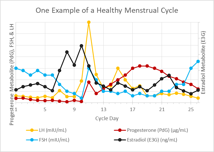
hormones across menstrual cycle
Figure 1. Hormone concentration throughout the menstrual cycle.

In Summary

This very quick overview of the menstrual cycle (aka ovulation cycle) forms the basis of every single fertility awareness method (FAM) today. Whether the method involves monitoring changes in cervical mucus, cervical position, basal body temperature, electrolyte composition of salivary/vaginal secretions, and/or at-home urinary hormone monitoring, these methods are highly reliable for predicting ovulation and are so reliable that their efficacy for avoiding unplanned pregnancy vies that of hormonal birth control.

These methods are also invaluable for shining light on a woman’s reproductive health and elucidating where hormonal imbalance lies within her cycle when things are a bit off. FAMs also provide real time data for women who are tracking their cycles so that you are able to adjust diet and lifestyle to support hormonal balance.

I will refer back to this article often in future posts on FAMs and hormonal health.

We Need Your Help

More people than ever are reading Hormones Matter, a testament to the need for independent voices in health and medicine. We are not funded and accept limited advertising. Unlike many health sites, we don’t force you to purchase a subscription. We believe health information should be open to all. If you read Hormones Matter, and like it, please help support it. Contribute now.

Yes, I would like to support Hormones Matter.   

Photo by Towfiqu barbhuiya on Unsplash.

Share

Hormones, Birth Control, and Insulin Resistance

1773 views

Little known fact. Your reproductive hormones influence how your body responds to insulin. The artificial hormones in hormonal birth control also play a huge role in how your body responds to insulin. And, your body’s response to insulin determines how well you are able to use glucose to supply your daily energy needs.

In this article, we will discuss the basics of how your body creates energy. In this first section, we will unpack:

  • How your body creates energy from glucose
  • Glucose vs. fatty acids as an energy source
  • How insulin resistance impacts the shift between glucose burning and fat burning
  • How glucose enters your cells to become fuel for energy
  • How insulin resistance interferes with the transfer of glucose into your cells

Then, we will tie in how your natural reproductive hormones, estradiol and progesterone, impact your body’s use of glucose as a fuel source and discuss how hormonal birth control disrupts this natural balance.

How the Body Creates Energy From Glucose

Many of your cell types are designed to run on glucose, a metabolic product of carbohydrates, as their main source of energy, and in fact, certain cells that don’t contain mitochondria (or contain very few mitochondria) like red blood cells and cells of certain parts of your eye (lens, retina, and cornea) rely either exclusively (as is the case for red blood cells) or primarily on glucose as an energy source.

The reason for this is that mitochondria are responsible for aerobic (oxygen required) energy creation processes within your body, and cells with no or very few mitochondria rely mostly on anaerobic (no oxygen required) energy creation by glycolysis in the cytoplasm of the cell. As we will discuss in more detail later, when your body uses fatty acids as a fuel source, this pathway is purely aerobic, so it is not possible for fatty acids to be used in anaerobic energy creation processes within your cells.

When you eat a meal containing sugar (sucrose) or carbohydrates, enzymatic processes begin breaking the sugar and carbs down into their basic structures within your digestive tract. The structure of both sugar and carbs contain glucose.

Glucose fuels the creation of ATP in a process known as glycolysis, which happens within the cell, and through oxidative phosphorylation (OXPHOS), which happens within the mitochondria (substructures within the cell). When ATP is broken down within your cells, it releases energy, which is harnessed to power your mitochondria and other important cellular functions. The by-products of that ATP creation (pyruvate and ATP) fuel additional energy production cascades within the cell.

How the Body Switches From Glucose to Fatty Acids for Energy

Even when particular cell types prefer carbs (glucose) as their energy source rather than fatty acids, most cell types are capable of using either of these macronutrients (and also, when necessary, amino acids) as a fuel in order to survive periods of fasting (including overnight fasting).

Insulin plays a key role in regulating whether your body uses glucose (glycolysis in the cell’s cytoplasm and OXPHOS in the mitochondria) or fatty acids (lipolysis in the cell’s cytoplasm and fatty acid oxidation in the mitochondria) as its preferred fuel source. This is because insulin impacts the ratio of two key enzymes (malonyl Coenzyme A and acetyl CoenzymeA) that determine which of these energy pathways is preferred (here and here). The ratio of these enzymes is dynamic, changing throughout the day in response to when and what you eat, and in response to this fluctuating ratio, your body preferentially uses carbs (glucose) or fatty acids as its fuel source.

In an insulin resistant state, your body does not easily shift between glycolysis/OXPHOS (glucose as fuel) and lipolysis/fatty acid oxidation (fatty acids as fuel) and instead remains in a state of using fatty acids as fuel. We will talk about why this is the case in the next section.

How Glucose Gets Inside Cells

The glucose released in your digestive tract from the food you eat is absorbed into your bloodstream, and when your blood glucose levels start to rise following a meal (or any drink containing carbs or sugar), it signals your pancreas to release insulin.

Insulin is the messenger that lets your cells (specifically, your skeletal muscle, fat, kidney, and liver cells) know there is glucose available in your bloodstream.  Insulin does this by binding to the cellular membrane, and this activates glucose transporters on the cellular membrane.

Once blood glucose levels start to drop, a healthy body clears insulin fairly quickly so that it can maintain adequate blood sugar levels. Insulin must be cleared so that blood sugar doesn’t drop too low.

What Is Insulin Resistance?

A number of factors influence how your cells respond to insulin. External influences (like stress, diet, and lack of sleep) along with internal factors (hormonal fluctuations) play a role in how the cells respond to insulin. And, different types of cells respond differently to insulin. Skeletal muscle cells are the most sensitive to insulin. Fat cells and liver cells are also sensitive to insulin, and so these cell types (skeletal muscle, fat, and liver) are the quickest to take up extra glucose from the bloodstream.

When your body becomes more insulin resistant, the cells are not as able to respond to insulin. My favorite analogy for this is to imagine that you are at a rock concert. You cannot easily hear the person next to you because the volume in the venue is so loud that your ears are overloaded by the background noise. In order to carry on a conversation, you must move to a quieter place. In this scenario, insulin is the background noise or the decibel level. When you are insulin resistant, your pancreas releases extra insulin to try to get your body’s cells to respond. This would be the same as somebody yelling at you in a concert hall so that you are able to hear them speak.

When you restore insulin sensitivity, it is like taking your body out of that loud concert hall and placing it somewhere quiet. Now, you are able to hear and carry on a conversation without any problems. When you restore insulin sensitivity, the cells are capable of responding to a much lower amount of insulin much more quickly and take the action of absorbing glucose from the bloodstream.

Insulin Resistance Begets Insulin Resistance

With insulin resistance, the cells are used to the high insulin environment (partially deaf to insulin), so they stop responding to insulin’s call. This prompts the pancreas to release more insulin in order to get your cells to hear the message to soak up the extra glucose circulating in the bloodstream. When insulin is unable to be heard because of the high background noise (because there is so much circulating insulin the cells are deaf to it), then glucose isn’t taken up by the cells. This then creates the false message from your cells to key organs to start releasing stored glucose (in a process called gluconeogenesis) to supply the body’s energy needs.

When we are talking about diabetes, this feedback loop often, but not in everyone with diabetes, results in a perfect storm of upward spiraling blood sugar levels.


insulin resistance cycle common in diabetes showing increased insulin resistance triggering gluconeogenesis resulting in higher blood sugar levels which increases insulin resistance
Figure 1. Insulin resistance begets more insulin resistance.

Even in conditions besides diabetes where blood sugar levels are dysregulated, you might have one condition (for example, insulin resistance), without the other (increased release of glucose from your body’s reserves).

With all of that in mind, let us take a look at how reproductive hormones impact insulin resistance and gluconeogenesis, the process of releasing glucose from stored reserves.

Estradiol, Synthetic Estrogens, and Insulin Resistance

Reproductive hormones play a key role in insulin resistance. Most scientific studies agree that estradiol (the endogenous estrogen produced primarily in the ovaries throughout the reproductive years) boosts the release of insulin from the pancreas. While at first glance, this looks like estradiol might contribute to insulin resistance because it prompts release of extra insulin, the opposite is actually true.

Estradiol is widely accepted as a potent compound to restore insulin sensitivity. Whether this is because of upregulation of insulin from the pancreas or whether it is also because of the influence estrogen has on the cells when it binds to estrogen receptors or a combination of both of these is not clear. What is clear, is that estradiol encourages cellular uptake of glucose and more rapid reduction of blood glucose levels after a meal. Estradiol also reduces gluconeogenesis in the liver suppressing the release of free glucose into the bloodstream from the body’s reserves, and this supports healthy blood sugar levels (here and here).

Estrogen Concentrations and Insulin Resistance

How estradiol affects insulin resistance is concentration dependent. Estradiol concentrations in the bloodstream within the normal circulating range (not more than 1 nanomolar abbreviated 1 nM) are associated with healthy insulin sensitivity and healthy blood sugar levels while concentrations higher than 1 nM are associated with insulin resistance. This may be why gestational diabetes is a common condition during pregnancy with up to 10% of pregnant women in America developing gestational diabetes. Progesterone also plays a key role in gestational diabetes as we will discuss in more detail below.

Non-bioidentical Estrogen and Insulin resistance

Ethinyl estradiol, the most common synthetic estrogen used in hormonal contraceptives here in America, also impacts insulin resistance, but like endogenous estradiol, the relationship is not straightforward. Ethinyl estradiol has been shown to impact insulin sensitivity and gluconeogenesis differently depending on:

  • its concentration in the hormonal birth control
  • what progestin (synthetic progesterone) it is paired with

Just as high concentrations of endogenous estradiol increase the chances of dysregulated blood glucose control, the synthetic estrogen, ethinyl estradiol, also increases chances of dysregulated blood glucose control. Chemical diabetes caused by hormonal birth control is also well documented in the literature. This is one of the reasons why, since the 1960s, the concentration of artificial estrogens in combined oral contraceptives has been dramatically reduced from upwards of 60 micrograms per pill to as low as 10 micrograms. Currently, most birth control options contain from 20 to 35 micrograms of ethinyl estradiol per pill.

Estrogen Binds to Insulin Receptors Affecting Insulin Resistance

Estrogens, whether synthetic or endogenous, affect blood sugar regulation differently at different concentrations because of their ability to bind to insulin receptors. This concentration-dependent effect of both endogenous estradiol and synthetic estrogens is often overlooked in the conversation regarding the impact of hormonal contraceptives on blood sugar control. Inasmuch as estrogens play a role in insulin sensitivity, insulin secretion, and in gluconeogenesis, and because estrogens are combined in hormonal contraceptives with a wide range of synthetic progestins, the effects on blood sugar regulation are quickly compounded and convoluted.

Progesterone, Progestins, and Insulin Resistance

As with estradiol, the concentration of progesterone also impacts whether progesterone improves or diminishes insulin sensitivity. It is generally accepted that higher concentrations of progesterone during pregnancy are a major contributor to gestational diabetes. Similarly, high concentrations of progesterone, even after menopause, are linked to an increased risk of developing type 2 diabetes.

The actions of progesterone on glucose metabolism is very much related to carrying a pregnancy to term, promoting glucose storage (rather than consumption of glucose for fuel) and promoting ketogenesis (fat burning) within the body. Even when not pregnant, progesterone is the dominant hormone during the luteal phase (second half of your cycle), and this effects how your body uses glucose and its sensitivity to insulin. This ties into common experiences during the second half of your cycle including carb cravings, potentially diminished appetite (if you are like me), and also weight gain.

Unlike artificial estrogens, of which there is only one used in the combined hormonal contraceptives available in the United States, for progestins, the synthetic forms of progesterone, there are four generations of progestins, with each generation containing progestins of different molecular structures. The class of molecules used in synthetic progestins are similar in structure to the endogenous progesterone molecule, but they are not the same. In other words, they are non-bioidentical.

Progestins bind differently to the progesterone receptors within the body (and also bind to a variety of other receptors), than the endogenous progesterone and their specific structure contributes to how much and whether insulin resistance increases. The molecular structure also affects how the body conserves glucose (increases glucose storage) or uses glucose (in the process of gluconeogenesis). It is generally believed that the androgenic nature of progestins determine their role in reducing insulin sensitivity (here and here).

Hormones and Body Composition

An interesting note, whether we are talking about natural reproductive hormones, estradiol and progesterone, or artificial hormones, ethinyl estradiol and the various progestins, these are all fat-soluble hormones. That means, these hormones may be stored in, and thus, impact the behavior of fat cells. One study evaluated the response of fat cells (adipocytes) in the presence or absence of treatment with artificial hormones and found that in the presence of artificial hormones, the adipocytes were more insulin resistant. This suggests that fat cells may serve as a reservoir for artificial hormones and endogenous hormones alike. They essentially soak up circulating hormones from the bloodstream, and these absorbed hormones in turn impact how the fat cells behave.

This finding means that body composition affects how you respond to hormones, whether endogenous or synthetic, and vice versa. It also suggests that, among other things, we ought to consider dosing hormonal contraceptives relative to body composition. Women with higher body fat may store more of the hormones than those with lower body fat and this may initiate or exacerbate insulin resistance.

Summary

In summary, reproductive hormones are intricately intertwined with metabolism, both with how the body creates energy and how it stores fats and carbs to meet energy demands between meals. Hormonal birth control impacts this finely choreographed dance between reproductive hormones and insulin sensitivity, and this seemingly small influence has a dramatic ripple effect. Insulin sensitivity dictates things like weight gain, oxidative stress, and even, as we will discuss in the next article, susceptibility to UTIs and UTI like symptoms.

Before you go, is there someone you know who needs to hear this message? Share this post with them.

We Need Your Help

More people than ever are reading Hormones Matter, a testament to the need for independent voices in health and medicine. We are not funded and accept limited advertising. Unlike many health sites, we don’t force you to purchase a subscription. We believe health information should be open to all. If you read Hormones Matter, like it, please help support it. Contribute now.

Yes, I would like to support Hormones Matter. 

Photo by isens usa on Unsplash.

Share

Connecting the Dots: Health Problems, Hashimoto’s, and Hormonal Birth Control

1847 views

I have always found it curious that many health-conscious women will pay more for meat and dairy products that promise “No Artificial Hormones,” but then don’t think twice about taking the powerful artificial hormones in birth control.

Based on observations from a recent work trip, I began wondering if this paradox could be just a strange quirk of human nature. I was working with a man who obsessed over everything he put in his body. He intently read nutrition labels to compare juices, perused the ingredients before purchasing a protein bar, and asked waiters at restaurants about their food preparation.

It is difficult to eat healthy when you are living on the road, and I was impressed by his commitment to doing so. He really took his health seriously. Then, one afternoon, he said he was going to take a break, picked up his laptop bag, and pulled out a pack of unfiltered Camels.

I wondered what kind of compartments must exist in his brain for this to make sense, and it brought my mind back to the women who make a concerted effort to avoid artificial hormones… except when they don’t.

The Perfect Example

I might have also wondered how my brain could be so de-compartmentalized that his smoking immediately triggered thoughts of birth control, but this isn’t about me. So, I’ll save that for the therapist.

Meanwhile, his dichotomy of action had piqued my interest, and I was unsure where my curiosity would lead me. Then, I met the perfect woman to help me take a deep dive into the topic.

Brandy Searcy has worked as a developmental scientist for pharmaceutical companies for over a decade. As the daughter and granddaughter of nurses, she grew up immersed in conversations centered around healthcare. So, pursuing a PhD in organic synthesis seemed almost a natural extension of her genetics and heritage.

Through her work, which has included forays into cancer research and pesticide development, she honed a keen understanding of endocrine disruptors. Her concern over xenoestrogens in health and beauty products led her to develop Rain Organica, a line of skin care products designed specifically for women looking to detox their lives.

I met Brandy when she invited me on her podcast to talk about my book.

Birth Control and the Compartmentalization Conundrum

After we finished recording, Brandy mentioned that she could not believe how long it took her to connect the dots and realize that so many of her problems were linked to hormonal birth control. This opened the door to a fascinating discussion.

As you might imagine, her family was deeply vested in Western medicine. So, when she began to battle acne at around the age of 14, her mother did what any loving mother would do. She drove her around the state of Georgia trying to find a dermatologist who would conjure up a magic potion to make her acne worries vanish.

After a few years and some bad experiences with Accutane, Brandy’s mindset began to shift. It was around the age of 20 that she decided that she would “treat my skin as an organ to be loved rather than as a battleground.”

Although she had identified the problems with Accutane, it would take another 20 years for her to recognize the role hormonal birth control was playing in her health struggles. Consequently, this would become the first of many milestones she would later identify as missed opportunities to connect the dots.

Living Both Sides of the Coin

“It’s almost like there were two of me. One side was touting this new, healthy approach to life, and the other side was completely ignoring the effects of hormonal birth control on my body.”

When Brandy reflects back on those days before the blinders came off, you can see clouds of guilt and maybe a hint of embarrassment cross her eyes. She says there were any number of events that should have been enough to make her see the light earlier. Like the time red flags and sirens went off in her head when her doctor suggested a form of birth control because the “hormones were localized.”

Looking back at it now, she laments, “If she (the doctor) thought hormones can be localized, why didn’t I question her wisdom on prescribing me birth control in the first place?”

Beyond the common misrepresentations by doctors, Brandy can pinpoint some very specific, significant events in her personal and professional life that she believes should have been enough for her to walk away from hormonal birth control.

Missed Warning Signs

“It’s mind-blowing to me that I couldn’t let myself connect the dots. How I couldn’t see it is beyond me.”

Brandy still feels overcome with dismay as she recounts the significant events, the missed warning signs. Here is her summary of those key events:

2008 – Right leg numbness – The doctor thought she might be experiencing transient ischemic attacks (TIA) caused by the synthetic estrogens in her birth control. He told her to stop taking it until they could identify the culprit. The issues turned out to be structural rather than a stroke, and she returned to The Pill without a second thought.

2012 – Lyme disease – Brandy became very ill. As they worked through the process of diagnosing her illness, the doctor told her to stop taking birth control for six months. During the course of testing, they learned that her ANA and CRP levels were high. Ultimately, she was diagnosed with and treated for Lyme disease. Once again, feeling better, the diagnosis was taken as an exoneration of hormonal birth control. She forgot all about concern for her ANA and CRP levels, and started right back on The Pill.

2012 – Literal warning signs – That same year, she visited a facility that previously manufactured synthetic estrogens. As she walked through the plant, she noticed the bright red “Carcinogen” signs everywhere – on the walls, on the pipes – literally everywhere. Even as one of her co-workers told her this is where estrogens used to be made, she never connected the danger and all these literal warning signs to the same little pill she was taking every day.

2016 – No periods – Brandy was already experiencing gall sludge when her gynecologist recommended a different birth control formulation that, when taken continuously, would allow her to never have a period again, right up until menopause. She loved the idea of eliminating her period and didn’t even make the connection when signs of Hashimoto’s thyroiditis began almost immediately after switching to this brand.

2017 – Gallbladder disease – She had to have her gallbladder removed. While Brandy was still unaware of hormonal birth control’s link to gallbladder issues, she also had a family history of gallbladder disease that kept her from even considering The Pill’s role in her gallbladder’s demise.

2018 – Hashimoto’s diagnosis – After two years of tests, Brandy was diagnosed with Hashimoto’s thyroiditis, yet another disease that has been linked to birth control use. This was the event that would finally open her eyes, but the realization still took a circuitous route as it wasn’t the diagnosis itself that helped her make the connection.

When Western medicine told this self-described type-A control freak that there was no cure, she began digging for herself and discovered a book on treating your thyroid using Ayurveda techniques.

Ayurveda is an alternative form of medicine originating from Asia, which focuses on the necessary balance of internal and external influences to maintain proper health. And, it provided the shift in mindset that finally caused Brandy to question birth control.

Looking back at everything now, Brandy says, “We are not made to live in a diseased state. We are made to be healthy, and if we aren’t healthy, it isn’t because our body is broken, it’s because we are putting something in that is making us not healthy.”

Seeing the Light

I asked what she might tell other young women to help them wake up to the dangers of The Pill, or at least give more thought to its potential risks. This led to another interesting rabbit hole as we discussed the various factors that prevent young women from truly contemplating the dangers. Here are some of the variables we discussed:

Lack of reproductive education – Young women aren’t taught about the phases of their cycle, nor how its ebbs and flows can actually help them monitor their health, nor are they educated on how their cycles may change over time.

In Brandy’s case, she had very heavy, irregular, and painful periods as a young girl. No one ever told her this was common when going through menarche. As a result, she said The Pill gave her a false sense of control. She had fully bought into a false narrative that periods should be embarrassing and that they serve no useful function. At some level, she believed that completely stopping her menstruation with potent chemicals might actually be better for her than respecting her body’s natural processes. This did not change even after two doctors had her stop hormonal birth control for health concerns.

Western medicine – We tend to give doctors an inordinate authority over our health decisions to the point of almost idolizing them. This is reinforced by a notion that they have taken the Hippocratic Oath, promising to first, do no harm. However, only slightly over half of all physicians today have taken the oath, and that percentage drops with each new graduating class.

The more entrenched a young woman’s faith in Western medicine the less likely she is to question birth control.

Addiction – Some women seem to develop a type of addiction to hormonal birth control. The mere suggestion that they should look for another option is enough to create severe anxiety.

Stockholm syndrome – Closely related, some women may take on a type of Stockholm syndrome that prevents them from connecting the dots. Stockholm syndrome is described as a coping mechanism that some victims of an abusive situation develop in which they actually grow fond of the abuser.

Brandy recalled, “In a lot of ways, if feels like I was in an abusive relationship, but I wasn’t able to see how abusive it was until I stepped away.”

Withdrawal – Beyond the addictive nature, quitting any synthetic steroid cold turkey can be hard on the system. There’s a reason doctors taper you off of prednisone and other steroids.

Many women experience withdrawal symptoms when they try to stop, and this is enough to drive them right back to The Pill.

Little support – Historically, there has been a lack of support for women coming off these potent synthetic hormones – some after decades of use. Even the medical professionals who prescribe the drug are woefully undertrained on dealing with the detoxification process necessary for a healthy transition off of The Pill. Actually, that is an understatement. Most doctors have not even contemplated the effects of coming off the synthetic steroids in birth control. They act is if you just stop and your body returns to normal.

When Brandy came off The Pill, she immediately began to see and feel changes in her body, including her first UTI, at the age of 40. This was the lightbulb moment when she realized how much impact the synthetic steroids had been having on her body. Despite having made it through 40 years with no UTIs, two of her doctors, who are still clearly wearing their birth control blinders, told her it sounded like she had poor hygiene habits. Somehow, in their eyes, I guess it took 40 years for those bad habits to catch up to her.

By the way, Brandy recently developed a course to help women through the transition off of hormonal birth control.

A Unique Formula

Clearly, there are lots of variables that can influence the way a woman perceives and judges birth control.

Every woman is different. Each has her own unique body chemistry. That is why a birth control formulation that seems harmless to one woman can be deadly for the next.

Brandy mused that the way women weigh their thoughts on The Pill is equally idiosyncratic. There is no one phrase or thought that will lead women to suddenly see the realities of hormonal birth control. Each woman has to hear the right message at the right time to help her properly weigh the benefits and risks for her situation. I say “properly” because the system is so stacked against women getting accurate information about this potent drug.

Brandy added this last thought related to one of the first big hurdles that women encounter – the overwhelming tendency to mitigate and downplay side effects. She advised, “The subtle symptoms are the first indicators. Don’t dismiss them because they seem insignificant. They are frequently pointing to something bigger.”

#1
In the Name of The Pill

37 customer reviews

In the Name of The Pill*

by Mike Gaskins

The FDA approved The Pill despite it not being proven safe. Today, it has been linked to everything from blood clots and cancer to lupus and Crohn’s disease — and still has not been proven safe.
This book explores the medical and historical disconnects that brought us to this point.


 Price: $ 17.95

Buy now at Amazon*

Price incl. VAT., Excl. Shipping

Last updated on October 21, 2023 at 9:38 pm – Image source: Amazon Affiliate Program. All statements without guarantee.

We Need Your Help

More people than ever are reading Hormones Matter, a testament to the need for independent voices in health and medicine. We are not funded and accept limited advertising. Unlike many health sites, we don’t force you to purchase a subscription. We believe health information should be open to all. If you read Hormones Matter, like it, please help support it. Contribute now.

Yes, I would like to support Hormones Matter.

Photo by Almos Bechtold on Unsplash.

Share

Why Aren’t Women Tested for Factor V Leiden and Other Clotting Disorders?

14479 views

When I had a stroke at age 28, my doctors did some tests and found that I have a fairly common clotting disorder called Factor V Leiden. They told me that this, combined with birth control pills, are what caused me to have the cerebral venous thrombosis (stroke). It didn’t occur to me then to ask what Factor V Leiden actually was. Or to ask why I hadn’t been testing for inherited clotting disorders before I was put on medication that increased my risk for blood clots. These things didn’t occur to me until much later, after I learned to walk again.

I spent most of the first two years after my stroke getting on with my life. It wasn’t until I was searching for a topic for my thesis that I revisited what happened to me. I had no idea that birth control pills could be so dangerous and I certainly didn’t know that I could have an inherited genetic condition which would make them exponentially more dangerous for me. “How many women have the same condition?” I wondered. “Why don’t we test them before they are put on hormones?” These are some of the questions I sought to answer with my research.

What is Factor V Leiden?

Factor V Leiden (FVL) is a 20,000-year-old mutation common in the general population and a major genetic risk factor for thrombosis. It’s the most common genetic clotting disorder, accounting for around half of all cases. It’s most commonly found in Caucasians (3-8%).

Patients with Factor V Leiden can be either:

  • Heterozygous: inherited one mutated gene from a parent

or

  • Homozygous: inherited two mutated genes, one from each parent

What Does It Do?

As my hematologist described, FVL doesn’t cause blood clots but once activated, it dangerously accelerates clotting. Researchers aren’t clear on why some people with FVL activate and others don’t but there is almost always a precipitating factor—surgery, trauma, immobility, use of hormones, etc.

According to a review in Blood, the journal for the American Society of Hematology, women with heterozygous FVL who also use oral contraceptives have an estimated 30 to 50-fold increased risk of blood clots, while women with homozygous FVL have a several hundred-fold increased risk.

It is the most common genetic cause of primary and recurrent venous thromboembolism in women.

We know that taking estrogen can increase the risk of blood clots, stroke, and heart attack in women. And estrogen, when taken by someone with FVL, can significantly increase the risk of blood clots. Whether women are taking synthetic estrogen in the form of oral contraceptives, or hormone replacement therapy or have increased concentrations of the endogenous estrogens due to pregnancy, they are at much greater risk of clotting.

FVL accounts for 20-50% of the venous thromboembolisms (VTE) that are pregnancy related. In the United States, VTE is the leading cause of maternal death. In addition to causing VTE in pregnant women, FVL has been linked to miscarriage and preeclampsia.

Perhaps the women most at risk for blood clots are those that have been placed on hormone replacement therapy (HRT). A recent review of data from several studies found that women taking hormone replacement therapy were at an increased risk of blood clot and stroke. Worse yet, women with FVL who are also on HRT were 14-16 times more likely to have a VTE.

Despite these risks, women are not systematically tested for FVL before they are prescribed oral contraceptives, before or during pregnancy, or before commencing HRT.

What Women Know about Birth Control and Blood Clots

Part of my thesis research included a survey to assess what women understand about the risks of birth control pills and clotting disorders. Over 300 women who had taken birth control pills participated. What I found was that most women do not understand the side effects of hormonal birth control, nor are they familiar with the symptoms of a blood clot.

As for clotting disorders, nearly 60% of the women surveyed had no knowledge of these conditions. When asked whether they knew about clotting disorders BEFORE they took birth control pills that number increases considerably.

Over 80% of women were taking a medication without the knowledge that they could have an undiagnosed genetic condition that would make that medication exponentially more dangerous.

This shouldn’t come as much of a surprise give that this information is not found in advertisements for birth control pills, on non- profit websites about birth control pills and their risks, or on literature provided with the prescriptions.

Why Aren’t Women Tested for Clotting Disorders?

The most common reason I found in my research for not testing women were cost-benefit analyses measured in cost per prevention of one death.

Setting aside the moral argument that you cannot put a price on a human life, because clearly the government and corporations do just that. (It’s $8 million in case you were wondering.) The cost of taking care of taking care of victims of blood clots is not insignificant.

Each year thousands of women using hormonal contraceptives will develop blood clots. The average cost of a patient with pulmonary embolism (PE) is nearly $9,000 (for a three-day stay not including follow-up medication and subsequent testing).

A hospital stay as a stroke patient is over twice that at nearly $22,000 (not including continuing out-patient rehabilitation, medications, testing, etc.). As a stroke survivor, I can tell you that the bills don’t stop after you leave the hospital. I was incredibly lucky that I only needed a month of out-patient therapy. Most patients need considerably more and will require life-long medication and testing. It’s important to note that due to the increasing cost of healthcare, the figures in these studies (PEs from 2003-2010; strokes from 2006-2008) would be exponentially higher now.

I’m not a statistician but I can do some basic math and while I wasn’t able to find data for the United States (surprise, surprise), the health ministry in France recently conducted a study that showed that the birth control pill causes 2,500 blood clots a year and 20 deaths.  The United States has 9.72 million women using the pill compared to France’s 4.27 million. This doesn’t include the patch, ring, injectable, or hormonal IUD, but for the sake of keeping things simple, let’s just use the pill. So we have over twice the pill-users as France, which means twice the blood clots (5,000) and twice the deaths (40). If we assume that half of the blood clots are PE and half are stroke, we come up with a whopping $77.5 million in hospital bills for these blood clots (not counting life-long treatment). Now adding the cost-of-life determined by the government (40 women times $8 million= $320 million) and we end up with nearly $400 million a year in damages caused by the pill. For the cost of only one year of damages, all 10 million women could have a one-time $40 blood test which would result in considerably fewer blood clots.

Furthermore, the research in my thesis shows that women would be willing to not only take these tests, but also to pay for them!

Of the 311 who answered the question, 82.3% (or 256) said they would be willing to take the test. Only 7.2% said no, with the other 10.6% “not sure.” More than 60% of respondents would be willing to pay for the test (up to $50).

In addition, the cost of a blood test is directly proportional to how frequently it is performed. An increase in testing will result in a decrease in the cost of testing.

Women Deserve Better

Putting aside the monetary costs for a moment, what about the emotional and physical toll for women who suffer these dangerous and debilitating blood clots? There is no excuse for women to suffer strokes, pulmonary embolisms, DVTs, multiple miscarriages, and still births because they have an undiagnosed clotting disorder.

That said, requiring a test before prescribing hormones to women would raise awareness of the dangers of these drugs and may reduce the overall number of women using them. Which leads one to wonder if the absence of testing for women is really just a public relations strategy.

Perhaps one of the most devastating cautionary tales of not testing for clotting disorders comes from Laura Femia Buccellato. Her daughter Theresa was 16 years old when she was killed from a blood clot caused by (undiagnosed) Factor V Leiden and birth control pills. Would Theresa be with us today if she had had a simple blood test? Would I have had a stroke? When we will demand better?

We Need Your Help

More people than ever are reading Hormones Matter, a testament to the need for independent voices in health and medicine. We are not funded and accept limited advertising. Unlike many health sites, we don’t force you to purchase a subscription. We believe health information should be open to all. If you read Hormones Matter, like it, please help support it. Contribute now.

Yes, I would like to support Hormones Matter.

This article was first published in September 2016.

Share

Women in Clinical Trials

6021 views

Do Hormones Matter with Clinical Trials Research?

I am repeatedly struck by the avoidable ignorance that surrounds this industry. Women’s hormones are considered a fringe science by many, despite the fact that hormones modulate all bodily functions and impact all aspects of pharmacokinetic and pharmacodymanic response, the key variables evaluated in clinical trials research. Repeatedly, we face the assumption that hormones make no difference to women’s health – quite ironic since simultaneously hormones are blamed for everything from migraines to mental health.

And then there is the long held notion that we don’t need any more data in women’s health – quite striking considering only 30% of Ob/Gyn clinical practice guidelines are based on data. Just the other day, a well-positioned female physician, one of the fewer than 4% female healthcare executives, argued that while what we doing was interesting,  it was unnecessary because the 1993 Gender Guidelines ‘mandated’ women be included in clinical trials.

Perhaps some history is in order. Until 1993, women of ‘childbearing potential’ were prohibited from participating in clinical trials research. This means that all of the drugs developed before 1993 (most are still being used today) were not tested on women; any potential dosing differences, sex- specific side-effects could not be found until after the drug reached the market and even then it was and is difficult to ascertain because sex-specific analytics were not, and are still not, routinely performed. This makes proving sex-based adverse events all but impossible.

In 1993, thanks in large part to efforts of the first large group of women to enter the US Congress (women in Congress matter); the FDA removed its prohibition against women with childbearing potential participating in clinical trials. The regulations, however, were not established until 1998 and what was implemented, though an improvement over the earlier prohibition of women in clinical trials was less specific than the original guidelines and resulted in less than satisfactory results. When those regulations are evaluated alongside industry trends over the last decade, it becomes abundantly clear why women suffer disproportionately from adverse events compared to men and why everyone in the healthcare industry should be concerned.

From Proposed Guidelines to Regulation

The 1993 guidelines recognized that women might process and react to drugs differently than men (indeed they do and this has been shown repeatedly) and therefore, it was recommended that pharmacokinetic studies be done to evaluate the difference in drug absorption, distribution, metabolism and excretion. It was further recommended that menstrual status, hormonal supplementation (oral contraceptives, HRT and the like) be evaluated.

The FDA was quite clear in its ability to mandate these changes – there was none.  Indeed, all that was possible was suggestion.

The agency recognizes that this change in FDA policy will not, by itself, cause drug companies or IRBs to alter restrictions they might impose on the participation of women of childbearing potential. We do not at this time perceive a regulatory basis for requiring routinely that women of childbearing potential be included in particular trials, such as phase 1 studies. However, careful delineation of drug effects by gender is expected by the agency and the FDA is determined to remove the unnecessary federal impediment to the inclusion of women at the earliest stages of drug development.  The agency is confident that the ethical, social, medical, legal and political forces will allow greater participation of women at the earliest phases of drug development.

So, the FDA was confident that ethical and social pressures would convince pharmaceutical companies to do the right thing. How well has that gone? The guidelines also indicated that it was not necessary to include women in phase 1 or 2 trials because there was no evidence to suggest sex differences in drug effectiveness:

Because documented demographic differences in pharmacodynamics appear to be relatively uncommon, it is not necessary to carry out separate pharmacodynamic – effectiveness studies in each gender routinely.

To summarize, data were never collected in the first place to suggest sex-differences might exist (remember before 1993, women were prohibited from participating in clinical research) and because there were no data to suggest a sex-differences, there was no need for additional data – makes perfect sense to me.

Fortunately, the National Institute of Health (NIH), the federal funding agency for health-related research, took the reins with a clear mandate – no funding unless the study included women in clinical research.  The NIH Revitalization Act of 1993, followed by updates and regulations in 1994, 2000 and 2011, mandated that all NIH funded clinical trials have sample sizes adequate to support ‘valid analysis’ of gender and racial subgroup effects. Unfortunately, however, NIH-sponsored clinical trials represent only 20% of all clinical trials. The remaining 80% are sponsored by pharma and fall under the FDA’s guidelines. And even though, by all accounts the NIH has done substantially better than the FDA, a recent report by the National Heart, Lung and Blood Institute (NHLBI) – a sub agency of the NIH, found that in clinical trials between 1997 and 2006 where the outcome of the study was stroke, myocardial infarction or death found that women represented only 27% of trial participants and only 13-19 of the studies included sex-based analyses.

Back to the FDA Regulations

In 1998 and 2000, the FDA officially instituted the Guidance for Industry, requiring all new investigation drug (IND) and new drug application (NDA) submissions include data on trial participation, efficacy and safety, be presented by age, race and sex. A report in 2001 by the General Office of Accounting (GAO) (no further GAO reports could be found on this topic) evaluated the success and failures of the gender guidelines. It wasn’t pretty:

The 1998 regulation has the force of law, but it is less specific than the 1993 guidance. The regulation required that safety and efficacy data already collected be presented separately for men and women in new drug application summary documents. It does not include criteria for determining the number of women to be included in clinical studies, nor does it require any analysis of the data presented. The 1998 regulation also requires the tabulation of the number of study participants by sex in investigational new drug annual reports. The regulation enacted in 2000 allows FDA to halt research programs for drugs for life-threatening conditions if otherwise eligible men or women are excluded from participation in studies based solely on their reproductive potential, but it does not require inclusion of any particular number of men or women.

How well did the FDA do in meeting and enforcing the guidelines? According to the GAO:

  • NDA and IND summary documents and annual reports often failed to meet the data presentation requirements
  • 30% the new drug application summary documents submitted to FDA by drug sponsors did not fulfill the requirements for the presentation of available safety and efficacy outcome data by sex
  • 39% of IND annual reports did not include demographic information
  • The FDA has the authority to suspend proposed research for life-threatening conditions if men or women are excluded, but has not yet done so

As a result of the non-enforcement, the GAO found that although the number of women in clinical trials now averaged 52%, most were enrolled in later stages of the trial. Women represented only 22% of early phase clinical trials. This is where most of the safety and dosing considerations are determined.

Finally, and perhaps the most troubling aspect of this report was that, the FDA had no procedures in place to evaluate, manage or enforce the regulations. As a result, they had no way of knowing whether women were included in the trials in sufficient numbers or whether the medications or devices gaining approval had gender-specific safety issues.

What does this mean?

For all drugs and devices approved before the 1998-2000 regulations and likely many years after, the safety and efficacy data were lacking for women. Despite the 52% female participation number that is bandied about as proof positive that women are represented sufficiently in clinical trials, that number reflects later phase trials, after safety and efficacy parameters are established. In the early phase trials, the number of female participants remains at a paltry 22%. Today, when large women-only (Women’s Health Initiative and Women’s Health Study) research are removed from the tabulations, the mean proportion of women included in all clinical trials hovers around 27%.

Most recently, the Institute of Medicine (IOM) Committee of Women’s Health Research reports a continued lack of

taking into account of sex and gender differences in the design and analysis of studies, lack of reporting on sex and gender differences, has hindered identification of potentially important sex differences and slowed the practice in women’s health research and its translation into clinical practice.

And although the IOM reports that the most progress has been made in cardiovascular research, the NHLBI and Cochrane Reports suggest otherwise. The NHLBI found female participation hovering around 27% in certain cardiovascular trials and Cochrane Reports found that out of 258 clinical trials, only 196 included women and only 33% of those reported sex or gender analytics.

Cardiovascular disease is the most common cause of death in American women and in recently recalled medications for heart disease there were disproportionately higher fatalities and serious adverse events in women than in men.

With high risk cardiac devices, a recent review of FDA pre-market approved devices from 2000 – 2007 (78) found significant gender bias in sampling and data reporting and significant lack of sex-specific safety data.

  • FDA summaries did not report gender data in 28% of studies examined
  • For studies reporting gender distribution, 67% of the participants were men

So, the suggestion that additional data in women’s healthcare are not needed is unquestionably false and dangerously ill-informed. The notion that hormones, which regulate every aspect of pharmacokinetics and pharmacodynamics are an irrelevant and a fringe science, is ignorant bordering in negligent. It is time for women to stand up and demand inclusion and analytics by sex for all drugs and devices.

Postscript: NIH Update 2014

Since this article was first published in February 2013, the NIH has made inroads towards more thorough assessment of the role of sex in basic, pre-clinical research. Recognizing the almost total reliance on male animals and cells in preclinical research obscures key sex differences that should guide clinical studies, the NIH instituted new guidelines in October 2014

…that require applicants [for NIH grant funding] to report their plans for the balance of male and female cells and animals in preclinical studies in all future applications, unless sex-specific inclusion is unwarranted, based on rigorously defined exceptions. 

It remains to be seen how rigorously these new guidelines will be enforced and whether they will impact health research in any discernible way.

Postscript: Update 2019

From a recent 10 year follow-up study assessing sex-inclusive research practices of journal articles published within nine of the biological disciplines, including pharmacology, researchers found some improvement in most of the disciplines, except pharmacology. Compared to 2009, where only 29% of the studies reviewed included male and female subjects, in 2019, 49% included both. In contrast however, pharmacology trended downward with only 29% of articles reporting the use of both sexes in 2019 compared to 33% in 2009.  Nevertheless, even though more women were included in more research studies across the disciplines reviewed, few researchers thought it was important to analyze sex based differences. Indeed, of the 49% of journal articles that included both sexes in 2019, only 42% analyzed data by sex, compared to 50% in 2009. Ironically, in pharmacology although the total number of women decreased in studies during this time period, analyses of sex based differences was more frequent increasing from 19% in 2009 to 48% in 2019.

Overall, it appears that we have yet to make much progress.

We Need Your Help

More people than ever are reading Hormones Matter, a testament to the need for independent voices in health and medicine. We are not funded and accept limited advertising. Unlike many health sites, we don’t force you to purchase a subscription. We believe health information should be open to all. If you read Hormones Matter, like it, please help support it. Contribute now.

Yes, I would like to support Hormones Matter.

This article was first published in 2013. 

Summer’s Best Bargain: Free Vitamin D

3471 views

The warmest season of the year is around the corner. Many of us are looking forward to school holidays, work vacations, and relaxing. And shopping often accompanies our summer fun. So take advantage of the best bargain of the season: better health – for free! That’s right, I am talking about sunshine: the light emitting from the fiery heart of our solar system. Ultraviolet B (UVB) rays from the sun provide us with an essential nutrient called vitamin D. Yes, this is the vitamin that not only strengthens our bones and muscles but may significantly protect us from a wide range of serious diseases including autoimmune disorders, cancer, contagious illnesses, diabetes, and heart disease, according to a plethora of credible medical studies from around the world.

Many people—across generations and geographical locations—suffer from low vitamin D levels from lifestyles that do not include unprotected sunbathing. Since the late 1980’s, the medical community has emphasized the need to “shun the sun” to avoid skin cancer. Consumers have embraced this advice by spending billions of dollars so they can slather chemical-laden lotions with exponentially increasing sun protection factors all over their bodies. This behavior has resulted in a vitamin D deficiency epidemic.

Moderate sun exposure is healthy for most individuals. Our bodies possess an inherent mechanism to process only the necessary intake of sun rays, about 20,000 international units of vitamin D. After our skin is exposed to direct sunlight under optimal conditions for about 20 minutes, its safety mechanism turns off the initial production of vitamin D. For many folks, it is then time to move to the shade or don additional protection to reduce the risk of sunburn.

Optimal conditions to enjoy summer’s vitamin D depend on a number of factors that we can, and in some cases, cannot control. These factors include:

Geographic location. The closer you are to the equator and the higher your altitude the better your opportunity to acquire vitamin D-rich sunlight.

Time of day. The window of sunlight between 10:00 in the morning and 2:00 in the afternoon is optimal. If your shadow is shorter than your height, you are in the potential vitamin D-producing time frame.

Sky clarity. An azure sky is highly preferable to cloud cover. UVB light is decreased by about 50 percent when penetrating clouds. Ozone pollution absorbs UVB rays before they reach your skin.

Skin. The less clothing, makeup, and sunscreen you wear, the better the odds that your skin can produce vitamin D. It also is important to understand that melanin, the pigment in your skin, absorbs UVB rays. The lighter your skin, the better chance you can make vitamin D more efficiently.

Age. Youth trumps older ages because the concentration of the vitamin D precursor in our skin, called 7-dehydrocholesterol, decreases with age.

Weight. Less weight means typically more vitamin D production from the sun. As vitamin D is fat-soluble, the body’s fat cells more rapidly absorb vitamin D, decreasing its availability to organs, tissues, and cells.

You may be thinking, “I live near sea level, far from the equator, in mostly cloudy conditions with cool summer temperatures; work full-time during the day; and am dark-skinned and overweight. How on earth (literally) can I get any measurable vitamin D from the sun?” Take advantage of sunny weather by enjoying an outdoor lunch break. Remove that hat, roll up your sleeves, and soak in the sun. Ten minutes of sun exposure is better than none.

Each individual’s options for absorbing nature’s gift of vitamin D may differ.* Fortunately, widely available sources of vitamin D including vitamin D3 supplements may be highly effective in raising your body’s D levels to protect you from a wide array of medical conditions. The information about, and benefits of, vitamin D could fill a book. In fact, I am so impressed with vitamin D’s health benefits that I recently published a book called Defend Your Life to encourage people to improve their health by taking vitamin D.

Happy summer, and happy health!

*Persons who have developed sarcoidosis, specific granulomatous diseases, and rare cancers may experience hypersensitivity to sunlight exposure.

Copyright © 2013 by Susan Rex Ryan
All rights reserved.

Image by pixel2013 from Pixabay .

Share

Share Your Hysterectomy Experience

4372 views

The hysterectomy and oophorectomy industry continues to flourish. There are over 600,000 hysterectomies every year. The oophorectomy rate is about 70% of the hysterectomy rate, some performed at the time of hysterectomy and others as separate surgeries. About 90% of these surgeries are for benign conditions (elective). These are merely estimates based on samples of inpatient data from short-term, acute-care, nonfederal hospitals along with hysterectomy and oophorectomy outpatient percentage estimates. According to the aforementioned publication, the rate of outpatient (ambulatory) hysterectomies increased from 14% in 2000 to 70% in 2014. Outpatient oophorectomies increased from 57% to 84% over the same time period. The Centers for Disease Control (CDC) reports only inpatient hysterectomies which is why hysterectomy rates are typically understated by the media. A 70% understatement is a gross misrepresentation and outpatient hysterectomies may now exceed 70%.

Hysterectomy is seen as panacea for a multitude of women’s health issues. Unfortunately, it is not, and yet, this perception that hysterectomy is a cure-all survives, largely because of false information from gynecologists, gynecologic oncologists, other medical professionals, hospitals, surgical centers, the media, and women who have had the surgery(ies).

The prevalence of hysterectomy and oophorectomy leads the public to mistakenly believe that a woman’s sex organs are disposable. In many cases, gynecologists fail to provide their patients with the necessary factual information to make an informed decision about these surgeries, leaving women and their partners to learn about the side effects after the fact. Some of the more commonly reported side effects include: bladder and bowel dysfunction, skeletal and figure changes, sexual dysfunction, emotional emptiness, and impaired ovarian function. Although some may believe these side effects are rare and thus rarely discussed pre-surgery, comments on these hysterectomy articles indicate they must be more common than many realize.

What makes these side effects even more troubling, is the fact these procedures are rarely needed. Women are coaxed into the surgery under the false pretense of cancer or pre-cancer or told it is their only or best option. Finally, many women’s organ(s) are removed despite having specifically told their surgeons that organ(s) should not be removed. Here is just one of those stories.

In light of the problems with hysterectomy, the HERS Foundation is collecting stories of post-hysterectomy problems. We are supporting that effort. If you would like to share your story, consider participating in the “In My Own Voice” project. To learn more, click here.

If you would like to share your story here on Hormones Matter, please contact us here.

Thank you in advance for sharing your hysterectomy experience.

We Need Your Help

More people than ever are reading Hormones Matter, a testament to the need for independent voices in health and medicine. We are not funded and accept limited advertising. Unlike many health sites, we don’t force you to purchase a subscription. We believe health information should be open to all. If you read Hormones Matter, like it, please help support it. Contribute now.

Yes, I would like to support Hormones Matter.

Share

Promiscuous Hormones and Other Fun Facts

6148 views

Every time I read about an environmental endocrine disrupting chemical, a medication or even sometimes actual endocrine research, I am amazed, dumbfounded by the lack of up-to-date information about how hormones and receptors work. Indeed, much of the research still operates on what is called the ‘classical’ view of hormone-receptor activity. And while, those functions are still accurate, there is so much more going on that merits understanding and investigation. Here are some basics that will blow your mind.

Understanding Hormones and Receptors

Hormones are gods. Like the gods of Greek mythology, hormones control everything. There is a not a physiological system that a hormone does not impact in some manner or another.  And this is why ignoring hormones in health research is so bloody dangerous and just downright stupid.

Hormones bind to receptors. This isn’t mind-blowing, but is the basis for all endocrine and in fact chemical activity in the body – compound A has to talk to body part B. The conversation is mediated through receptor binding. The shape of the hormone determines which receptors and how strongly the hormone fits or binds. Much like a lock and key, only certain keys open certain locks.

Hormones are promiscuous. Hormones sleep around. Hormones like binding with other hormone receptors and are often similar enough in structure to do so, especially at higher concentrations. This means that estrogens will bind with estrogen receptors but also androgen receptors. Progesterone binds with estrogen receptors but also gluccocorticoid (cortisol) and mineralocorticoid receptors, and so on. Because of this cross-binding, the measure of interest becomes the hormone’s ‘affinity’ for a certain receptor. How much does hormone A like receptor B. The stronger the affinity, the longer it stays bound and the more intracellular effects it exerts.

The most shapely hormones bind more. Like mating, shape matters. The more shapely hormones bind to more receptors, more tightly. For example, the shape of MBP, the metabolite of BPA fits into the estrogen receptor better than BPA. In fact, MBP is a 1000 times stronger than BPA in its affinity for the estrogen receptors (ERs). If both MBP and BPA were competing for the affections an ER, MBP would win. This happens with all hormones and hormone-like chemicals, the more shapely the compound, the better it fits with a receptor and the longer it stays there.

Promiscuous hormones spread the love. The effects of an endocrine disruptor, even an estrogenic one may not be limited to the reproductive classics. Estrogen receptors are located all over the body and the brain, so too are androgen, progesterone, glucocorticoid, mineralocorticoid and thyroid receptors. So any time we talk about a hormone’s impact on health, we have to look at where its receptors are located. Ditto for chemical that binds to hormone receptors. And to make it even more complicated, because these hormones bind promiscuously, one also has to consider the fact that the hormone may act at other, non-similar receptors. Hormone promiscuity makes endocrine research infinitely more complex than asking whether hormone A binds to hormone receptor A or how long a substance takes to clear from the body.

Hormones beget more hormones. That’s right, hormones have babies and lots of them. Called metabolites, the offspring of one hormone can lead to a whole family of other hormones, each promiscuously binding with receptors. It’s a veritable orgy in there. How many and what type of hormone offspring depends entirely on the enzymes the hormone meets as it travels through the body. This means, it’s not enough to simply measure the effects of the parent hormone on health, but one has to look at its offspring or metabolites as well.

Take Home

Hormones systems are complicated. Hormone disrupting chemicals likely disrupt a myriad of physiological systems, often transgenerationally. One off, linear, cause-effect, dose-response studies cannot begin to understand the system changes involved with endocrine modulation. This requires large scale, data modeling tasks, that have yet to be designed.  For the time being, any study that claims to have proven that a man made, endocrine disrupting chemical  or hormone altering medication is completely safe or works only on the intended physiological system, is likely lying. They just haven’t found how or where the chemical is wreaking havoc. I’d err on the side of caution.

Image credit: Jan Saenredam, Public domain, via Wikimedia Commons

We Need Your Help

More people than ever are reading Hormones Matter, a testament to the need for independent voices in health and medicine. We are not funded and accept limited advertising. Unlike many health sites, we don’t force you to purchase a subscription. We believe health information should be open to all. If you read Hormones Matter, like it, please help support it. Contribute now.

Yes, I would like to support Hormones Matter.

This article was first published May 3, 2013. 

Share
1 2 3 11