mitochondrial damage

A Rant about Lupron and Oophorectomy, Some Mechanisms and Clues to Recovery

10513 views

A Rant

Women who have been given Lupron (leuprolide) and the other GnRH agonists and antagonists and women who have had their ovaries removed are thrust violently into menopause. Overnight. There is no gradual decline of ovarian hormones that allows for molecular adaptations to the new state of chemical senescence. No, none of that. Just the chemical trauma of the loss of hormones.

The experience is akin to castration; something that should never be considered as a viable treatment option for any benign disease process, especially in young people. Yet millions of young women and children (Lupron for precocious puberty, gender dysphoria, and now autism) are prescribed these drugs and undergo these procedures annually without as much as the slightest recognition that there might be negative sequelae. Indeed, women are told routinely that what they experience with Lupron, its analogs, or upon oophorectomy is not real, that it does not exist, and that their symptoms are no more than some form of psychosomatic stress – the long vestige of Freudian acquiescence called hysteria. It is not. The symptoms these women experience is real and directly related to how the loss of estradiol and other ovarian hormones damage the mitochondria.

Some Mechanisms

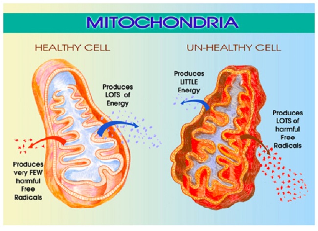
Estradiol regulates mitochondrial energetics via multiple mechanisms both directly and indirectly. I have written about the mitochondrial damage provoked by the loss of estradiol previously, see here, here and here. In brief, the loss of estradiol fundamentally changes the shape and functionality of the mitochondria, effectively disabling not only their ability to produce ATP (cellular energy), but also, reducing their capacity to perform the myriad of vital functions for which the mitochondria are responsible. The reduction of ATP is alone sufficiently damaging and I would argue the root of all disease. Without sufficient ATP all sorts of vital functions grind to a halt, but when one considers that the mitochondria are responsible for managing immune and inflammatory signals, the production of steroids, the sequestration of Ca+ (cell excitability), the removal of toxicants, both endogenous and exogenous, and even cell life/death cycles, the logic behind using a form medically induced castration as a treatment for any disease becomes suspect.

For all the young women who suffer through the loss of estradiol, either via Lupron, its analogs or via oophorectomy, the damage is believed to be irreparable, mostly because there is neither recognition of the ill-effects nor any research into possible recovery options. Some of the damage may be permanent, unfortunately. It is impossible to tell at this point. However, and this is a big however, the human organism is remarkable in its ability to heal and sustain life despite our best efforts to the contrary. I believe strongly that the body can recover from just about anything, save except death. One just has to give it what it needs to heal. That is, if we provide the core substrates, the appropriate fuel sources and nutrients and if we remove the toxins, healing can occur. It will not be quick and it may not be complete, but it will happen. This is where basic research comes into play. Though not ideal, it can guide us. I will explain, but first, let us review some components of mitochondrial illness.

Understanding Mitochondrial Damage

Mitochondria are central to cell survival, and thus, our survival. Mitochondria take the foods we eat and through a series of enzymatic reactions convert the food into chemical energy called ATP. ATP fuels everything. Without ATP, cells struggle to function, become hypoxic, protein synthesis and repair processes falter producing aberrantly folded proteins, until the damage becomes too great, overwhelming their capacity to function. Cells die, tissues and organs die, and eventually, we die. Before we die, however, a whole host of seemingly random and complex illnesses and symptoms emerge as a direct result of the diminished ATP. That is, in an effort to keep us alive, the mitochondria and the cells in which they reside initiate survival cascades, key among them, inflammation and immune reactivity. These cascades, if left unchecked, evoke even more damage, illness, and eventually death. Yes, mitochondrial damage can precipitate death or a life so painful that death may seem preferable. The pain and suffering these women experience is real.

What Causes Mitochondrial Illness?

Mitochondrial illness can be initiated via a whole host of interacting variables. Genetics play a role, but so too do epigenetics – environmental stressors that activate or deactivate genes. Diet is a huge contributor. Too much sugar, processed food, alcohol and not enough nutrients are key variables determining mitochondrial illness or health. Nearly every, if not every, pharmaceutical damages mitochondria via one mechanism or another. All environmental and industrial chemicals damage the mitochondria. Hormones too influence mitochondria. Estradiol is top among them, but likely not the only hormone influencing mitochondria, only the most frequently studied.

Presentation of Mitochondrial Illness

Mitochondrial illness breaks all the rules of modern medicine. The symptoms are highly varied and individual. They do not fit into our discretely compartmentalized view of illness, and how could they? Mitochondria are the engines of every cell in the body, powering all life sustaining functions, from the brain and nervous system to the heart, the GI system to the musculature and everything in between. So when the mitochondria struggle, we have symptoms everywhere, in every compartment of the body, but exactly how those symptoms present is as individual as we are.

When the mitochondria are struggling, the systems that require the most energy are taxed most, but depending upon the individual’s genetic/epigenetic and nutritional liabilities, all sorts of weird symptoms may emerge due to the lack of ATP, the survival cascades initiated, and the other mitochondrial functions that now struggle. In that regard, even folks with the same constellation of genetic variables, express their symptoms differently. The wild variability in symptom expression makes mitochondrial disorders simultaneously the most difficult and the easiest to diagnose; most difficult if one subscribes to a compartmentalized, organ specific form of medicine and easiest if one looks to root causes. At the root of every disease process, whether cause or consequence, are struggling mitochondria. All disease begins and ends in the mitochondria. As such, unless and until the needs of the mitochondria are addressed, healing cannot occur. The flipside, of course, is if we support mitochondria, healing becomes possible.

Mitochondria, Estradiol and the Problem with Medically Induced Menopause

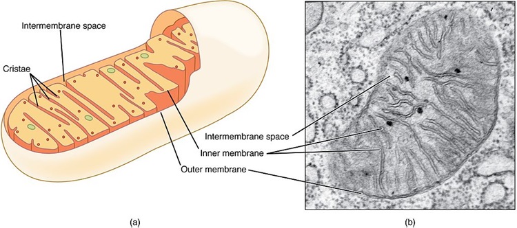
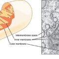
As mentioned previously, when the mitochondria are deprived of estradiol, the membranes surrounding the mitochondria become deformed. The increased permeability of these membrane causes all sorts problems, but top among them, the transfer of nutrients is less efficient. Mitochondria require at least 24 vitamins and minerals to efficiently convert the food we eat into ATP.  Though it is not clear what happens first, whether the enzyme machinery responsible for processing ATP diminishes heralding the increased permeability and deformation of the mitochondrial membranes or whether the loss of estradiol deforms the membranes first and that then results in a loss of ATP, it is clear that the loss of estradiol severely constricts mitochondrial functioning. In both cases, the loss of functionality initiates the survival cascades, and what many call the death spiral, begins.

Clues to Recovery

The easy answer would be to replace the lost estradiol. The problem with that, however, is that the ovaries synthesize a whole host of hormones, not just estradiol, and our ability to replicate those hormones in the appropriate concentrations is just not there yet. Nevertheless, working with a physician or pharmacist experienced in bioidentical hormone replacement may be helpful, at least in the transition period while the body is adapting to the new state of chemistry. The use of synthetics, however, are likely to do more damage than they are worth.

I believe a more prudent approach would be to tackle the health and efficiency of the mitochondria themselves; an approach that had it been undertaken prior to the use of Lupron or oophorectomy may have been able to reduce the impetus for these procedures in the first place. In fact, these recommendations are not specific to Lupron or oophorectomy, but carryover to any type of mitochondrial illness.

Vitamins, Minerals, and Diet

Considering the mitochondrion’s primary purpose is to convert the food we eat into life sustaining ATP, the simplest and most often ignored component of recovery, is to feed the mitochondria what they need to function and avoid the stuff they do not. In other words, eat well and avoid chemical toxicants. In practice, however, this seems to be exceedingly difficult for most of us. Decades of processed food marketing has skewed our preferences away from ‘real food’, and in many ways, disconnected us from the purpose of eating – to fuel our bodies.

Mitochondrial Nutrients to Rescue Mitochondria

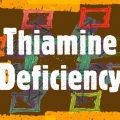
There are 24 of vitamins and minerals required to power the mitochondrial machinery responsible for converting food into ATP. Ensuring an adequate supply of nutrients is critical to repairing mitochondrial damage, perhaps more important than any other aspect of recovery. Top among them are thiamine and magnesium. We have written about this extensively on Hormones Matter and in our book. Thiamine is the gatekeeper to the mitochondrial factory, involved in the initial enzymatic reactions required to convert consumed carbohydrates, fats and proteins into ATP and just about every enzyme reaction throughout the process. More so than any other nutrient, thiamine is critically important to mitochondrial functioning, but its deficiency is least likely to be recognized, even though it causes a host of serious disease processes including death. Magnesium is an essential cofactor for thiamine. Deficiencies in thiamine and magnesium may well account for the vast majority of modern illnesses.

The other B vitamins are also important, as are a variety of minerals. Which ones and in what doses are required for recovery is individual, but often, the dosages are far higher than what is proposed by the RDA and in combinations that are not typically found in standard, over-the-counter (OTC) supplements or even in marketed mitochondrial cocktails, though some of these products are considerably better than the OTCs. In the case of mitochondrial recovery, vitamins and minerals are used at pharmacological doses in order to kick start, and in many cases, compensate for the increased nutrient demands caused by the damage and/or by genetic or epigenetic variables. For individuals with severe deficiencies and/or poor absorption, repeated intravenous vitamins/minerals may be warranted. The details of dosing are covered in our book.

The Mitochondria Diet: Eat Real Food

The second component of healing mitochondria involves diet more broadly. That’s right, what we eat can harm or heal us. Processed foods must be eliminated, as should alcohol and other toxicants. Diets should be high in protein and good fats and low in carbohydrates, and to the extent feasible, organic. Why proteins and fats? Chemistry. Health demands that protein synthesis outpace protein breakdown. That requires a ready supply of protein from the diet and the appropriate nutrients listed above to process them. There is much debate about how much protein, but broadly, and depending upon level and type of illness, one must consider.08 – 1.5kg of protein per kg of weight per day. In research of critically ill patients, higher protein consumption (~1.5kg/kg/day) is associated with better outcomes. With our current predilection for excess carbohydrates, most folks are nowhere near even the lower end of these requirements and that creates a barrier to healing.

The integrity of mitochondrial and all cellular membranes require fatty acids. For women recovering from the loss of estradiol, and the subsequent deformation of mitochondrial membranes, a diet poor in essential fatty acids would be doubly debilitating. Indeed, for most people, an increase in fatty acids would improve health considerably. Fatty acids are also a key fuel substrate for mitochondrial ATP (which just so happens to be thiamine dependent as well).

In general, lower carbohydrate intake is warranted. High carbohydrate intake exacerbates thiamine deficiency and in fact, by itself, with no other risk variables, can induce thiamine deficiency. Researchers from USC have shown that immediately following the loss of estradiol, brain glucose transporters begin to downregulate forcing the brain into ketosis.  That is, at least in mice, the loss of estradiol forces a metabolic shift towards using ketones as the primary fuel source to produce ATP. This means that our traditionally carbohydrate dense diets may not meet the brain’s energetic demands when estradiol is absent. Granted, this research used mice and is preliminary, but it suggests an additional route to healing post Lupron and oophorectomy would be to increase dietary fat. Ketosis may be an option to repair mitochondrial damage.

Having said that, there are disorders of fatty acid metabolism which make lower carbohydrate diets very difficult and sometimes even dangerous. Full blown manifestations of these disorders usually occur in infancy or childhood, but more subtle manifestations brought on by single nucleotide polymorphisms (SNPs) in key genes may remain somewhat latent until triggered. Anecdotally, the inability to metabolize fats seems relatively common among the folks who interact with us, manifesting in what can only be described as energetic collapse post ingestion of fats and proteins. In the cases where SNP analyses are available, the difficulties are related to what have been traditionally considered ‘rare’ variances in key enzymes. Nevertheless, the parameters of these recommendations hold true for most people. Eat real, nutrient dense foods. The specifics of the diet, like the specifics of the nutrient replacements is all that varies.

Avoid Toxicants

Finally, with poorly functioning mitochondria, taxing these organelles further by ingesting the chemically laden foodstuffs produced by conventional agriculture and livestock practices makes healing that much more difficult. To the extent dietary toxicants can be avoided, they should. Similarly, since most, if not all medications damage mitochondria and deplete vital nutrients by one mechanism or another, medication use should be evaluated thoughtfully assessing potential benefits against likely risks.

Is it Really That Simple?

Yes and no. In many ways, it is very simple. Feed the mitochondria and they will do the rest, but in other ways, it is not so simple. Diet is absolutely critical, but mitochondrial recovery also includes using supplements at pharmacological doses, sometimes intravenously, to rescue damaged mitochondria and induce a more favorable mitochondrial replication process. This is difficult for some folks to access. Few physicians have any background in this and many simply do not believe that mitochondrial damage exists or evokes illness. Worse yet and despite evidence to the contrary, many physicians are steadfast in their belief that pharmaceuticals have no bearing on mitochondrial function, and thus cannot possibly be responsible for these illnesses. This means that to the extent pharmaceuticals do not treat and often make matters worse in these cases, fuel to the ‘it-must-be-hysteria-or-psychosomatic’ fire is added. In many ways, the biggest stumbling block is the notion that mitochondrially-mediated disease processes exist at all. A close second, is that nutrients can be used to recover mitochondria and in many cases, override even genetic defects.

Making matters even more complicated, there is no one-size-fits-all diet or supplement protocol. While it is true that thiamine is critical and must be addressed if there is any hope of recovery, everything else downstream must be considered on an individual basis. We each carry a host of unique genetic, epigenetic and environmental exposures that combine to make a complicated chemistry; one that no one wants to untangle and few have the skills to do so. As a result, it is much easier to dismiss the symptoms as psychosomatic and prescribe an antidepressant or other pharmaceutical. I would also mention, even among those of us who have a background in this stuff, we do not know everything and cannot know everything. Untangling these patterns takes time, effort from the patient, lots of research from both parties, and a fair degree of trial and error. Finally, recovery is neither a straight line nor an absolute. There are setbacks, sometimes serious ones, and health has to be actively managed from this point forward. Even with all of these complications, however, better health is possible if one tends to the mitochondria.

We Need Your Help

More people than ever are reading Hormones Matter, a testament to the need for independent voices in health and medicine. We are not funded and accept limited advertising. Unlike many health sites, we don’t force you to purchase a subscription. We believe health information should be open to all. If you read Hormones Matter, like it, please help support it. Contribute now.

Yes, I would like to support Hormones Matter. 

This article was published originally on August 6, 2018. 

Food Composition and Hyperglycemia

5177 views

Over the last few months, I have written a number of white papers on thiamine for contract. They may or may not be published in part or in full at some future date. Among them, I was contracted to write separate papers about thiamine in diabetes, cardiovascular disease, and Alzheimer’s disease. As I began writing the first article, I realized that these were not separate topics. Rather, each disease process was simply a different manifestation of the same core problem: persistent hyperglycemia. This, in turn, was a direct response to our current ultra-processed, chemically-laden, refined sugar, garbage-food environment; a problem we all seem reticent to confront.

The garbage foods that we consume lead to metabolic dysfunction marked by, among other things, hyperglycemia. Hyperglycemia, in turn, leads to specific metabolic adaptations that result in the inability to efficiently convert consumed foods, not just sugars, but amino and fatty acids as well, into energy. (See here for details.) Poor energy metabolism then drives cravings and overeating as a compensatory reaction to increase metabolic energy, which in turn, further entrenches hyperglycemia and its metabolic cascades. It is a deadly spiral, the likes of which are evident in skyrocketing rates of metabolic ill-health. A recent study found that only 12% of the population, 20% if the authors were generous in their description, could be considered metabolically healthy.

From my perspective, it is this shift in metabolic capacity, in the pathways used to metabolize food that drives much, if not all, modern illness. Importantly, many of the disease processes we now consider to be separate entities, like diabetes, the various cardiovascular diseases, the neurodegenerative diseases like Alzheimer’s and dementia, cancer, and even the litany of chronic autoimmune, inflammatory, or pain and fatigue related disease processes, may not be separate at all. They may just represent the way the consumption of ultra-processed foods and the resulting hyperglycemia mix with the individual’s unique genetic and environmental circumstances to form disease. In other words, food provides the spark, hyperglycemia is the kindling, and how and where the flame burns is determined by the individual’s genetics and the totality of his or her life, lifestyle, and environmental exposures. It all begins with food though.

What Are Ultra-processed Foods?

Just about everything in the middle aisles of a super market or purchased from a fast food establishment would be considered ultra-processed. These products are:

…formulations of several ingredients which, besides salt, sugar, oils and fats, include food substances not used in culinary preparations, in particular, flavours, colours, sweeteners, emulsifiers and other additives used to imitate sensorial qualities of unprocessed or minimally processed foods and their culinary preparations or to disguise undesirable qualities of the final product.

In other words, most of the American diet. These products are highly palatable, densely caloried (because of all of added sugars and fats), and loaded with synthetic chemicals, but have no discernable endogenous nutrient content. Sadly, almost 60% of the American diet for adults and close to 70% for kids aged 2-19 years is comprised of ultra-processed food products.

Processing is not the only problem though. Conventionally grown and raised food and livestock have all but bred out of their products any semblance of nutrition in favor of bigger, faster-growing, and more attractive products. In the place of nutrients, we get excess sugars (yes, conventionally grown produce has a higher sugar content than organic or that was grown in the past), along with lots of herbicides, pesticides, hormones, antibiotics and veritable laundry list additional mitochondrial poisons. From farm to table, the composition of modern food products is lacking nutrients while rich with potential anti-nutrient and toxicant compounds. Is it any wonder only 12-20% of the population can be considered metabolically healthy or that hyperglycemia drives modern illness?

Why Hyperglycemia?

Backing up just a bit, let us talk about how discussions of hyperglycemia are framed conventionally and what that has to do with the composition of the foods we ingest. Most discussions of hyperglycemia involve either the absence of sufficient insulin as in the case of Type 1 diabetes or a developed resistance to insulin as in the case of Type 2 diabetes. In either case, there is insufficient insulin available, either absolutely or relative to need, to transport glucose from the bloodstream into the cells and this results in hyperglycemia. Much of the research involves defects in pancreatic islet cell function, glucose receptors and transporters relative to these diseases. In general, diet exacerbates hyperglycemia. With type 2 diabetes, however, diet accounts for almost all of the disease process itself. In many, but not all cases of type 2 diabetes, diet also induces obesity and may provoke a host of additional disease process affecting the heart and the brain. Indeed, Alzheimer’s disease is now considered an outgrowth of persistent hyperglycemia and has been categorized as type 3 diabetes.

This linkage of diabetes with obesity leads many to conclude that if the individual just reduces his/her calories and/or increases activity and loses weight, the diabetes, the obesity, and the assortment of other disease processes that ensue, would resolve and/or be prevented. For some this may be true, but if the persistent rates of obesity, despite reductions in caloric intake are any indicator, this aspect of diet is only indirectly related to the disease at hand. My research involving the some of the metabolic pathways associated with hyperglycemia, leads me to believe that hyperglycemia represents more than just an excess of calories, carbohydrate or otherwise, and that changes to pancreatic islet function, and glucose receptors and transporters are simply adaptive response to ailing mitochondrial metabolism. What is causing metabolism to fail? The American diet of ultra-processed food-like products that are high refined sugars, trans fats and chemical toxins, but low in usable macronutrients and micronutrients – that is the root of these illnesses.

Micronutrient Deficiency Underlies Hyperglycemia

Adenosine triphosphate (ATP), the fuel source for cellular function, the energy currency that all organisms require to survive, is derived entirely from food. The foods we eat provide the macronutrients – protein, fats, and carbohydrates, and the micronutrients –vitamins and minerals – that, with a little oxygen, are then processed by the mitochondria into ATP. Absent frank starvation, the key variables in this process are the micronutrients. Thiamine and its activating partner magnesium are especially important because they manage the gates to this process. Micronutrients derived from foods allow for the catabolism of consumed macronutrients so that it may be turned into ATP. Vitamins and minerals fuel the enzymatic machinery that allows energy factory to work. Insufficient micronutrients slow down enzyme capacity (the energy machinery), causing a backup of macronutrients (a supply excess), at the gates. That excess has to be dealt with. Some of it is forced through alternate pathways that, through a variety processes, break down and salvage some of the macronutrients as a way to temper the backup, but most of the excess either just floats around in the blood or is stored in the fat cells. The glucose that floats around in the blood and desensitizes the glucose receptors and transporters and re-regulates pancreatic islet function – that is hyperglycemia. The glucose that is stored as fat – that is obesity.

Those macronutrients that cannot be processed because of absent micronutrients, not only lead to the hyperglycemia cascades and the various diseases processes associated therewith, but their consumption produces little to no energy or ATP and, in most cases, consumes it. In other words, despite ingesting an excess of calories, the mitochondria, and thus the human in which they reside, are starving. If macronutrients cannot get into the factory, the factory cannot produce ATP. The result is cravings and overeating, which no amount of willpower will overcome. This is why a simple reduction of caloric intake, absent recognition of food composition, does not work for many with type 2 diabetes. They are already starved for energy. Proteomic studies in rodents fed comparable diets illustrate this pattern of poor energetic capacity with reduced expression of the proteins involved in energy metabolism and increased expression of those marking oxidative stress and aberrant cell proliferation (cancer pathways).

A Technical Aside

In more technical terms, when the excess sugars cannot be processed via oxidative phosphorylation or through the pentose phosphate pathway – processes that ultimately produce ATP and other important substrates – they are diverted through salvage pathways like the polyol/sorbitol, hexosamine, diacylglycerol/PKC, AGE pathways. This leads not only to decrements in ATP production but the macro- and microvascular cell damage associated with persistent hyperglycemia leading to heart disease and neurological dysfunction.

Similarly, in the absence of sufficient micronutrients, thiamine in particular, the catabolism of branched chain amino acids suffers, resulting in increased branched chain keto acids, especially short and medium chain acylcarnitines. Surplus acylcarnitines then overwhelm the b-oxidation pathway involved in fatty acid metabolism. This, in turn, leads to incomplete fatty acid metabolism (dyslipidemia) and the formation of the pro-inflammatory diacylglycerol and ceramides associated with metabolic dysfunction. The hyper-activation of ceramide synthesis expedites cell death, blocking complex 3 of the electron transport chain in the mitochondria.

Inadequate micronutrient availability, and again, thiamine and magnesium especially, further imperials the alpha oxidation of fatty acids. This is the step before beta-oxidation. Poor alpha-oxidation results in increased phytanic acid and disrupted sphingolipid homeostasis; two patterns with linked with a variety of neurological sequelae. All of this is linked to persistent hyperglycemia, which evolves from inadequate micronutrient content relative to demands.

Coincidently, COVID death is linked to both increased ceramide synthesis and disturbed sphingolipid homeostasis.

We postulate that SARS[1]CoV-2 causes endothelial damage by binding ACE2 and misbalancing the renin-angiotensin pathway, dysregulating sphingolipids and activating the ceramide pathway, known to mediate endothelial cell apoptosis in the setting of radiation damage. Such injury also generates reactive oxygen species, vasoconstriction and hypoxia, and ultimately the deposition of platelets on an exposed vessel basement membrane initiating the intravascular coagulopathy and multi-organ failure, pathognomonic of severe COVID-19 and death.

Underlying both processes are micronutrient deficient patterns of hyperglycemia, e.g. insufficient thiamine, magnesium and likely other nutrients, but most have not been investigated. Inasmuch hyperglycemia accounts for much of the risk for COVID severity, it is difficult not wonder if these pathways were not already entrenched pre-virus and the virus simply escalated the negative adaptations beyond rescue.

Food Composition Matters More Than Caloric Intake

From this perspective, it is clear that it is not solely an excess of calories that causes hyperglycemia, or even an excess of carbohydrates, although both play a large role. It is the quality or composition of the food that is the problem. Modern foods are calorie dense, sure, primarily because of the use of refined sugars and added fats. They are also loaded with chemical poisons, which we all seem to disregard as important. Carbohydrates derived from natural, organic, and unadulterated fruits, vegetables and grains, carry with them vitamins, minerals, fiber, and proteins that allow the body to convert the macronutrient substrates into useable energy. Indeed, a diet rich in these types of foods is unlikely to induce hyperglycemia or obesity. In contrast, processed foods, while high in carbohydrates, fats, and chemicals that are toxic to the mitochondria, carry few to no micronutrients, little to no fiber, or other compounds that can be used by the body to produce ATP all the while carrying an abundance of chemical toxins. From a metabolic standpoint, ultra-processed foods are nothing more than edible poisons. They demand more energy to process than they add and wreak havoc with far more systems than were illustrated here. The hyperglycemia and associated damage that ensues is evidence of this process. If we are to tackle these health issues, the entirety of modern food landscape relative to metabolic health must be addressed.

We Need Your Help

More people than ever are reading Hormones Matter, a testament to the need for independent voices in health and medicine. We are not funded and accept limited advertising. Unlike many health sites, we don’t force you to purchase a subscription. We believe health information should be open to all. If you read Hormones Matter, like it, please help support it. Contribute now.

Yes, I would like to support Hormones Matter. 

Image created using Canva AI.

This article was published originally on October 28, 2021. 

Beriberi: The Great Imitator

33207 views

Because of some unusual clinical experiences as a pediatrician, I have published a number of articles in the medical press on thiamine, also known as vitamin B1. Deficiency of this vitamin is the primary cause of the disease called beriberi. It took many years before the simple explanation for this incredibly complex disease became known. A group of scientists from Japan called the “Vitamin B research committee of Japan” wrote and published the Review of Japanese Literature on Beriberi and Thiamine, in 1965. It was translated into English subsequently to pass the information about beriberi to people in the West who were considered to be ignorant of this disease. A book published in 1965 on a medical subject that few recall may be regarded in the modern world as being out of date and of historical interest only, however, it has been said that “Those who do not learn history are doomed to repeat it”. And repeat it, we are.

Beriberi is one of the nutritional diseases that is regarded as being conquered. It is rarely considered as a cause of disease in any well-developed country, including America. In what follows, are extractions from this book that are pertinent to many of today’s chronic health issues. It appears that thiamine deficiency is making a comeback but it is rarely considered as a possibility.

The History of Beriberi and Thiamine Deficiency

Beriberi has existed in Japan from antiquity and records can be found in documents as early as 808. Between 1603 and 1867, city inhabitants began to eat white rice (polished by a mill). The act of taking the rice to a mill reflected an improved affluence since white rice looked better on the table and people were demonstrating that they could afford the mill. Now we know that thiamine and the other B vitamins are found in the cusp around the rice grain. The grain consists of starch that is metabolized as glucose and the vitamins essential to the process are in the cusp. The number of cases of beriberi in Japan reached its peak in the 1920s, after which the declining incidence was remarkable. This is when the true cause of the disease was found. Epidemics of the disease broke out in the summer months, an important point to be noted later in this article.

Early Thiamine Research

Before I go on, I want to mention an extremely important experiment that was carried out in 1936. Sir Rudolf Peters showed that there was no difference in the metabolic responses of thiamine deficient pigeon brain cells, compared with cells that were thiamine sufficient, until glucose (sugar) was added. Peters called the failure of the thiamine deficient cells to respond to the input of glucose the catatorulin effect. The reason I mention this historical experiment is because we now know that the clinical effects of thiamine deficiency can be precipitated by ingesting sugar, although these effects are insidious, usually relatively minor in character and can remain on and off for months. The symptoms, as recorded in experimental thiamine deficiency in human subjects, are often diagnosed as psychosomatic. Treated purely symptomatically and the underlying dietary cause neglected, the clinical course gives rise to much more serious symptoms that are then diagnosed as various types of chronic brain disease.

  • Thiamine Deficiency Related Mortality. The mortality in beriberi is extremely low. In Japan the total number of deaths decreased from 26,797 in 1923 to only 447 in 1959 after the discovery of its true cause.
  • Thiamine Deficiency Related Morbidity. This is another story. It describes the number of people living and suffering with the disease. In spite of the newly acquired knowledge concerning its cause, during August and September 1951, of 375 patients attending a clinic in Tokyo, 29% had at least two of the major beriberi signs. The importance of the summer months will be mentioned later.

Are the Clinical Effects Relevant Today?

The book records a thiamine deficiency experiment in four healthy male adults. Note that this was an experiment, not a natural occurrence of beriberi. The two are different in detail. Deficiency of the other B vitamins is involved in beriberi but thiamine deficiency dominates the picture. In the second week of the experiment, the subjects described general malaise, and a “heavy feeling” in the legs. In the third week of the experiment they complained of palpitations of the heart. Examination revealed either a slow or fast heart rate, a high systolic and low diastolic blood pressure, and an increase in some of the white blood cells. In the fourth week there was a decrease in appetite, nausea, vomiting and weight loss. Symptoms were rapidly abolished with restoration of thiamine. These are common symptoms that confront the modern physician. It is most probable that they would be diagnosed as a simple infection such as a virus and of course, they could be.

Subjective Symptoms of Naturally Occurring Beriberi

The early symptoms include general malaise, loss of strength in knee joints, “pins and needles” in arms and legs, palpitation of the heart, a sense of tightness in the chest and a “full” feeling in the upper abdomen. These are complaints heard by doctors today and are often referred to as psychosomatic, particularly when the laboratory tests are normal. Nausea and vomiting are invariably ascribed to other causes.

General Objective Symptoms of Beriberi

The mental state is not affected in the early stages of beriberi. The patient may look relatively well. The disease in Japan was more likely in a robust manual laborer. Some edema or swelling of the tissues is present also in the early stages but may be only slight and found only on the shin. Tenderness in the calf muscles may be elicited by gripping the calf muscle, but such a test is probably unlikely in a modern clinic.

In later stages, fluid is found in the pleural cavity, surrounding the heart in the pericardium and in the abdomen. Fluid in body cavities is usually ascribed to other “more modern” causes and beriberi is not likely to be considered. There may be low grade fever, usually giving rise to a search for an infection. We are all aware that such symptoms come from other causes, but a diet history might suggest that beriberi is a possibility in the differential diagnosis.

Beriberi and the Cardiovascular System

In the early stages of beriberi the patient will have palpitations of the heart on physical or mental exertion. In later stages, palpitations and breathlessness will occur even at rest. X-ray examination shows the heart to be enlarged and changes in the electrocardiogram are those seen with other heart diseases. Findings like this in the modern world would almost certainly be diagnosed as “viral myocardiopathy”.

Beriberi and the Nervous System

Polyneuritis and paralysis of nerves to the arms and legs occur in the early stages of beriberi and there are major changes in sensation including touch, pain and temperature perception. Loss of sensation in the index finger and thumb dominates the sensory loss and may easily be mistaken for carpal tunnel syndrome. “Pins and needles”, numbness or a burning sensation in the legs and toes may be experienced.

In the modern world, this would be studied by a test known as electromyography and probably attributed to other causes. A 39 year old woman is described in the book. She had lassitude (severe fatigue) and had difficulty in walking because of dizziness and shaking, common symptoms seen today by neurologists.

Beriberi and the Autonomic Nervous System

We have two nervous systems. One is called voluntary and is directed by the thinking brain that enables willpower. The autonomic system is controlled by the non-thinking lower part of the brain and is automatic. This part of the brain is peculiarly sensitive to thiamine deficiency, so dysautonomia (dys meaning abnormal and autonomia referring to the autonomic system) is the major presentation of beriberi in its early stages, interfering with our ability for continuous adaptation to the environment. Since it is automatic, body functions are normally carried out without our having to think about them.

There are two branches to the system: one is called sympathetic and the other one is called parasympathetic. The sympathetic branch is triggered by any form of physical or mental stress and prepares us for action to manage response to the stress. Sensing danger, this system activates the fight-or-flight reflex. The parasympathetic branch organizes the functions of the body at rest. As one branch is activated, the other is withdrawn, representing the Yin and Yang (extreme opposites) of adaptation.

Beriberi is characterized in its early stages by dysautonomia, appearing as postural orthostatic tachycardia syndrome (POTS). This well documented modern disease cannot be distinguished from beriberi except by appropriate laboratory testing for thiamine deficiency. Blood thiamine levels are usually normal in the mild to moderate deficiency state.

Examples of Dysfunction in Beriberi

The calf muscle often cramps with physical exercise. There is loss of the deep tendon reflexes in the legs. There is diminished visual acuity. Part of the eye is known as the papilla and pallor occurs in its lateral half. If this is detected by an eye doctor and the patient has neurological symptoms, a diagnosis of multiple sclerosis would certainly be entertained.

Optic neuritis is common in beriberi. Loss of sensation is greater on the front of the body, follows no specific nerve distribution and is indistinct, suggestive of “neurosis” in the modern world.

Foot and wrist drop, loss of sensation to vibration (commonly tested with a tuning fork) and stumbling on walking are all examples of symptoms that would be most likely ascribed to other causes.

Breathlessness with or without exertion would probably be ascribed to congestive heart failure of unknown cause or perhaps associated with high blood pressure, even though they might have a common cause that goes unrecognized.

The symptoms of this disease can be precipitated for the first time when some form of stress is applied to the body. This can be a simple infection such as a cold, a mild head injury, exposure to sunlight or even an inoculation, important points to consider when unexpected complications arise after a mild incident of this nature. Note the reference to sunlight and the outbreaks of beriberi in the summer months. We now know that ultraviolet light is stressful to the human body. Exposure to sunlight, even though it provides us with vitamin D as part of its beneficence, is for the fit individual. Tanning of the skin is a natural defense mechanism that exhibits the state of health.

Is Thiamine Deficiency Common in America?

My direct answer to this question is that it is indeed extremely common. There is good reason for it because sugar ingestion is so extreme and ubiquitous within the population as a whole. It is the reason that I mentioned the experiment of Rudolph Peters. Ingestion of sugar is causing widespread beriberi, masking as psychosomatic disease and dysautonomia. The symptoms and physical findings vary according to the stage of the disease. For example, a low or a high acid in the stomach can occur at different times as the effects of the disease advance. Both are associated with gastroesophageal reflux and heartburn, suggesting that the acid content is only part of the picture.
A low blood sugar can cause the symptoms of hypoglycemia, a relatively common condition. A high blood sugar can be mistaken for diabetes, both seen in varying stages of the disease.

It is extremely easy to detect thiamine deficiency by doing a test on red blood cells. Unfortunately this test is either incomplete or not performed at all by any laboratory known to me.

The lower part of the human brain that controls the autonomic nervous system is exquisitely sensitive to thiamine deficiency. It produces the same effect as a mild deprivation of oxygen. Because this is dangerous and life-threatening, the control mechanisms become much more reactive, often firing the fight-or-flight reflex that in the modern world is diagnosed as panic attacks. Oxidative stress (a deficiency or an excess of oxygen affecting cells, particularly those of the lower brain) is occurring in children and adults. It is responsible for many common conditions, including jaundice in the newborn, sudden infancy death, recurrent ear infections, tonsillitis, sinusitis, asthma, attention deficit disorder (ADD), hyperactivity, and even autism. Each of these conditions has been reported in the medical literature as related to oxidative stress. So many different diseases occurring from the same common cause is offensive to the present medical model. This model regards each of these phenomena as a separate disease entity with a specific cause for each.

Without the correct balance of glucose, oxygen and thiamine, the mitochondria (the engines of the cell) that are responsible for producing the energy of cellular function, cannot realize their potential. Because the lower brain computes our adaptation, it can be said that people with this kind of dysautonomia are maladapted to the environment. For example they cannot adjust to outside temperature, shivering and going blue when it is hot and sweating when it is cold.

So, yes, beriberi and thiamine deficiency have re-emerged. And yes, we have forgotten history and appear doomed to repeat it. When supplemental thiamine and magnesium can be so therapeutic, it is high time that the situation should be addressed more clearly by the medical profession.

We Need Your Help

More people than ever are reading Hormones Matter, a testament to the need for independent voices in health and medicine. We are not funded and accept limited advertising. Unlike many health sites, we don’t force you to purchase a subscription. We believe health information should be open to all. If you read Hormones Matter, like it, please help support it. Contribute now.

Yes, I would like to support Hormones Matter.

This article was published originally on November 4, 2015. 

Digging Deeper into Mitochondrial Dysfunction

15087 views

When you have a hammer, everything becomes a nail, or so they say. I worry about this as I dig deeper into mitochondrial dysfunction. Could all of these disparate symptoms and conditions have their roots in the mitochondria? Could it be that simple? Perhaps. More and more, as I search for explanations for the devastating symptoms that so many of our readers report, the research I find points to mitochondrial dysfunction. Sure, changes in gut microbiota and function are apparent and often related and certainly immune dysregulation is a component of these illnesses, but the underlying connection among these disturbances seems inevitably and inextricably linked to dysfunctional mitochondria as the central hub of illness. Heal the mitochondria, heal the body is quickly becoming my new mantra.

Mitochondria as Danger Sensors

Some researchers argue that the mitochondria are the danger sensors for host organisms; having evolved over two billion years to identify and communicate signs of danger to the cells within which they reside. The signaling is simple and yet highly refined, involving a series of switches that control cellular energy, and thus, cellular life or death. When danger is present, energy resources are conserved and the immune system fighters are unleashed. When danger is resolved, normal functioning can resume.

If the danger is not resolved and the immune battles must rage on, the mitochondria begin the complicated process of reallocating resources until the battle is won or the decision is made to institute what can only be described as suicide – cell death. Cell death is a normal occurrence in the cell cycle of life. Cells are born and die for all manner of reasons. But when cell death occurs from mitochondrial injury, it is messy, and evokes even broader immune responses, setting a cascade in motion that is difficult to arrest.

And, if the on the battlefield, the host army is understaffed and under-resourced, no matter how hard the immune fighters battle, the fight will be lost, maybe not immediately, but eventually. All sorts of mechanisms will be employed to reallocate and reinforce needed battlements, but they will be for naught, further depleting already scarce host resources, until the decision is made, within the mitochondria, to begin pulling back, withdrawing, and ultimately casting the final orders of cell death.

It’s not Autoimmunity, but Impaired Immunity

I never much liked the war model of health and disease, but it seems to work well as metaphor for immune functioning, as it is far more illustrative and useful than the self-versus non-self-characterization. Really, what army with two billion years of experience, one that contains all of the memories and skills of battles past, would misidentify itself and begin broad scale fratricide  – kill itself and its brethren for no other reason but mistaken identity and do so for years on end?  Sure, there can be errors, over compensation and other weaknesses in the immune system, but not continued aggression towards itself in some maladaptive response. That makes no sense and contradicts the very notion and function of an immune system – to keep the host organism alive and well. Indeed, when we consider the trillions of microbes – clear non-self entities – that live inside and upon us, the idea that the immune system evolved simply to kill the non selves seems laughable. And so, I reject the concept of autoimmunity, not because the patients who suffer from continued immune system activation are not ill, they are, but because the concept of autoimmunity belies the very nature of immune function and severely limits possible approaches to recovery.

The Naming of Things

Many of you might be thinking ‘what the heck does what we name things have to do with understanding illness?’  Well, the language and the characterization of disease impacts therapeutic choices. In a system where autoimmunity dominates the discussion, survival is predicated on suppressing the invading immune army. Consequently, most therapeutic options for autoimmune disease are immunosuppressant, and mostly they fail. In contrast, if one characterizes immune function by its ability to protect and sustain life by fending off dangers or threats to survival, be they self or non-self, it does not matter, then we can be open to finding causes for those failed battles. We can ask questions like: what resources are missing that would allow the immune army to fend off the danger once and for all or what could heal the damaged cells, scavenge toxicants and oxidants or re-calibrate mitochondrial energy production? When we re-frame the discussion in this way, we open the door to a deeper understanding of health and disease. It is from this perspective, one that says chronic immune activation is not a disease itself but a symptom of an on-going and failing immune battle, that we can get to the mitochondria as the central hub for chronic ill-health.

Evolution and the Mighty Mitochondria

Mitochondria are interesting little buggers, having evolved from the very parasites our immune system sought to protect us against. Called symbionts, the mitochondria were microbial intruders swallowed by the host. In a brilliant move of survival, they somehow convinced the host organism not only not to kill them but to let the mitochondria, a parasitic intruder, run the host’s energy supply. The mitochondria proved their utility and developed a symbiotic relationship with the cells within which they resided. Over time, mitochondria developed a myriad of intricate communication and resource allocation mechanisms to ensure not only their survival but that of their host organism. And so, in many ways, the mitochondria evolved as part of a cooperative and collaborative ecosystem; one in which they sense and communicate danger to the rest of the organism, and if need be, initiate the final death programs; something, they should be loath to do, since their survival depends entirely on host survival.

Clearly though, and from the very beginning, the mitochondria positioned themselves as the brains of the operation. Mitochondria control energy. There is no other resource more important to the living organism than energy. Consider the most consistent sickness behaviors across all illness include, lethargy, fatigue, sleepiness, often followed by muscle and body aches, anorexia or the loss of interest in eating. The reduction in energy is purely a function of mitochondrial resources. The achy muscles are moderated by mitochondrial retractions of energy and the loss of appetite too, mitochondrial diminishments – recall the orexin/hypocretin system. By whatever pathway, declining mitochondrial energy production arises when danger signals, or more appropriately, cell damage signals are communicated. It is then that the immune armies are activated and inflammation sequences unleashed.

What Does Mitochondrial Dysfunction Look Like?

Everything and nothing at the same time. Mitochondrial dysfunction doesn’t lead to one, clear cut disease, even when there are clear genetic markers, but predisposes one to everything. Where mitochondrial damage is felt and the subsequent immune events present is complex and dependent upon the interactions among the host organism’s innate predispositions, environmental exposures and nutritional status, with the latter two significantly influencing each other. The microbial composition of the host, especially in the gut, but also on the skin and the various mucous membranes that interface with the outside world, can also moderate or trigger the danger signals that lead to mitochondrial dysfunction. More often than not though, mitochondrial damage is felt where oxygen demands are greatest, the brain, the heart, the gut, the muscles. Diseases that are currently identified discretely might all have common symptoms – the mitochondria.

Certainly, chronic fatigue should be considered mitochondrial in nature. I don’t think there is a more clear-cut example of mitochondrial dysfunction than severe fatigue, muscle pain and weakness. The question becomes, from where does the dysfunction originate and how can it be fixed or healed?

Migraine, seizures, ataxias and other neurological disorders are emerging as mitochondrial, particularly was more work is done on the hypocretin/orexin system.

Autonomic dysregulation, recognized under the umbrella as dysautonomias are mitochondrial in nature.

Thyroid dysfunction is likely mitochondrial in nature; the interaction between thyroid hormones and mitochondria is direct. Given the mitochondria’s role in steroidogenesis, other hormone systems are likely modulated by mitochondrial functioning.

Research is emerging suggesting that gastrointestinal disturbances, particularly those of dysmotility like IBS, gastroparesis, constipation and pseudo obstruction but also anorexia are mitochondrial in nature. Indeed, the GI system has its own nervous system, called the enteric nervous system. Only 10-15% of GI motility is controlled from brain’s autonomic system. The rest is controlled on site by the enteric system, mostly from cells called the interstitial of cells Cajal – the smooth muscle cells that propagate contractility and rhythm and form the gut barrier between the inside contents and the rest of the body.  The mitochondria control energy usage and production here. Mess with these cells, diminish oxygen usage, pull back energy production and all sorts of things go wrong. We can get ill-timed contractions or no contractions at all, making the movement of food stuffs through the GI impossible. Poor absorption and metabolism of nutrients, and increased permeability of the tight junctions allowing for the leaky gut scenario common in many chronic conditions, become prominent and are also symptoms of mitochondrial dysfunction. Even anorexia, the will to eat, can be disturbed significantly by mitochondrial damage.

And I suspect, although I have no evidence to support this claim, in women, mitochondrial damage can express itself in the reproductive organs, especially when oxygen demands are greatest, menstruation and pregnancy. Consider the increasingly painful muscle contractions for some women involved in shedding the uterine lining during menstruation, just as diminished oxygen and energy in other muscles begins the cycle of lactate production and buildup that initiates pain, so too might this happen in the uterus. As mitochondrial deficiencies persist, uterine dysfunction would grow and compensatory immune system mechanisms increase until the compensatory mechanisms take a life of their own. When we consider the mitochondrial influence on GI motility, their influence on uterine function is not difficult to imagine. I have an inkling that endometriosis, the excessive growth of endometrial cells first within the uterus and then in regions of the body where they ought not be, is a protective mechanism, albeit an aberrant and problem causing one, that indicates increased mitogenesis and cell growth as a compensatory reaction to some original mitochondrial inadequacy. How this might happen molecularly provides some intriguing possibilities.

Immune function and inflammatory reactions are directly controlled by mitochondrial signals of danger and so to the extent we see chronic inflammatory conditions, one can look towards mitochondrial resources and the ensuing danger signals for clues towards reducing these reactions. While much of the clinical research is nascent, more and more clinicians, often from disparate specialties and sometimes without recognizing the immune-mitochondrial connections, have made great inroads towards healing and restoring mitochondrial function through diet and nutritional supplementation paired with the reduction and removal of environmental and medical toxins and dietary inflammasomes.

Is Everything a Nail, When One has a Hammer?

Maybe, but I can’t help but thinking that this mitochondrial hammer might be the one to hit the nail on the head and finally make some inroads towards reducing the suffering and burden of chronic disease. Only time will tell. For the moment, however, this is a hammer that deserves more recognition and whether it turns out to be the final clue or not one thing is clear, mitochondrial health is critical for human health. Deny the mitochondria their due and chronic, complicated illness will persist.

We Need Your Help

More people than ever are reading Hormones Matter, a testament to the need for independent voices in health and medicine. We are not funded and accept limited advertising. Unlike many health sites, we don’t force you to purchase a subscription. We believe health information should be open to all. If you read Hormones Matter, like it, please help support it. Contribute now.

Yes, I would like to support Hormones Matter. 

Image credit: OpenStax, CC BY 4.0, via Wikimedia Commons

This article was published previously on Hormones Matter in June 2014.

Thyroid Hormones, Mitochondrial Functioning, and Hair Loss

28557 views

Hair loss is a common symptom of thyroid disease. In our research, hair loss, changes in color or luster, and skin changes are regularly reported as one of the first symptoms noticed in an emerging non-allergenic adverse reaction to a medication or vaccine. These symptoms often coincide with unexplained fatigue and muscle pain. Given that many patients who develop the more chronic, multi-symptom medication or vaccine reactions also develop thyroid disease and frequently exhibit signs of mitochondrial damage, I wondered if somehow the hair and skin changes could be early warning signs of diminished mitochondrial functioning. I also wondered if all of these variables were connected. It turns out that not only are they connected, but incredibly interdependent.

What are Mitochondria?

Recall from high school biology, the mitochondria are those bean-shaped organelles inside cells that are responsible for cellular respiration or energy production. Through a variety of pathways, the mitochondria provide fuel for cell survival. In addition to cellular energy production, mitochondria control cell apoptosis (death), calcium, copper, and iron homeostasis, and steroidogenesis. In essence, mitochondria perform the key tasks associated with cell survival, and indeed, human survival. Damage the mitochondria and cellular dysfunction or death will occur. Damage sufficient numbers of mitochondrion and chronic, multi-symptom illness arises.

How to Damage Mitochondria

Mitochondria are remarkably resilient given the proper nutrients, but without those nutrients, they can be highly susceptible to damage. Mitochondrial damage can be inherited via mutations in maternal DNA (mtDNA) or nuclear DNA and present at birth or remain latent until triggered later in life, as in the case of mitochondrial endocrinopathies. Mitochondria are also susceptible to epigenetic changes, which can be heritable and acquired and remain latent until triggered.  Finally, mitochondrial impairment can derive from pharmaceutical or environmental exposures and nutrient or cofactor deficits. The sheer number of mechanisms that can influence mitochondrial functioning and heritability make diagnosing and predicting mitochondrial dysfunction difficult at best, particularly acquired or functional mitochondriopathies that are not evident from genetic or epigenetic testing. It is precisely those acquired mitochondriopathies, particularly those seemingly triggered by pharmaceutical reactions, that we are most interested in here at Hormones Matter. Indeed, acquired mitochondrial damage represents a nascent and emerging field in medicine, particularly in toxicology, as many drugs and vaccines damage mitochondrial functioning both directly and indirectly.

What Mitochondrial Damage Looks Like

Mitochondrial damage presents in a highly diverse, multi-organ, multi-symptom manner. On the surface, patients with mitochondrial dysfunction will appear to have multiple, unconnected diagnoses, from gastrointestinal distress to cognitive deficits, from cardiac arrhythmias to multiple sclerosis-like symptoms, and everything in between and beyond. According to Dr. Richard Boles, an expert on mitochondrial dysfunction:

“Mitochondrial dysfunction doesn’t really cause anything, what it does is predisposes towards seemingly everything. It’s one of many risk factors in multifactorial disease. It can predispose towards epilepsy, chronic fatigue, and even autism, but it doesn’t do it alone. It does it in combination with other factors, which is why in a family with a single mutation going through the family, everyone in the family is affected in a different way. Because it predisposes for disease throughout the entire system.”

This is partially because the human body contains over a billion mitochondria which are essential to cellular functioning in every cell of the body. Where the dysfunction emerges is dependent upon where the impaired mitochondria reside, by what mechanism the mitochondria are damaged, and how intervening variables, such as overall health, nutrition, and environment come into play. Given the mitochondrion’s role in energy production, highly energy-dependent tissues such as the brain, the heart, the liver, and even muscles, are most susceptible to direct mitochondrial damage. And considering the mitochondrion’s role in cellular energetics, fatigue is almost always present with mitochondrial dysfunction.

Hormone Synthesis and Mitochondrial Functioning

Adding yet another layer of complexity, mitochondria also control steroid production in the adrenal glands, ovaries, testes, and thyroid. Any impairment of mitochondrial functioning can have a significant influence on hormone production and regulation. Since hormones, like the mitochondria, also impact all facets of biological homeostasis, energy, and metabolism, damage to endocrine mitochondria can represent a double-hit and begin a cascade of endocrine ill-effects that are difficult to control. This is particularly true of the thyroid gland.

Thyroid Hormones and Mitochondrial Functioning

The cells within the thyroid gland are dependent upon proper mitochondrial functioning to maintain health and proper mitochondrial functioning is dependent upon thyroid hormones to manage cellular energy production. This reciprocal and interdependent relationship makes the thyroid especially susceptible to a mitochondrial spiral. Both thyroid and mitochondrial damage have been observed in our medication and vaccine adverse reaction populations.

Thyroid hormones regulate mitochondrial functioning. Triiodothyronine (T3) in particular is considered one of the major regulators of mitochondrial activity stimulating mitochondrial biogenesis (the birth of new mitochondria) both directly (genomic), indirectly (non-genomic), and epigenetically.

T3 is responsible for increasing cellular heat production and oxygen consumption, core activities of mitochondrial metabolism. In hypothyroid states, heat and oxygen are reduced, whereas, in hyperthyroid states, the two are increased. Here the intracellular patterns of heat and energy production correspond to the clinical symptoms of hypo- and hyperthyroid states. Other thyroid hormones along  the hypothalamus – pituitary – thyroid axis (HPT) and the other iodothyronines within the thyroid hormone metabolic pathway influence mitochondrial functioning. Remove or reduce the presence of the thyroid hormones and mitochondria produce less energy and eventually die. With them, the cells in which they reside die too. Conversely, as mitochondria within the thyroid become less efficient, smaller concentrations of thyroid hormones are produced.  With reduced thyroid hormones, mitochondrial efficiency continues to decline and so on, and so on.

Hair Follicles: Mini – HPTs

German researchers recently identified multiple mechanisms by which human hair follicles are responsive to thyroid hormones. Their research showed that human skin and hair follicles possess an equivalent peripheral HPT axis with all of the corresponding hormones such as the central HPT. It turns out that hair follicle mitochondria are differentially responsive to each of the thyroid hormones along that axis and are responsive to other iodothyronines not typically considered bioactive, such as diiodothyronine (T2).

An interesting finding, related specifically to the hair follicle, and perhaps other mitochondria, thyroid hormones were protective against reactive oxygen species (ROS) production via multiple mechanisms. ROS, also called free radicals or oxidants, are natural by-products of oxygen (energy) metabolism important to a number of basic cell and life processes, like signaling and the defense against pathogens, but ROS levels must be kept in strict balance. Too much or not enough ROS and health goes awry. In the case of adverse fluoroquinolone reactions, increased ROS production is implicated.  According to the hair-follicle study, thyroid hormones protect against ROS production and regulate the enzymes that scavenge for and eliminate free radicals – our own internal antioxidants. If this function is conserved throughout the body, it provides one more reason to investigate and appropriately manage thyroid damage in medication adverse reactions.

Hair Loss and Mitochondrial Damage

Skin and hair follicles are dense with mitochondria and highly regulated by thyroid hormones such that the mechanism for hair loss in some individuals can be attributed to either diminished thyroid hormones and/or damaged mitochondria. Since the relationship between thyroid hormones and mitochondria is reciprocal, it is difficult to tell which impairment comes first. However, given what we know about hair growth cycles and what we know about thyroid hormones and mitochondrial functioning, it is possible to speculate and backdate a chemical insult precipitating sudden and unexplained hair loss. For more incipient reactions, it is a bit more difficult. Regardless, however, it appears that unexplained hair loss is a sign of poor mitochondrial functioning.

Hair growth occurs in phases. The anagen phase is the growth cycle where hair follicles grow about 1 cm per day for 28 days. This growth phase lasts for 2-7 years. The exact time frame is genetically, or more specifically, epigenetically determined by factors associated with the health of the maternal grandmother. After the anagen phase, the hair follicles reach a transitional, quiescent period lasting approximately 2-3 weeks. This is then followed by the telogen phase where hair begins to fall out. At any given time, up to 90% of hair follicles are in the anagen or growth phase while the remaining follicles are either catagen (10-14%) or telogen phases (1-2%).

Chemo Induced Hair Loss: Answers in the Mitochondria?

With chemotherapy, hair loss begins 2-4 weeks after treatment begins and although multiple mechanisms have been investigated, none have been able to explain or treat effectively chemo-induced hair loss. I would suspect that given the time frame, the toxic insult of chemotherapy, the role of mitochondria in hair growth, and the connection to thyroid damage, that chemo-induced alopecia is representative of mitochondrial damage. The ability to maintain hair growth during chemo may be related to supporting mitochondrial and/or thyroid health.

In the case of other presumed less toxic or at least less directly toxic chemical insults such as medication or vaccine adverse reactions, the initial loss of hair that begins either in the weeks preceding the full onslaught of symptoms or coincident with those symptoms, marks a decline in mitochondrial functioning and likely an impending decline in thyroid functioning.

Hair Loss: A Reallocation of Mitochondrial Resources

Considering, that hair generation is an energy (read mitochondrial) intense process, sudden hair loss could be an early marker that mitochondrial resources are limited and being reallocated towards more critical operations like brain and heart functioning. When the components for proper mitochondrial functioning are absent, be it the thyroid hormones or the co-factors necessary for cellular energy (ATP) production, the first wave of resource allocation might be to cease non-essential activities. The non-essential activities would include hair growth (and wakefulness in general – read Medication and Vaccine Adverse Reactions and the Orexin – Hypocretin Neurons). Sudden or unexplained hair loss could indicate mitochondrial impairment. Backdate the hair loss 2-4 weeks and an illness, a medication, vaccine, or environmental exposure could be the culprit. Whatever the cause, the thyroid and mitochondrial health should be considered and treatment initiated accordingly because if the disease process continues, the symptoms will expand beyond the hair, potentially to every tissue and organ in the body. Concurrently, investigate and amend nutritional status. Mitochondrial functioning is critically dependent on proper nutrients. Deficits in important nutrients, like thiamine, can have severe repercussions.

Feed your thyroid. Feed your mitochondria.

We Need Your Help

More people than ever are reading Hormones Matter, a testament to the need for independent voices in health and medicine. We are not funded and accept limited advertising. Unlike many health sites, we don’t force you to purchase a subscription. We believe health information should be open to all. If you read Hormones Matter, and like it, please help support it. Contribute now.

Yes, I would like to support Hormones Matter.

This article was first posted on Hormones Matter in May of 2014.

COVID Notes: Considering Drug Induced Mitochondrial Damage

4959 views

Much has been written about the associations between COVID severity, chronicity, and pre-existing conditions. Top among those conditions include cardiovascular disease and diabetes, likely type 2, but both are lumped together. What has not been discussed is why this would be the case. On a basic level, fighting two illnesses takes more energy than fighting one. This is obvious. What is not obvious is that many modern illnesses, especially cardiovascular disease and type 2 diabetes, begin in the mitochondria as a consequence of diet and lifestyle. Statistically, 80% of cardiovascular disease and 78-83% of type 2 diabetes can be traced back to longstanding dietary, lifestyle and environmental issues* that effectively diminish mitochondrial energetic capabilities and disrupt metabolic flexibility; and the remainder that did not originate from diet and lifestyle are certainly affected by these variables.

To function effectively and to convert the foods we eat into energy or ATP, the mitochondria require sufficient vitamins and minerals, 22 of them, in fact. Western diets, while high in calories, are woefully low in these micronutrients, even when fortified, creating what we refer to as high calorie malnutrition. Against this dietary backdrop, reduced ATP then leads to a constant, low level molecular hypoxia. This is not a hypoxia of obstruction or exertion, but more fundamental. For without proper nutrients, mitochondria can neither utilize oxygen effectively to create ATP, nor do they have sufficient ATP to traffic the O2 into the hemoglobin where it can be pumped into circulation to feed tissues and organs. It is a subtle desaturation, at least initially, but one that initiates all sorts of compensatory reactions to mitigate risk; reactions that are necessary and lifesaving in the short term but become increasingly harmful as time passes.

With insufficient ATP, inflammatory and immune reactions become disrupted and even seemingly chaotic; hormone and electrolyte regulation becomes imbalanced and organ and brain function diminishes. We get disrupted autonomic function (dysautonomia), which cycles back and further disrupts everything else. Depression, anxiety and other mental health issues are also common. This underlying mitochondrial distress is part of the reason why patients with comorbid conditions are at increased risk of not only developing but succumbing to COVID, or really, any virulent pathogen. Their mitochondria are already taxed. They are already carrying low-level hypoxia and, in a very real way, they simply do not have the energy to mount or manage a successful defense.

Now, to add insult to already injured mitochondria, we prescribe medications to manage these conditions rather than correct the root cause, which remember is mitochondrial distress. These medications, while they effectively provide the semblance of health, likely cause more damage to an already damaged system. That is, we get more normal labs, or in the case of antidepressants or anxiolytics, we may feel better, but they do nothing to correct the problem. They only exacerbate it further.

An Unappreciated Factor in COVID Severity and Chronicity

A little appreciated fact in medicine, all pharmaceuticals damage mitochondrial function by some mechanism or another. I have published extensively on this topic here on HM and in our book. Sometimes they deplete critical micronutrients and other times they directly distress, damage and/or deform the mitochondrial membrane by forcibly overriding the regulation of key enzymes involved in ATP production. This, of course, is often compounded by poor nutrition and nearly continuous exposures to chemical toxicants in the environment. It is a perfect cycle of destruction. Poor nutrition causes poorly functioning mitochondria, which decreases ATP while increasing cell level hypoxia, which then initiates inflammation and alters immune reactivity, and rather than correct this, we prescribe medications to override what are necessary reactions to poor nutrition and environmental exposures. These medications then elicit additional damage, further decreasing mitochondrial efficiency and ATP, which necessitates extra nutrients to maintain ATP and stave off more damage.

When we consider the association between COVID severity and comorbid health issues, it must be against the backdrop of nutrition and pharmaceutically and environmentally induced mitochondrial damage. The only variables we can control directly are nutrition and pharmaceutical exposures. We can add more nutrition and we can apply medications more cautiously, but more often than not, we choose to do neither. We ignore nutrient status and stack medications on top of each other endlessly, all the while wondering why the patient’s health continues to decline.

Common Drugs Block Vitamins B1, B9, B12, and CoQ10

To illustrate the state of drug-induced mitochondrial hypoxia that plague so many of the patients threatened by COVID, let us look one common medication that as of 2017, 78 million Americans were taking: metformin. Metformin damages the mitochondria by multiple mechanisms that ultimately lead to reduced ATP, entrenched molecular hypoxia, inflammatory cascades and altered immune reactivity. This, of course, is in addition to the neurological sequelae.

Perhaps the most critical nutrient for in mitochondrial health is thiamine. Thiamine, is blocked by metformin. Metformin blocks vitamin B1 – thiamine – uptake  by multiple mechanisms. When metformin is present, a set of transporters that normally bring thiamine into the cell to perform its task as a cofactor in the machinery that converts carbs to ATP, brings metformin into the cell instead, replacing thiamine altogether. The transporters involved are the SLC22A1, also called the organic cation transporter 1, [OAT1] and the SLC19A3. Metformin also blocks the lactate pathway and acetyl coenzyme A carboxylase (an enzyme necessary to process fatty acids into fuels). Thiamine is critical for mitochondrial function and its position as gateway substrate into the each the of the pathways leading to the electron transport chain, means that insufficient or deficient thiamine limits ATP production, induces cell level hypoxia and all of the inflammatory cascades that go with this process.

Metformin also depletes vitamins B12 and B9, which are responsible for hundreds of enzymatic reactions and particularly important in central nervous system function including myelination (how many cases of diabetic neuropathy or multiple sclerosis are really vitamin b12 deficiency?) One study found almost 30% of Metformin users were vitamin B12 deficient. For the US alone, that’s 26 million people who could be vitamin B12 deficient and likely do not know that they are deficient. What happens when one is B12 deficient? Inflammation increases, along with homocysteine concentrations, which is a very strong and independent risk factor for heart disease (the very same disease metformin is promoted to prevent).  What else happens when B12 is deficient? Poor iron management, better known as pernicious anemia.

Metformin tanks CoEnzyme Q10 which effectively cripples mitochondrial ATP production even further, by as much as 48% in muscles. Imagine having to function in such a reduced capacity. Now imagine having to fight a deadly virus or recover from one. Finally, if the reductions in nutrients and ATP weren’t sufficiently troubling, metformin also interferes with the body’s innate toxicant metabolism pathways, the P450 enzymes, rendering those who use this drug less capable of effectively metabolizing a whole host of other medications and environmental toxicants.

This is one medication. Very few adults who go down this pathway are prescribed just one medication. With metformin, one is likely also to have a statin, perhaps a blood pressure medicine, and if the patient is a women, some form of birth control or hormone replacement. Many are also on antidepressants or anxiolytics. Statins, for example, severely deplete CoQ10, further crippling the electron transport chain. Synthetic hormones deplete a whole host of nutrients (thiamine, riboflavin, pyridoxine, folate, vitamin B12, ascorbic  acid, and zinc) while damaging mitochondria via multiple mechanisms.

Long COVID and Medication

Just as the use of medications leading up to and during the illness impact the functioning of one’s mitochondria, the use of medications across time, as one recovers from the infection, will negatively impact mitochondrial capacity as well. This, of course, is in addition to the demand COVID itself places on mitochondrial energy capacity. Data suggests that at least 10% and upwards of 80% of COVID survivors have lingering symptoms. Among the most common are fatigue, brain fog, muscle pain and weakness, and breathing difficulties along with an array of dysautonomias.  These are classical indicators of ailing mitochondria and yet common treatment protocols involve more of the same medications and none of the nutrients needed to support them. As we go forward and recover from the COVID pandemic, I think it is incumbent upon us to look at mitochondrial health more closely.

We Need Your Help

More people than ever are reading Hormones Matter, a testament to the need for independent voices in health and medicine. We are not funded and accept limited advertising. Unlike many health sites, we don’t force you to purchase a subscription. We believe health information should be open to all. If you read Hormones Matter, like it, please help support it. Contribute now.

Yes, I would like to support Hormones Matter. 

*Environmental issues should be considered as the totality of chemical exposures from environmental, agricultural, industrial, and pharmaceutical sources. Environmental exposures damage mitochondria and should not be excluded as contributing factors to illness.

Adult Onset TMAU: Intense Fishy Body Odor Syndrome

21612 views

I am 24 year old male from the UK who has developed trimethylaminuria (TMAU). TMAU is a condition where the liver enzyme called Fm03 fails to oxidize the smelly chemical compound trimethylamine (TMA), resulting in a smell of rotting fish, fecal material, and rotting eggs combined that leaves the body via breath and bodily fluids. I learned recently that I am heterozygous for this condition and although the research suggest only those who are homozygous develop symptoms, I believe that my poor diet and lifestyle that included heavy alcohol use, recreational drugs, a variety of prescription drugs, including potent antibiotics, long term antacids and a brief stint with anabolic steroids, combined to trigger this disease process. TMAU is rare disorder and there are no clear treatments. I am writing this in the hopes that someone can help me to put my life back together.

Early History: Setting the Foundation Ill-health

My diet for young lad was not great. I ate a lot of processed foods. I don’t believe I had any childhood illnesses, but I got my doctors medical records and I believe I was on and off antibiotics as a young infant. I have a hiatal hernia but that came later on around 19 years of age. My lifestyle wasn’t great either. Growing up I smoked a lot of weed daily, and on the weekends I would drink. I thought I was healthy though. I had a good life growing up no worries in the world, played high level sports, and had some good friends.

Things slowly started changing for me when I was 17 or 18 years of age. I noticed my stomach was in bad pain 24/7 especially in the morning, and would throw up and be sick after eating. I got acid reflux pretty bad as well. I saw my doctor, and of course, they did no investigating. They just shoved me on acid reducing drugs (proton pump inhibitors –PPIs). I took PPIs for two years approximately, and they did wonders for my reflux, so I thought they were pretty good. I clearly failed to realize the importance of stomach acid in the body.

I developed chronic constipation. I wouldn’t have bowel movements for at least a week at a time. I had inflamed hemorrhoids that seemed to prolapse. I got major brain fog, and after eating, I would become extremely tired and would bloat. I also developed bad breath, and after exercise or anytime I would need to use my muscles for lifting heavy objects at work, I would get painful radiating aches all in my joints, especially elbow and shoulders. I knew things weren’t right but I was so uneducated about everything that I didn’t even realize any importance of gut health. Becoming sick was the only reason I stumbled across this unknown world.

The Decline

When I was 19 years old, I had an infected tooth. I ended up having a root canal and the tooth extracted. I took strong antibiotics at that point. I don’t remember which ones. Before it healed though, I went on a lads’ holiday and drank heavily. Thinking back, I can’t believe how stupid I was.

I also took strong antibiotic several times for reasons I cannot remember, including metronidazole (Flagyl) and amoxicillin. Sometime between the ages of 18-20, I also had inflamed ball/tonsil on one side of my throat and ended up having that removed for reasons I cannot remember. Just looking back at everything, it is clear that I put my body under major stress.

Fast forward a year, I split up with my girlfriend of three years. This was a very stressful time. I took it upon myself to take some steroids as my close mate at the time was doing it and seeing results in the gym. So stupidly, I organized and put it upon myself to experiment. I thought I had nothing to lose as I was already feeling sorry for myself. The anabolic steroid I was taking was Anavar. I was 22 at this point and I took the steroids for only a month, but looking back, this may have been the last straw. While I was taking them, I carried on my normal activities of drinking on the weekends with these steroids still in my system.

By taking these steroids, my breath odor got worse by tenfold. People two meters away from me would cover their noses when I spoke. I was shocked and baffled on how this could actually happen. It was humiliating. By this time my hair started falling out and thinning. It still happens to this day. I finally did some research, and boom. I found that if one is predisposed to the male pattern baldness and take steroids, the baldness gene is activated early. The conversion of testosterone to the hormone DHT, attacks your hair follicles. At age 22, I had bad breath, severe stomach issues and was going bald.

After I found the steroid hair loss connection, I spent the next few months vigorously searching for answers. I scoured the internet, and fell into a depression. I overwhelmed myself into trying to figure out what was going on inside my body. The stress of this was crippling as I wanted to avoid everybody. Things got even worse as my breath odor slowly transformed into body odor as well, especially after sweating.

Intense Body Odor: A Clear Sign of TMAU

I first noticed after a long 90 minute football match, people were avoiding me, and holding their breath when they walked past me. I could not understand why. I’ve not long come out the shower, surely it can’t be me? Can it? My head became a complete mess. I thought I was going crazy. Fast forward a few months, and my friends asked me to go to a music festival. I reluctantly accepted as I had been cooped up in my bedroom for too long. I was very stressed over my socially debilitating situation, so desperately purchased some Chlorella supplements from Holland and Barrett, as I found a small print on the internet that they freshen you from the insides.

During this festival I ended up taking around 12 tablets of chlorella whilst I was there, hoping for some sort of reduction of symptoms. Since being at a festival, I drank and took some narcotics. A few hours passed and then I suddenly realized people around me was “reacting” to me. I started to part crowds like the river Nile. As the horrible cold feeling of me becoming a human sewage tank dawned over my whole body. I couldn’t smell a single bad thing off me, but the way everyone was holding their noses and pointing at me confirmed the nightmare is actually happening and I’m living it. The ONLY positive thing about this awful situation is the fact it confirmed for me that all this isn’t in my head and it’s actually happening. I wanted the ground to swallow me up I couldn’t take the humiliation and degrading feeling anymore. I ended up running two miles out the festival and locked myself in my hotel room in a flood of confusion and tears.

I noticed that I was making people cough, and clear their throats and also made peoples noses run. So whatever my body was emitting was obviously an irritant to everyone else. Whilst I lay there in my bed trying to get my head around this disturbing nightmare I’m living in, people in the next room were coughing profusely and shouting what is that smell. So whatever I was emitting was penetrating through walls and causing people to have allergic reactions.

Finding the Strength to Discover a Cause

By the time I made it home, I had completely hit rock bottom. I became a hermit, I never wanted to leave the house, I had no one to speak to, and no doctor wanted to listen. Suicidal thoughts raced through my mind every day and the thought of death felt pleasant, as I would not have to continue living this nightmare. I somehow found the determination to dive my head into overwhelming research. My eyes wouldn’t leave my laptop screen throughout the day. It became an obsession, and I would wake up and go to bed with my head dived into the internet. All this information got way too much for me to handle and started to take a step back.

I ended up obtaining certain tests to help paint a picture of what the hell is going on. I spent a bit of money on these tests what I will list here.

  • Organic acid test (Oat)
  • GI map test
  • 23andme genetic test
  • SIBO
  • Candida test
  • Heavy metal test

I noticed on the GI map tests, it shows gut dysbiosis. I had low good bacteria and high bad or opportunistic bacteria. I also had H Pylori, leaky gut and low IgA levels.  The heavy metals showed high arsenic levels and the OAT test showed that everything was out of balance.

The Source of the Foul Smell: Trimethylaminuria or TMAU

With research, I discovered the condition called trimethylaminuria -TMAU. TMAU is a condition where the liver enzyme called Fm03 fails to oxidize the smelly chemical compound trimethylamine (TMA), resulting in a smell of rotting fish/fecal and rotting eggs and more to leave the body via breath and bodily fluids. TMA is produced in the gut. I always thought I could not have this condition because you are born with it, and I most definitely didn’t think I was born with this. So this is where the genetic test came into play. I looked at what genetic variants are associated with TMAU, and I found that I had the genetic variants, but they were highlighted in yellow, meaning I only have them from one parent.

Genetic TMAU 1 is diagnosed by receiving both faulty genes from both parents. I am thinking that maybe I have an underactive enzyme, that works at maybe 50 percent, but that is overridden by the excess TMA in my gut and which it cannot keep up. Also, I would like to refer back to the steroid period. As I stumbled across a research article on doctors injecting mice with the hormone DHT and it was said that it reduced the FM03 enzyme by 90 percent, and it seems very coincidental this condition peaked while taking steroids.

Where I am Now

A year has passed since the festival, and my life has never been the same since. The only emotions I have felt are sadness, anxiety and hopelessness. Everyone treats me like a piece of trash, the constant comments I hear behind my back, and having to stand there in a group of people noticing them silently taking the piss out of me takes its toll. I can no longer go and exercise or play football with a group of people because of this condition and I used to play semi-professional. The days I muster up the strength to go to work as a plumber if end up sweating, I get reactions and comments making me feel like I don’t deserve to even be there.

Everything in this life what I used to take for granted, and also what around 80 percent of the population do, are the small things: socially meeting up with your friends, speaking to people face to face without having crippling social anxiety, going out for meals, bonds of friendships and relationships what have now been destroyed with TMAU. This condition has taken everything away from me. It has taken my dignity, my confidence, my motivation, my happiness, my self-esteem, my football what lived for, and now it is destroying any strong bonds I had.

My mum has been diagnosed with cancer, and this hit me hard, and she is the main reason why I am writing this post. I cannot bare for her to look at me anymore wasting my life in sadness. I believe I may have been a catalyst in her cancer, diagnosis as I definitely put her under a lot of stress over the years due to this sudden onset of this condition; and she is the reason I have motivation to try and tackle this, and seek any sort of help. It is my last ditch attempt at trying to beat TMAU. I will not let her live the rest of her remaining precious years watching me in the gutter. I need her to see me back to my old self and back on my feet. I need for her to see me succeed.

We Need Your Help

More people than ever are reading Hormones Matter, a testament to the need for independent voices in health and medicine. We are not funded and accept limited advertising. Unlike many health sites, we don’t force you to purchase a subscription. We believe health information should be open to all. If you read Hormones Matter, like it, please help support it. Contribute now.

Yes, I would like to support Hormones Matter. 

Chemistry Versus Philosophy: Where Rubber Meets Road in Diet Debates

5288 views

At least once a week, I have a conversation involving all the reasons why someone cannot/does not/will not consider changes to their diet a necessary step to health. Sometimes the conversation is more about the difficulties of overcoming lifelong bad habits. For those folks, change is difficult but not impossible. Sometimes the conversation involves appreciating that diet and nutrients actually matter. For these folks, change is possible but considerably more difficult. It often does not occur until their health hits rock bottom and they have nothing else to lose by addressing diet. This, of course, makes healing that much more arduous.

Inevitably, however, there are folks for whom eating a particular way is a deeply entrenched philosophical decision. There, I am of no help. Nothing I say and no research I provide will convince them that their body chemistry does not care about their food philosophy. Maintaining that philosophical fortitude is all that matters, health be damned and it often is, sometimes quite severely. These are the conversations that simultaneously infuriate me and break my heart. To watch someone’s health degenerate, knowing all-the-while it does not have to, is perhaps one of the more painful aspects of my work.

It seems that food is no longer valued for its nourishment potential. Instead, it has become a religion of sorts, one that is wrapped tightly in emotion. It is our reason for pleasure and pain, stress, and in many cases, though we don’t like to admit it, no small amount of self-loathing. It seems no matter what we eat, we feel guilt and then, as if to bury that guilt, we give ourselves a reason to eat more of the very foods we know we should not eat. It is a vicious cycle. With all of these emotional tags to food, it is difficult to acknowledge that food, or good food rather, is a necessary component of health. What is even more difficult to acknowledge is that unhealthy foods or even just the wrong foods, can induce disease.

Food, Mitochondria and Energy

A fundamental, though unrecognized, component of health is mitochondrial functioning. As the producers of cellular energy and regulators of a host of other important functions, mitochondria determine how well our bodies respond to stressors. And let’s face it, everything in life is a stressor requiring some amount of energy to resolve. Living itself requires energy. Living in a toxic, ramped up world is a big stressor, requiring more energy. Illness is a stressor, chronic illness even more so. The medications used to treat most illnesses are stressors, damaging the mitochondria by a myriad of mechanisms including depleting vital nutrients. Those nutrients have to come from food, real food, not the processed, sugary, food-like substances we crave. Sometimes, the extra energy needed to fight illness requires supplements, at pharmacological doses, but, and this is important, supplements will never compensate for a bad diet. Ever.

A Healthier Way to Think about Food

What we ingest and how well we metabolize those foods determines to what degree and whether the mitochondria function. In that regard, food is the very foundation of health or disease. It can heal us or harm us based upon its chemistry and ours. For all the complexity of nutrition, it is really quite simple: does the chemistry of the food you eat match the needs of your chemistry? If it does not match, no matter what else you do to improve your health, there will always be something lacking. This is a critical point that is frequently ignored in modern medicine.

Folks often ask me what they should eat and while I cannot recommend a particular diet, here are three questions to evaluate the ‘healthiness’ your diet. Is the inherent chemistry of the food you eat well-suited to your body’s chemistry? Does what you eat provide your body with the necessary macro- and micronutrients it needs to function efficiently? Does what you eat reduce or induce stress in your the body?

How do you know the answers to these questions? Simple. Ask yourself, are you healthy? Are you doing all that you want to do without pain and without medications? Do you have what you consider an appropriate amount of energy? If the answer is yes to each of these questions, then congratulations, you are among the healthy and maybe there is no need to look at diet. For most folks, however, the answer is no to one or all of these questions. In fact, for most of the folks I interact with, energy levels are suboptimal, pain and other issues are present, and medications are used chronically to subsist. This is where diet matters most, and sadly, this is also where dietary changes are often the most difficult.

If one is chronically ill, using multiple medications, chances are the chemistry of the food consumed does not match the nutritional demands. Sometimes the diet is too toxic – e.g. conventionally grown, raised or processed foods. Other times, the diet simply does not provide sufficient macronutrients (protein and fat) and/or micronutrients (vitamins and minerals) to meet the body’s energetic demands. This effectively starves the mitochondria, evoking the reactions involved in chronic disease: inflammation, immune and metabolic dysfunction. Reactions, that no amount of medication can resolve.

Still Don’t Believe Diet Impacts Health?

Perhaps one of the clearest examples of the effects of diet on health can be seen below. Dr. Wahls was essentially chair/bedridden, crippled by multiple sclerosis until she addressed her diet. Sadly, none of her physicians suggested addressing diet. She, like so many others, had to come to this recognition on her own and figure out what her body needed to heal.

If you have not seen this, take 20 minutes to watch it.

And while the Wahls’ diet may not work for everyone, the point it makes is clear. Diet and nutrients matter. Chemistry matters. One’s philosophical or emotional ties to food do not.

If you are suffering from a complex or chronic condition, consider how what you are eating affects your health. Put aside your philosophical views on food and just look at the chemistry.

We Need Your Help

More people than ever are reading Hormones Matter, a testament to the need for independent voices in health and medicine. We are not funded and accept limited advertising. Unlike many health sites, we don’t force you to purchase a subscription. We believe health information should be open to all. If you read Hormones Matter, like it, please help support it. Contribute now.

Yes, I would like to support Hormones Matter.

Photo by George Huffman on Unsplash.

This article was published originally on March 7, 2018.