mitochondrial damage - Page 2

Thoughts on Inflammation, Vaccines and Modern Medicine

6619 views

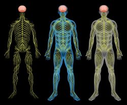
One of the core components of an HPV vaccine adverse reaction inevitably includes some degree of seemingly unexplainable but observable brain inflammation and white matter disintegration. The brain inflammation falls under a number of different names and diagnoses, some are regionally specific, cerebellar anomalies for example, while others demarcate a more diffuse injury including, acute disseminated encephalomyelitis (ADEM), myalgic encephalomyelitis (ME), sometimes known as chronic fatigue, multiple sclerotic (MS) type lesions and, the newest and perhaps more prescient among them, a set of conditions designated as Autoimmune/Inflammatory Syndrome Induced by Adjuvants or ASIA that denote chronic inflammation both centrally and peripherally relative to vaccine adjuvant exposure.

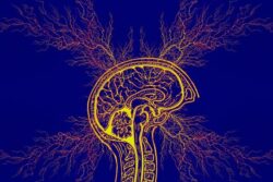
Is the Brain Immune Privileged?

Despite the observance of brain inflammation in many post HPV vaccine victims, many practitioners, and indeed, the FDA and CDC, seem loathe to recognize that an aluminum lipopolysaccharide adjuvanted virus vector might induce a neuro-inflammatory response, leaving patients with little recourse post injury. The difficulties attributing brain inflammation to a vaccine reaction stem from a long held belief that the blood brain barrier is stalwart in its protection against peripheral trespassers.  The brain has long been considered, ‘immune privileged’ having little to no communication with peripheral immune function. Indeed, the perceived impenetrableness of the blood brain barrier is so extensive that brain-body separation might as well be complete, with a brain in bottle and a decapitated body.

Logically, we know this cannot be true. There must be crosstalk between the immune systems of brain/central nervous system and that of the body. How else could we survive if the two modalities were segregated so completely? It turns out, that logic may be prevailing. A decade of research suggests that the long held notion brain immune – privilege is completely and utterly incorrect. Indeed, the immune system not only guides early neurodevelopment (and so mom’s immune function matters) but communicates and affects brain morphological changes chronically. Likewise, signals from the brain continuously influence peripheral immune function.

The immune system appears to influence the nervous system during typical functioning and in disease. Chronic infection or severe illness may disrupt the balance of normal neural–immune cross-talk resulting in permanent structural changes in the brain during development, and/or contributing to pathology later in life. The diversity, promiscuity, and redundancy of “immune” signaling molecules allow for a complex coordination of activities and precise signaling pathways, fundamental to both the immune and nervous systems. 

It should not be surprising then, that nutrient status and toxicant exposures in the periphery, in the body, affect central nervous system function and are capable of inducing brain inflammation and vice versa. And yet, it is; perhaps even more so than any of us realize.

Re -Thinking Brain Inflammation

When one reads through the definitions, research and case reports of ADEM, ME, MS or other instances of brain inflammation, the notion that biochemical lesions in the periphery are linked to observed neuro-inflammatory reactions is far from center stage. Nevertheless, if we can accept the premise what happens in and to the body does not stay in the body, then we can begin to re-frame our approach to brain inflammation. Specifically, we can look at inflammation more globally and ask not only what triggers inflammation, but allows inflammation to persist chronically, regardless of its location. If there is an on-going peripheral inflammatory response, is it not prudent to suspect that a similar response might be occurring within the central nervous system, even if our imaging tools are not yet capable of visualizing the inflammation; even if it is too premature to observe demyelination, neuronal, axonal swelling or other telltale signs of chronic brain inflammation?  I think it is.

Vaccine Adjuvants: A Pathway to Brain Inflammation

With the HPV vaccine, and indeed, any vaccine, the deactivated viral vectors come with a cocktail of additional chemical toxicants and a metal adjuvant to boost the recipient’s immune response, as measured by the increase in post vaccine inflammatory markers. It is believed that without these adjuvants (and data back this up), the recipient’s immune response is insufficiently activated to merit ‘protection’ against the virus. The strength or size of the immune response is then equated with success and protection.

By this equation, an excessive immune response that continues chronically and is eventually labeled ‘autoimmune’ as innate systems begin to fail, is in some way not a failure or side effect, but an example of extreme success; the larger the immune response, the stronger the vaccine. And so, skewed as this observation may seem, within the current vaccine-paradigm there can be no ‘side-effects’, not really. By design, there should be inflammation, even brain inflammation; the more the better. Also by design, metal, lipid soluble, adjuvants cross the blood brain barrier and directly induce brain inflammation. To say vaccines don’t or somehow couldn’t induce brain inflammation is ignorant, if not, utterly negligent, and quite simply, defies logic. Again, for prudence and safety, shouldn’t we assume that an inflammatory reaction in the body might also ignite some concordant reaction in the central nervous system?

Why Aren’t We All Vaccine Injured?

What becomes apparent though, is even with exposure to the most toxic brew of vaccines, not all who receive vaccines are injured, at least observably. (I would argue, however, even those who appear healthy post vaccine, had we the tools to observe brain inflammation more accurately, would show a central inflammatory response, at least acutely, and likely, progressively). So what distinguishes those individuals who seem fine post vaccine, particularly post HPV vaccine, from those who are injured severely and sometimes mortally?

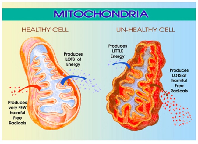
More and more, I think that the fundamental differences between vaccine reactors and non-reactors rest in microbial and mitochondrial health. Indeed, all vaccines, medications, and environmental toxicants damage mitochondria, often via multiple mechanisms, while altering microbial balance. Whether an individual can withstand those mitochondrial insults depends largely upon a balance struck among three variables: 1) heritable mitochondrial dysfunction, genetic and epigenetic; 2) the frequency and severity of toxicant exposures across the lifetime; and 3) nutrient status. Those variables then, through the mitochondria, influence the degree and chronicity of inflammation post vaccine. With the HPV vaccine in particular, the timing of the vaccine, just as puberty approaches and hormone systems come online, may confer additional and unrecognized risks to future reproductive health.

Mitochondria and Microbiota

The mitochondria, as we’ve written about on numerous occasions, control not only cellular energy, but cell life and death. Every cell in the body, including neurons in the brain, require healthy mitochondria to function appropriately. Healthy mitochondria are inextricably tied to nutrient concentrations, which demand not only dietary considerations but balanced gut microbiota. Gut bacteria synthesize essential nutrients from scratch and absorb and metabolize dietary nutrients that feed the mitochondria. Indeed, from an evolutionary perspective, mitochondria evolved from microbiota and formed the symbiotic relationship that regulate organismal health. Disturb gut bacteria and not only do we get an increase in pathogenic infections and chronic inflammation, but also, a consequent decrease in nutrient availability. This too can, by itself, damage mitochondria.

When the mitochondria are damaged, either by lack nutrients and/or toxicant exposure, they trigger cascades of biochemical reactions aimed at conserving energy and saving the cell for as long as reasonably possible. When survival is no longer possible, mitochondrial sequestration, and eventually, death ensue, often via necrosis rather than the more tightly regulated apoptosis. Where the mitochondria die, cells die, tissue dies and organ function becomes impaired. I should note, as steroid hormone production is a key function of mitochondria, hormone dysregulation, ovarian damage and reduced reproductive capacity may be specific marker of mitochondrial damage in young women.

Mitochondria and Inflammation

Mitochondria regulate immune system activation and inflammation and so inflammation is a sign of mitochondrial damage, even brain inflammation. Per a leading researcher in mitochondrial signaling:

The cell danger response (CDR) is the evolutionarily conserved metabolic response that protects cells and hosts from harm. It is triggered by encounters with chemical, physical, or biological threats that exceed the cellular capacity for homeostasis. The resulting metabolic mismatch between available resources and functional capacity produces a cascade of changes in cellular electron flow, oxygen consumption, redox, membrane fluidity, lipid dynamics, bioenergetics, carbon and sulfur resource allocation, protein folding and aggregation, vitamin availability, metal homeostasis, indole, pterin, 1-carbon and polyamine metabolism, and polymer formation.

The first wave of danger signals consists of the release of metabolic intermediates like ATP and ADP, Krebs cycle intermediates, oxygen, and reactive oxygen species (ROS), and is sustained by purinergic signaling.

After the danger has been eliminated or neutralized, a choreographed sequence of anti-inflammatory and regenerative pathways is activated to reverse the CDR and to heal.

When the CDR persists abnormally, whole body metabolism and the gut microbiome are disturbed, the collective performance of multiple organ systems is impaired, behavior is changed, and chronic disease results.

Reducing Inflammation

Instinctively, we think reducing inflammation pharmacologically, by blocking one of the many inflammatory pathways, is the preferred route of treatment. However, this may only add to the mitochondrial damage, further alter the balance of gut microbiota and ensure increased immune activation, while doing nothing to restore mitochondrial and microbial health. In emergent and acute cases, this may be warranted, where an immediate, albeit temporary, reduction in inflammation is required. The risk, however, is that short term gains in reduced inflammation are overridden by additional mitochondrial damage and increased risk of chronic and/or progressive inflammation. The whole process risks becoming a medical game of whack-a-mole; a boon to pharmaceutical sales, but devastating to those who live with the pain of a long-standing inflammatory condition.

In light of the the fact that damaged mitochondria activate inflammatory pathways and that vaccines, medications and environmental toxicants induce mitochondrial damage, perhaps we ought to begin looking at restoring gut microbial health and overall mitochondrial functioning. And as an aside, perhaps we ought to look at persistent inflammation not as an autoinflammatory reaction, but for what is it, an indication of on-going mitochondrial dysfunction.

We Need Your Help

Hormones Matter needs funding now. Our research funding was cut recently and because of our commitment to independent health research and journalism unbiased by commercial interests we allow minimal advertising on the site. That means all funding must come from you, our readers. Don’t let Hormones Matter die.

Yes, I’d like to support Hormones Matter.

Image by Pavlo from Pixabay.

This post was published originally on Hormones Matter on September 22, 2014.

Cognitive Testing Post Adverse Reaction: A Lost Opportunity

6173 views

In the not too distant past, before sophisticated brain imaging tests became available, it was the job of the neuropsychologist to assess brain function and brain damage based upon an array of cognitive and behavioral tests. These tests measured the functional capacity of different brain regions. They were entirely behavioral and performance based and could, with a fair degree of accuracy, identify whether and where a brain injury was located and the extent of the damage. Results from these tests could then indicate a need for surgical intervention and/or suggest a prognosis and therapeutic options; options that generally involved a cognitive therapy of sorts to retrain or regain lost capacities.

And then, technology caught up; brain imaging became possible and physicians no longer needed the neurocognitive assessment as a diagnostic, but only for rehabilitation purposes once the brain damage was identified. Non-invasive brain imaging was a remarkable technological advancement. How much better and more accurate diagnoses and interventions could be if physicians were able to see the damage in advance, and indeed, at every phase of treatment. No need to delineate the subtle behavioral signs linked to brain injury in order to diagnose, just scan the brain to rule in or rule out trauma and deal with the deficits after the fact.

Functional Cognitive Testing – A Missed Opportunity

I can’t help but wondering though, if we’ve lost something important by switching so completely to visually based diagnoses. For example, what if the damage is at the molecular level and unable to be detected via imaging, or even via current laboratory markers? How do we even know which lab markers to look for if we don’t ascertain that there are in fact decrements in functioning? Do we even recognize brain injuries as extant if they are not visible by current imaging or laboratory techniques?  I have a sense that we don’t. Cognitive deficits, especially those occurring in previously healthy individuals, following an illness, medication, vaccine or even post pregnancy, may be disregarded along with further diagnostic and therapeutic possibilities when the indices of injury exclude assessing functional capacity.

I was reminded of this recently from a patient story. She, and others like her, experienced a loss of reading comprehension post-fluoroquinolone reaction. Medication and vaccine induced cognitive disruptions are not uncommon. In elderly populations they are quite well documented. In the younger adult populations, however, the research is sketchy at best. In the case I mentioned, the patient was a previously healthy, active young woman. After taking a course of fluoroquinolone antibiotics, and in addition to a myriad of other side effects, she reports losing her ability to comprehend text; something that would be quite disabling in our current text-based world.

I lost a lot of my reading comprehension while I was floxed. I could still officially read – if you gave me a short memo that said, “buy milk,” or something like that, I could read it. But reading a novel or complex materials for work became really difficult. I lost track of the content of the beginning of a paragraph by the time I reached the end of the paragraph. I struggled to understand things that I used to be able to read with ease.

Another fluoroquinolone patient describes her deficit:

I remember going into a restaurant a few months after being floxed. I sat down, looked at the menu, and couldn’t understand a single thing. I couldn’t make sense of anything. It was as though trying to read a foreign language. I put it down, and wanted to stand up and start screaming, and breaking glasses and dishes.

Read any of the fluoroquinolone social media and these observations are not uncommon. Similarly, decrements in cognitive function have been reported in our research on the side effects of the HPV vaccine, during and after Lupron treatments, and even with oral contraceptives.

What I find both most interesting and most troubling is that the loss of attentional capacity, loss of short term memory and loss of language comprehension following the administration of a medication or vaccine may be indicative of a broader health issue; one that should be investigated further. No doubt in many patients these deficits were not explored, at least not functionally, as imaging tests are often negative. That is a shame. Functional cognitive assessments, like those common in clinical practice in the past, and yet still in academic research, would more finely delineate the patterns of medication induced cognitive disorders. These tests could tell us the brain regions susceptible to the medication-induced events in the absence imaging or lab markers. In fact, these tests might help us design more appropriate lab markers. More importantly, functional neurocognitive testing could provide clues about the patient’s overall health. Let me explain.

Linking Cognitive Performance to Overall Health

Each of the medications I mentioned above have distinctly different pharmacological mechanisms of action; so different, one might wonder why I would even consider looking for commonalities in their adverse reaction patterns. Initially, I didn’t. But then the data from our research began flowing in, and along with the data, patient stories began arriving. Slowly, pattern similarities began emerging; similarities that I could not explain by solely looking at the drug’s specific mechanisms of action. There had to be an underlying factor or factors that somehow connected these medications and vaccine reactions. What were they? And per the current topic at hand, how might have functional neurocognitive assessments inspired or expedited our understanding? Not all of the pieces to the puzzle are clear, but here are the clues thus far.

Clue 1. Three of the medications we study negatively affect the thyroid (Lupron, Fluoroquinolones and Gardasil). Thyroid influence on central nervous system functioning, cognitive and behavioral performance is well known.

Clue 2. Thyroid damage is linked to cerebellar ataxia, acute and chronic, via white matter demyelination. Cerebellar ataxia has been noted post fluoroquinolone, post Gardasil and post Lupron.

Clue 3. Thyroid damage is linked to peripheral demyelination. Again, all three medications include demyelination syndromes as part of their reaction profiles.

Thyroid dysfunction alone, without any other intervening variables could explain the cognitive and many of the neurological symptoms we were seeing, but was it sufficient to explain all of them? Probably not, there must something else at play. What could it be?

Clue 4. Each of these drugs are linked to mitochondrial damage (mitochondria are an unrecognized target for many pharmaceuticals and environmental agents). These drugs increase the production of reactive oxygen species (ROS) and decrease cellular energetics via changes in mitochondrial functioning. Mitochondrial damage evokes multi-system, seemingly disparate illnesses, much like what we are seeing. Cerebral mitochondrial dysfunction can cause serious cognitive and behavioral symptoms.

Clue 5. Thyroid and mitochondrial health are reciprocally connected. Damage the thyroid and mitochondrial functioning diminishes. Damage the mitochondria and thyroid functioning diminishes. We have two factors that are inherently related.

Thyroid and Mitochondrial Functioning

What factor could initiate a thyroid – mitochondrial cascade and connect completely dissimilar drugs to these reactions; reactions which are often complex, affect multiple physiological systems, but are also integrally dependent upon proper thyroid and/or mitochondrial function (because of their reciprocal relationship)?  Could there be such a connection?  A few more clues.

Clue 6. A heartwrenching patient story: A Long and Complicated History Topped by Levaquin, highlights a particular set of neurological symptoms that every neuropsych student should immediately recognize.

Clue 7.   Patients from the post fluoroquinolone and the Gardasil groups have been identified clinically with thiamine deficiency. I suspect post Lupron patients may also have thiamine deficiencies, but none have been tested yet.

Clue 8. Both the fluoroquinolones and Gardasil increase thiaminase, an enzyme that blocks thiamine. Higher thiaminase means lower thiamine. Oral contraceptives are believed to increase thiaminase and so women using oral contraceptives in combination with a fluoroquinolone and/or the HPV vaccine Gardasil or Cervarix would be at higher risk for thiamine deficiencies.

Drug Induced Thiamine Deficiency, Cognitive Deficits – The Mechanism

It turns out, thiamine deficiency, or more specifically, a medication induced blockade of thiamine may be at the root of these adverse reactions. Thiamine is a co-factor in mitochondrial and cellular energy, the currency of which is adenosine triphosphate (ATP).  Without thiamine, the mitochondria become defunct, as do the cells in which they reside, and they eventually die. High energy organs like the brain, the heart and the GI tract are often affected dramatically. Similarly, given the reciprocal relationship between the thyroid and mitochondrial functioning and their combined influence on cerebral, cardiac and metabolic homeostasis, diminished drugs that attack the thyroid and diminish thiamine may be doubly dangerous.

In most recent work, thiamine deficient syndromes have been expanded to include five conditions, with fair degree of overlap between them.

  1. Gastrointestinal beriberi: abdominal pain, lactic acidosis, vomiting.
  2. Neuritic beriberi: sensorimotor polyneuropathy, peripheral neuropathy (likely multiple B vitamins involved).
  3. Dry beriberi: high output cardiac disruption without edema
  4. Wet beriberi: high output cardiac disruption with edema (dysautonomias, including POTS)
  5. Wernicke’s encephalopathy: mental status changes, ocular abnormalities, gait ataxia

Given the current nutritional trends with high intake of sugar, fats and processed foods, it is likely that when these medications directly block thiamine production, they do so against the backdrop of already suboptimal thiamine intake. When we consider that oral contraceptives block also block thiamine and that women are more likely to already suffer from low thyroid function, the effects of either the fluoroquinolones or Gardasil on the mitochondrial thiamine could be devastating. How many other medications or vaccines affect mitochondrial functioning and/or thyroid health? How many other medications or vaccines contain anti-thiamine components and diminish this critical mitochondrial co-factor?

Loss of Reading Comprehension and Other Missed Opportunities

Thiamine deficient cognitive decline is well characterized and includes the loss of language comprehension, in more severe cases, deficits in language production, cerebellar ataxia, tremors and as it progresses, seizures, coma, and death. All reversible with thiamine replacement. The cognitive deficits reported by patients, post medication or vaccine reaction, when observed alone but especially when taken in combination with the other tell tale signs of incipient thiamine deficiency, could have lead researchers or clinicians to these diagnoses. At the very least, it should have lead clinicians to thyroid dysfunction, but more often than not, this was not the case.

Cognitive deficits in previously high functioning individuals are reported regularly after medication or vaccine reactions. Almost to a tee, most are ignored once imaging tests rule out blatant injury, but they shouldn’t be. These deficits, when functionally assessed, would provide valuable clues regarding the regions of the brain most susceptible to medication or vaccine induced injuries; clues that could identify damage and disease processes well before detected by imaging tests. By dismissing patient complaints of cognitive deficits we lose valuable research, diagnostic, and therapeutic opportunities. And perhaps, even more importantly, when we segregate symptoms by organ or body part and fail to see the inherent connections among symptoms and physiological systems, we miss the opportunity to help patients heal.

We Need Your Help

Hormones Matter needs funding now. Our research funding was cut recently and because of our commitment to independent health research and journalism unbiased by commercial interests we allow minimal advertising on the site. That means all funding must come from you, our readers. Don’t let Hormones Matter die.

Yes, I’d like to support Hormones Matter.

Image created using Canva AI.

This post was published originally on Hormones Matter on May 21, 2014.

The Fluoroquinolone Time Bomb – Answers in the Mitochondria

16629 views

Two of the more perplexing features of Fluoroquinolone Toxicity (an adverse reaction to a fluoroquinolone antibiotic – Cipro/Ciprofloxacin, Levaquin/Levofloxacin, Avelox/Moxifloxacin or Floxin/Ofloxacin) are delayed reactions and tolerance thresholds. Both of these features of Fluoroquinolone Toxicity can be explained by noting that fluoroquinolones have been shown to damage mitochondria and cause oxidative stress, and that delayed onset of a disease state, as well as tolerance thresholds, are features of illnesses brought on by pharmaceutical induced mitochondrial damage and oxidative stress.

Delayed Reactions and Tolerance Thresholds with Fluoroquinolone Reactions

By “delayed reactions” I mean that adverse reactions to fluoroquinolones can occur weeks, months or even years after administration of the fluoroquinolone has stopped.  For the lawsuit filed by Public Citizen on behalf of patients who tore or ruptured tendons after taking a fluoroquinolone, (a suit that prompted the addition of the black box warning on all orally and IV administered fluoroquinolones) notes that “Fluoroquinolones, including CIPRO®, are associated with an increased risk of tendinitis and tendon rupture in all ages. This risk is further increased in older patients usually over 60 years of age, in patients taking corticosteroid drugs, and in patients with kidney, heart or lung transplants”. Tendon tears and ruptures that occurred within one year of the patient taking the fluoroquinolone were accepted as being related to the patient’s fluoroquinolone use. Patient reports have noted that new adverse symptoms of fluoroquinolone toxicity have occurred years after administration of the fluoroquinolone has ceased.

Many patients also experience a tolerance threshold for fluoroquinolone use.  A patient can tolerate fluoroquinolones well, experiencing few or no side-effects, until his or her threshold is reached.  After the patient’s tolerance threshold is reached, multisymptom systemic illness ensues. This patient’s story, found on the Fluoroquinolone Wall of Pain, illustrates the issue of tolerance thresholds:

On April 15, 2013 I was prescribed Avelox. I had been on this drug many times for chronic sinus infections. This time was different. Within 10 minutes of the first dose I went into anaphylaxis. I stopped breathing, had numerous convulsions and two grand Mal seizures. Since that day I have suffered with seizures, convulsions, tremors, debilitating fatigue, muscle weakness, vision loss, severe neuropathic pain, vomiting, nausea, lack of appetite, tendon, and vein problems.

This patient tolerated Avelox (moxifloxacin – a fluoroquinolone) well until her tolerance threshold was reached. Once her tolerance threshold was reached, she experienced multi-symptom systemic illness.

I personally experienced both a delayed reaction to Cipro/Ciprofloxacin (also a fluoroquinolone) and a tolerance threshold for it. I took 7 500-milligram pills of Cipro in 2009 without notable incident. I was even able to hike the entire 500-mile Colorado Trail in 2010 (no peripheral neuropathy or weakness were present at that time). When I took 7 more 500-milligram pills in 2011 I experienced a severe adverse reaction that began two full weeks after I was done taking the pills. I experienced multiple musculoskeletal (I couldn’t walk more than a block) and nervous system symptoms (I lost my memory and reading comprehension), and I would describe the reaction as feeling like a bomb had gone off in my body.

Fluoroquinolone Time Bomb: It’s All About the Mitochondria

My experience of a delayed onset of systemic health issues after having previously tolerated Cipro/Ciprofloxacin well, is typical of diseases that are brought on by a pharmaceutical causing mitochondrial dysfunction. (Multiple journal articles have noted that fluoroquinolones cause mitochondrial damage and oxidative stress.)

In “Mechanisms of Pathogenesis in Drug Hepatotoxicity Putting the Stress on Mitochondria” it is noted that:

…damage to mitochondria often reflects successive chemical insults, such that no immediate cause for functional changes or pathological alterations can be established. There is indeed experimental evidence that prolonged injury to mitochondria, such as that which typifies oxidative injury to mitochondrial DNA or to components of the electron transport chain (ETC), has to cross a certain threshold (or a number of thresholds) before cell damage or cell death becomes manifest.

Each time mitochondria is injured, the patient gets closer to his or her personal tolerance threshold for mitochondrial damage. Once the threshold is crossed, cell damage and apoptosis occur – which manifest themselves in various states of illness.

It is further explained in “Mechanisms of Pathogenesis” that:

…approximately 60% of mitochondrial DNA must be deleted from the mouse genome before complex IV activity is compromised and serum levels of lactate are elevated. This non-linear response can be explained upon consideration that the molecules that subserve mitochondrial function (e.g., mitochondrial DNA, mRNA, and ETC proteins) are present in excess of amounts required for normal cell function. This reserve (or buffering) capacity acts as a protective mechanism; however, at a certain stage of damage, the supply of biomolecules needed to support wild-type mitochondrial function becomes compromised.

The lay person’s summary of the above excerpts is that we have excess mitochondrial DNA and that excess mitochondrial DNA keeps each of us from developing a systemic multi-symptom illness whenever mitochondrial DNA is adversely affected (many pharmaceuticals and environmental toxins adversely affect mitochondrial DNA). However, when mitochondrial DNA is depleted sufficiently, cellular dysfunction, oxidative stress and cell death, ensue.

Multiple studies have noted that fluoroquinolones deplete mitochondrial DNA (here, here and here).  When enough mitochondrial DNA are depleted, adverse reactions that are systemic and include multiple symptoms simultaneously, occur.

Multi-Symptom Reaction: Look to Mitochondrial Damage

It is often difficult for the patient who is experiencing a systemic multi-symptom illness to connect his or her illness to the mitochondria damaging drug or toxin that hurt him or her because of the time delay between the cause (mitochondria damaging chemical) and the effect (bomb going off in body and mind). Though the delayed onset of fluoroquinolone toxicity and mitochondrial dysfunction symptoms are noted in many articles (here, here), the reason for the delayed onset of symptoms is not known.  In “Mechanisms of Pathogenesis” it is hypothesized that “an initial adaptive response was followed by a toxic response” when cells are exposed to a mitochondria damaging chemical. Perhaps the delay in adverse reaction onset is due to a toxic response taking time to develop.

Many pharmaceuticals damage mitochondria. Bactericidal antibiotics (including fluoroquinolones), Statins, acetaminophen, some chemotherapy drugs, vaccines, and many others, cause mitochondrial dysfunction, oxidative stress and cell death. Mitochondrial dysfunction and oxidative stress are connected to a variety of ailments, from chronic fatigue syndrome to Alzheimer’s disease and obesity. However, the FDA and other drug regulatory agencies have systematically ignored damage to mitochondria caused by pharmaceuticals and “mitochondrial toxicity testing is not required by the US FDA for drug approval.”

The recognition of delayed adverse reactions and tolerance thresholds for mitochondrial damaging drugs and vaccines will go far in helping both doctors and patients to recognize mitochondrial damage related adverse drug reactions (and adverse vaccine reactions). Once the reactions are recognized, perhaps some pressure can be put on the FDA and/or the pharmaceutical companies to test how drugs affect mitochondria before they are released onto the market. After all, mitochondrial damage and oxidative stress are causally related to almost every chronic illness.  It would be nice if doctors, those in the pharmaceutical industry, the FDA regulators, and others, recognized the harm that drugs do to mitochondria, and the symptoms of iatrogenic mitochondrial dysfunction.

Information about Fluoroquinolone Toxicity

Information about the author, and adverse reactions to fluoroquinolone antibiotics (Cipro/ciprofloxacin, Levaquin/levofloxacin, Avelox/moxifloxacin and Floxin/ofloxacin) can be found on Lisa Bloomquist’s site, www.floxiehope.com.

Share Your Story

If you have suffered from a fluoroquinolone or any other medication reaction, please consider sharing it on Hormones Matter.

We Need Your Help

More people than ever are reading Hormones Matter, a testament to the need for independent voices in health and medicine. We are not funded and accept limited advertising. Unlike many health sites, we don’t force you to purchase a subscription. We believe health information should be open to all. If you read Hormones Matter, like it, please help support it. Contribute now.

Yes, I would like to support Hormones Matter.

Image was created using Canva AI.

This story was published originally on Hormones Matter in March 2014.


Testing for Cerebral Mitochondrial Dysfunction Post Medication or Vaccine Induced Damage

3804 views

Over the last several weeks I have been the beneficiary of a fair amount of synchronicity; seemingly random introductions to interesting people and research connections from disparate sources. Not the least of which was a recent introduction to the concept of lactate doublets. What the heck are lactate doublets?  I had no idea and I am certain most of you reading this do not know either. Well, it turns out lactate doublets may be a missing piece in the diagnostic puzzles that present post medication and vaccine adverse reactions. Let me explain, but first a bit of background.

Mitochondrial Dysfunction

If you’ve followed Hormones Matter for any length of time, you know that we cover complicated diseases and medication and vaccine reactions. Inevitably, these reactions include neurological and neuromuscular symptoms that, almost to tee, fail to show up on standard diagnostic testing. This is particularly troubling when the symptoms include seizures, migraines, tremors, ataxias, neuropathies, dysautonomias and other obvious signs of nervous system damage.

We’ve attributed many of these cases to mitochondrial damage and subsequent dysfunction but testing for damage has proved problematic in most cases. While genetic disorders of mitochondrial dysfunction are reasonably well documented, the more dynamic, environmental and even epigenetic functional changes suspected in toxin, medication or vaccine induced mitochondrial disease are neither well documented nor easily recognized. With no easy testing, folks whose mitochondria are functionally or operational deficient, suffer needlessly and chronically. So when I read the report about lactate doublets as evidence of mitochondrial dysfunction in autism, I was intrigued. And, since I had just attended a talk on the mechanics of measuring basal metabolic rate as indicator of mitochondrial respiration e.g. mitochondrial functioning, I was doubly intrigued.  Could we use these measures as pre-screening tools for mitochondrial damage in health and disease? After all, what could be more fundamental to overall health and cellular energetics than aerobic and anaerobic metabolism? Identify problems with energy metabolism and there’s a good chance something is hinky in mitochondria land.

The Wonders of Lactate

For those of you who are athletes, lactic acid and lactate are familiar topics. For those who are not athletes, consider the last time you exercised rigorously and that point at which the demand for oxygen and fuel for your muscles out-paced your body’s ability to adapt and provide that fuel. What happened? Your muscles began to fatigue, you slowed down, you may have begun to hyperventilate and eventually you hit the wall, exhausted.

If you’re an athlete, a large part of your training involves moving that exhaustion threshold, that wall, further and further away. You train to increase muscle strength – anaerobic capacity, cardiopulmonary functioning- aerobic capacity and the tolerance for pain. Lactate production and utilization is the key to training and athletic success. In the common vernacular, lactate is incorrectly referred to as lactic acid. They are two different molecules. Lactic acid is precursor to lactate and lactate is both a substrate and a product of anaerobic metabolism. Lactate is currency for energy metabolism between cells, tissues and organs. The point at which lactate production outstrips its utilization as fuel or energy is one of the first steps of fatigue in healthy, athletic individuals. In not so healthy individuals and many disease processes, excess lactate can be a marker for mitochondrial dysfunction. In not so healthy individuals where the mitochondria are functioning sub-optimally, that feeling of hitting the exercise wall, is continuous with normal activity.

Lactate and Mitochondria

Over the last several months we’ve discussed mitochondrial ATP production on a number of occasions, mostly in relation to the aerobic process of the energy production that converts glucose (sugars) to adenosine triphosphate (ATP), the cellular energy that fuels our existence. This process requires large amounts of oxygen and numerous chemical co-factors, thiamine being one of the more major among them. Diminish oxygen levels or one of the necessary co-factors and energy/ATP production is reduced.

Not to worry yet, we have a backup or secondary ATP production cycle involving lactate. Here glucose is converted to compound called pyruvate which then is either shuttled into the mitochondria to produce ATP or it remains in either the extracellular or  intracellular cytosolic (cell fluid) space and is converted to lactic acid and then to lactate. The pyruvate>lactate path is anaerobic. That is, it does not require oxygen. The heart, kidneys and liver prefer lactate for fuel and rely heavily on anaerobic metabolism. In our exercise model, anaerobic metabolism and increasing lactate production parallel increasing exercise intensity, though it is less efficient. In comparison to aerobic metabolism that produces 38 mol of ATP, the anaerobic pyruvate>lactate path produces only 2 mol ATP.

The connection between lactate and mitochondrial functioning was only recently discovered and remains hotly debated. For a long time, it was believed that lactate remained outside the mitochondrion. Now, evidence suggests that mitochondria can convert lactate to ATP and that lactate is shuttled in and out of mitochondria to be used when needed. Based upon this possibility, measuring lactate either alone or in combination with respirometry measures may provide an indirect marker of mitochondrial functioning. Here’s where it gets interesting.

Body and Brain Lactate

Lactate is a fuel used readily in the body. Lactate levels change relative to metabolic needs (exertion and stressors) and efficiencies (oxygen usage, co-factor availability). Exercise physiologist have been measuring lactate and other indicators of metabolic functioning and path (aerobic versus anaerobic) for decades using a variety of respirometry tools, from breathing apparatus to blood tests and tissue biopsies. In a grossly oversimplified manner, the extent to which one produces and utilizes lactate during training indicates one’s overall fitness. Could we use some of those same tools, the less invasive ones, to provide a broad indicator of mitochondrial health?  Yes, we can.

Indeed, those same aerobic/anaerobic processes that occur in our body occur in the brain. Even though the brain is a huge consumer of glucose as its preferred fuel, recent evidence suggests that it also produces and consumes lactate in parallel to the body during exercise and other stressors. Beyond just the production of lactate in brain trauma, where oxygen and glucose are depleted rapidly and chronically, brain lactate levels appear to correspond with shifts in aerobic/anaerobic metabolism.

My thought, and what began this entire adventure, couldn’t measures of aerobic/anaerobic metabolism be adapted to compare healthy mitochondrial functioning versus non-healthy mitochondrial functioning? Wouldn’t a skew towards anaerobic metabolism and excess lactate, especially during rest and low exertion, indicate, at least broadly, mitochondrial damage?  The answer is yes; measures of respirometry, along with body and brain lactate could broadly indicate mitochondrial functioning.

But wait, there’s more. Instead of tissue biopsies, and perhaps even instead of the blood tests, researchers have figured out how to image lactate metabolism within the brain and presumably the body. Enter the lactate doublet.

Imaging Lactate with Magnetic Resonance Spectroscopy

Magnetic resonance spectroscopy (MRS) is like a magnetic resonance imaging (MRI) except that instead of simply taking pictures of the tissue, through a myriad of complicated calculations, the MRS measures the relative concentrations of specific metabolites in the brain and other tissues. The MRS can measure lactate metabolism in the brain and researchers around the world have begun to look at brain lactate as markers of different disease process, like autism, aging and brain injury. It is not without technical difficulties (machine calibration is complicated) and controversy, especially around the clinical interpretation, but I surmise it will open up a whole new area of diagnostic possibilities once the early glitches are worked out.

What is a Lactate Doublet?

The lactate molecule has two, weakly coupled, signals or resonances. When viewed on the MRS the lactate doublet presents as a double peak in the signal algorithm.

Lactate Doublets in Cerebral Mitochondrial Dysfunction

The presence of lactate can be diagnostic of specific types of brain tumor or stroke. Since lactate is elevated in mitochondrial disease, evidence of lactate doublets from MRS, even when MRI imaging shows no irregularities, can point to cerebral mitochondrial dysfunction. More specifically, in patients with genetic mitochondriopathies researchers have been able to delineate the regional differences in brain lactate corresponding to the neurological and clinical symptomotology associated with each mitochondriopathy. As was indicated previously, lactate doublets have been recognized in autism, aging (although mechanisms remain contended) and other mitochondrial disease processes.

Using MRS and Respirometry to Detect Post Medication or Vaccine Induces Mitochondrial Damage

Dynamic and functional changes to mitochondria are emerging as culprits in the more complicated adverse reactions we observe post medication and post vaccine. Recognizing the potential for mitochondrial damage post reaction is difficult, especially those impacting the nervous system. Conventional MRI’s and other imaging tests rarely detect visible lesions that can be attributable to clinical symptoms and standard blood tests are often normal, leaving the doctor and the patient without recourse. In advance of genetic testing and the measurement of each co-factor required for proper mitochondrial functioning, respirometry and MRS could be used to identify potential deficits in mitochondrial functioning. Measuring respiratory efficiency and lactate production and usage efficiency could be an easily detectable marker to rule in or out mitochondrial dysfunction. Those results could then be used to determine the need for additional testing that would identify more specific causes of mitochondrial dysfunction.


Same Disease, Different Symptoms: It’s all in the Mitochondria

8619 views

Adverse reactions to fluoroquinolone antibiotics (Cipro/Ciprofloxacin, Levaquin/Levofloxacin, Avelox/Moxifloxacin and Floxin/Ofloxacin) can manifest in patients as a multi-symptom chronic illness that most resembles fibromyalgia, chronic fatigue syndrome / myalgic encephalopathy (ME) and/or an autoimmune diseases.  As is the case with those chronic multi-symptom illnesses, the symptoms of fluoroquinolone toxicity vary greatly from one individual to another. Though almost everyone who suffers from fluoroquinolone toxicity has some sort of musculoskeletal issues (fluoroquinolones have a black box warning about the risk of tendon rupture), neuropathy and autonomic nervous system dysfunction, those broad categories of symptoms are where the similarities between individuals affected end.  Some people suffering from fluoroquinolone toxicity have severe insomnia, others don’t. Some develop dietary intolerances, others don’t.  Some become anemic, others don’t. Some develop Raynaud’s, others don’t.  Some have urticaria, others don’t.  I could go on and on.

Why are there such vast differences between how fluoroquinolone toxicity manifests itself from one person to another?

Look Beyond the Disease Model of Medicine

The traditional approach to medicine, using our existing paradigms, would answer that question by saying that fluoroquinolone toxicity is only responsible for the musculoskeletal issues, neuropathy and autonomic nervous system dysfunction that are the common links between those who are suffering from it; and that insomnia, dietary intolerances, Raynaud’s, anemia, etc. are from something else.

I don’t buy that answer though. The people suffering from fluoroquinolone toxicity were healthy before they crossed their tolerance threshold for fluoroquinolones, and it was only after they were exposed to fluoroquinolones that any of their symptoms emerged.  The reports of thousands of people suffering from fluoroquinolone toxicity lead me to believe that fluoroquinolones cause a multi-symptom illness that can manifest itself in a variety of different ways.

Oxidatative Stress and the Mitochondrial Damage: Explaining Chronic Multi-Symptom Illness

Another possible answer to the question of why symptoms differ so much from one person to another, one that I think is closer to the truth, is that fluoroquinolones cause mitochondrial damage and that mitochondrial disorders can manifest themselves in a variety of different ways.  It is noted by Doctors Bruce H. Cohen, MD and Deborah R. Gold, MD, in Mitochondrial Cytopathy in Adults:  What we Know So Far, that:

“A problem that has vexed the study of mitochondrial diseases ever since the first reported case (in 1962) is that their manifestations are remarkably diverse.  Although the underlying characteristic of all of them is lack of adequate energy to meet cellular needs, they vary considerably from disease to disease and from case to case in their effects on different organ systems, age at onset, and rate of progression, even within families whose members have identical genetic mutations.  No symptom is pathognomonic, and no single organ system is universally affected. Although a few syndromes are well-described, any combination of organ dysfunctions may occur.”

Doctors Cohen and Gold go on to say that:

“symptoms (of mitochondrial damage) such as fatigue, muscle pain, shortness of breath, and abdominal pain can easily be mistaken for collagen vascular disease, chronic fatigue syndrome, fibromyalgia, or psychosomatic illness.”

Multiple studies have shown that fluoroquinolones deplete mitochondrial DNA and lead to an increase in oxidative stress and depletion of antioxidants within cells (source 1 and source 2).  Oxidative stress and mitochondrial dysfunction (OSMD) are almost certainly why fluoroquinolone toxicity manifests itself in the form of chronic multi-symptom illness (CMI).

Even though it has been shown that oxidative stress and mitochondrial dysfunction can cause chronic multi-symptom illness, the question still remains, WHY are there such vast differences between how mitochondrial damage manifests itself from one individual to another?

A possible answer to this question lies in the fact that reactive oxygen species (ROS) generated by damaged mitochondria are signaling mechanisms that control gene expression / epigenetics.

Please excuse the momentary pause while I point out how mind blowing and important that sentence is.  MITOCHONDRIAL PRODUCED REACTIVE OXYGEN SPECIES CONTROL GENE EXPRESSION.  It is a huge discovery that is just now being accepted and verified by scientists.  It is noted in the article Oxidative Stress and Oxidative Damage in Carcinogenesis that, “Through regulation of gene transcription factors, and disruption of signal transduction pathways, ROS are intimately involved in the maintenance of concerted networks of gene expression.”   Also, per Dr. Marcin Kaminski, “The notion that mitochondria can play a role in a cell as a generator of strictly regulated oxidative signals is more recent, and some 10 years ago was regarded almost as heresy.  Now the opinion has changed since a number of new observations have been made.”

Dr. Kaminski also pointed out in a personal conversation that topoisomerase enzymes, which are blocked by fluoroquinolones are also crucial for regulating gene expression.  According to the FDA warning label for Cipro/ciprofloxacin:

The bactericidal action of ciprofloxacin results from inhibition of the enzymes topoisomerase II (DNA gyrase) and topoisomerase IV (both Type II topoisomerases), which are required for bacterial DNA replication, transcription, repair, and recombination

Perhaps the differences in how individuals react to fluoroquinolones are due to the differences in which genes are triggered as a result of both mitochondrial damage (and resultant oxidative stress) and the influence of topoisomerase interrupters on gene expression.

Individual Susceptibilities Influence Mitochondrial Damage

To use myself as an example, my 23andme genetic test results showed that I had a genetic predisposition toward rheumatoid arthritis (RA), an autoimmune disease.  When I first was struck with fluoroquinolone toxicity, I was not aware that Cipro was the culprit behind the sudden deterioration in my health, and I thought that I had an autoimmune disease – with RA being the one that I suspected because my joints were swollen, inflamed and painful.  It turns out that I didn’t have RA, rather, I was suffering from fluoroquinolone toxicity.  But the symptoms manifested themselves in a way that made it look and feel very much like I had RA  Another example is of a gentleman who commented on a blog about fluoroquinolone toxicity, www.floxiehope.com, who noted that his hereditary haemochromatosis (excess iron in the body) was brought on (or at least worsened) by his adverse reaction to a fluoroquinolone.  I, on the other hand, was helped greatly by supplementing iron and suspect that I was anemic after having an adverse reaction to Cipro.

Even though there are genetic differences from person to person, and the expression of those differences may explain why the symptoms of fluoroquinolone toxicity syndrome differ from one individual to another, the entire chronic disease state – with all of the symptomatic differences between individuals, is brought on by fluoroquinolones and thus, despite the individual differences, the symptoms cumulatively should be considered to be part of fluoroquinolone toxicity syndrome.  Even though I had a genetic predisposition for R.A., it likely would have remained dormant (I don’t know of anyone in my family who has ever had R.A.) had it not been triggered, (along with musculoskeletal issues, neuropathy and autonomic nervous system dysfunction) if I had not taken Cipro and had not suffered through damage to my mitochondria.  I cannot be sure of that – it’s not possible for anyone to know at this point, but it is an interesting assertion to ponder.

My assertion, that fluoroquinolones cause changes in gene expression, and that the genes that are expressed determine what symptoms of fluoroquinolone toxicity present themselves, of course needs to be tested and verified before it is accepted as truth.  I hope that more scientists look into the adverse effects of fluoroquinolones and all other mitochondrial damaging pharmaceuticals.  After all, our mitochondria and the ROS that they produce affect our GENES, and our genes are pretty important.

Information about Fluoroquinolone Toxicity

Information about the author, and adverse reactions to fluoroquinolone antibiotics (Cipro/ciprofloxacin, Levaquin/levofloxacin, Avelox/moxifloxacin and Floxin/ofloxacin) can be found on Lisa Bloomquist’s site, www.floxiehope.com.

Participate in Research

Hormones MatterTM is conducting research on the side effects and adverse events associated with the fluoroquinolone antibiotics, Cipro, Levaquin, Avelox and others: The Fluoroquinolone Antibiotics Side Effects Study. The study is anonymous, takes 20-30 minutes to complete and is open to anyone who has used a fluoroquinolone antibiotic. Please complete the study and help us understand the scope of fluoroquinolone reactions.

Hormones MatterTM conducts other crowdsourced surveys on medication reactions. To take one of our other surveys, click here.

To sign up for our newsletter and receive weekly updates on the latest research news, click here.

What Else Can I Do To Help?

Hormones MatterTM is completely unfunded at this juncture and we rely entirely on crowdsourcing and volunteers to conduct the research and produce quality health education materials for the public. If you’d like help us improve healthcare with better data, get involved. Become an advocate, spread the word about our site, our research and our mission. Suggest a study. Share a study. Join our team. Write for us. Partner with us. Help us grow.

Please support Hormones Matter and our research projects. Contribute to our crowdfunding campaign – Crowdfund Us.

Your Mighty Mitochondria

7761 views

There are over ten million billion mitochondria in the human body (Lane p. 1). Each cell (with a few exceptions) contains an average of 300-400 mitochondria that are responsible for generating cellular energy through a process called ATP (Adenosine Triphosphate). Both oxidative stress and antioxidants are created within/by mitochondria. The oxidative stress (caused by reactive oxygen species – ROS) and antioxidant molecules regulate the aging process (Lane p. 4) and are also cellular signals that “regulate diverse physiological parameters ranging from the response to growth factor stimulation to the generation of the inflammatory response, and dysregulated ROS signaling may contribute to a host of human diseases.” (1)

Mitochondria are metabolic signaling centers that “influence an organism’s physiology by regulating communication between cells and tissues.” (2). Mitochondria regulate apoptosis – programmed cell death, as well as autophagy – the breakdown of cellular components during times of starvation. Mitochondria also play key roles in cellular processes including “calcium, copper and iron homeostatis; heme and iron-sulfer cluster assembly; synthesis of pyrimidines and steroids; thermogenesis and fever response; and calcium signaling” (3)

Are you confused? Do the above paragraphs sound like scientific gibberish to you leaving you wondering, “Yes, but what does that MEAN??”

The Importance of Mitochondria

Basically, it, along with the pages of information that I left out, means that mitochondria are really important to cellular functioning and health. They regulate energy production, aging, epigenetic signaling between and within cells and many other important functions. Proper functioning of mitochondria is vital, and when mitochondria are not operating properly, a wide range of disease states can ensue (2). It makes sense that if the energy centers of cells are not operating properly; the system (the body) starts to shut down in a variety of ways. “Mitochondrial dysfunction is associated with an increasingly large proportion of human inherited disorders and is implicated in common diseases, such as neurodegenerative disorders, cardiomyopathies, metabolic syndrome, cancer, and obesity.” (2) Additionally, there is significant evidence that many of the mysterious diseases of modernity, such as fibromyalgia, chronic fatigue syndrome / myalgic encephalomyelitis, Gulf War Syndrome, autism and many other chronic, multi-symptom illnesses, have their roots in mitochondrial dysfunction and resultant oxidative stress. (4)

The History of Mitochondria

The existence of mitochondria was discovered in the late 1800s. Their purpose was unknown until the 1950s when “it was first established that mitochondria are the seat of power in cells, generating almost all our energy.” (Lane p. 6) In 1967 Lynn Margulis proved the “existence of DNA and RNA in mitochondria.” (Lane p. 15) From 1967 through 1999, according to Immo Scheffler, “’Molecular biologists may have ignored mitochondria because they did not immediately recognize the far-reaching implications and applications of the discovery of the mitochondrial genes. It took time to accumulate a database of sufficient scope and content to address many challenging questions related to anthropology,biogenesis, disease, evolution, and more.’” (Lane p. 7) Almost everything that is known about the role of mitochondria in cellular signaling and gene expression (5), apoptosis, autophagy, metal metabolism, regulation of enzymes, and many other important functions, has been discovered since the turn of the century. Despite the fact that all eukaryotic organisms have (or at least once had) mitochondria, the realization that mitochondrial health is vital to over-all human health is a recent realization. The link between mitochondrial dysfunction and disease, especially chronic multi-symptom disease, is well documented in peer-reviewed journals, yet it is not an officially recognized cause of those diseases and they are considered by many to be mysterious.

Vulnerable yet Strong: Mitochondria and Tolerance Thresholds

The role of mitochondrial dysfunction in disease remains unacknowledged because of some fascinating features of mitochondria. Mitochondria are an interesting mix of vulnerable and resilient. Mitochondrial DNA (mtDNA) and mitochondrial genes are more vulnerable than nuclear DNA and nuclear genes to damage caused by chemical toxicants (like pharmaceuticals and environmental pollutants) because mitochondrial genes “sit on a single circular chromosome (unlike the linear chromosomes of the nucleus) and are ‘naked’ – they’re not wrapped up in histone proteins.” (Lane p. 15) Histone proteins protect nuclear DNA and because mtDNA isn’t wrapped in histone proteins, it is vulnerable. This vulnerability means that mtDNA is easily damaged. This slide describes additional factors that affect mitochondrial vulnerability to environmental pollutants:

Factors that affect mitochondrial vulnerability to environmental toxicants
Mitochondria as a Target of Environmental Toxicants. Permission to print graphic provided by Joel N. Meyer.

Despite its vulnerability, mtDNA is, at the same time, quite hearty and resilient. MtDNA can take a punch, and a threshold of damage must be crossed over before a disease state will ensue. In Mechanisms of Pathogenesis in Drug Hepatotoxicity Putting the Stress on Mitochondria it is noted that, “damage to mitochondria often reflects successive chemical insults, such that no immediate cause for functional changes or pathological alterations can be established. There is indeed experimental evidence that prolonged injury to mitochondria, such as that which typifies oxidative injury to mitochondrial DNA or to components of the electron transport chain (ETC), has to cross a certain threshold (or a number of thresholds) before cell damage or cell death becomes manifest.” The researchers go on to note that, “This non-linear response can be explained upon consideration that the molecules that subserve mitochondrial function (e.g., mitochondrial DNA, mRNA, and ETC proteins) are present in excess of amounts required for normal cell function. This reserve (or buffering) capacity acts as a protective mechanism; however, at a certain stage of damage, the supply of biomolecules needed to support wild-type mitochondrial function becomes compromised.” (6)

Pharmaceutical Safety and Mitochondria: No Testing Required

To put it simply, because of the tolerance threshold that mitochondria have to damage, the damage done to mitochondria will not show up as a disease until the threshold is crossed. This makes the testing of the deleterious effects of pharmaceuticals and environmental toxins on mitochondria difficult. The damage done by the chemical toxin doesn’t show up until multiple exposures to mitochondrial damaging toxins have been experienced (and it likely doesn’t need to be the same toxin – different mitochondrial damaging toxins can erode the mitochondria’s tolerance threshold). Also, mitochondria display an “initial adaptive response was followed by a toxic response” (6) to damaging toxins.

The mitochondrial tolerance threshold for damage would need to be taken into consideration when testing drugs or environmental pollutants for their adverse effects on mitochondria, IF drugs and pollutants were tested for their effects on mitochondria at all. Unfortunately, “mitochondrial toxicity testing is still not required by the US FDA for drug approval.” (7) The authors of Mitochondria as a Target of Environmental Toxicants note that, “growing literature indicates that mitochondria are also targeted by environmental pollutants” but the EPA does not require testing of environmental pollutants for their affects on mitochondria either.

Studies have shown that bactericidal antibiotics (including fluorouquinolones) (8), statins (9), chemotherapy drugs (3), acetaminophen (6), metformin (a diabetes drug) (10), and others, damage mitochondria. The environmental pollutants that have been shown to damage mitochondria include rotenone, cyanide, lipopolysaccharide, PAH quinones, arsenic, and others (3).

Though it’s not excusable, it’s understandable that the FDA and EPA have not historically required testing of pharmaceuticals or environmental pollutants for their effects on mitochondria. Until very recently, much of what is currently known about mitochondria was not yet discovered. The link between multi-symptom chronic illnesses (including autism) and mitochondrial dysfunction and damage (4) was not yet known when the vast majority of the drugs that are on the market were going through their initial testing and review. What is known now about the important role of mitochondria in epigenetic signaling was not known until recently – and almost all laymen and probably plenty of scientists still don’t realize how much the molecules generated in our mitochondria affect our genes. All of the drugs and environmental pollutants that are on the market have been put on the market without their effects on mitochondria being studied, or even noted by the regulatory agencies that are entrusted with protecting our health and safety. The ignorance of everyone involved would be less consequential if people weren’t so sick. In addition to being connected to the mysterious diseases of modernity, mitochondrial damage is also implicated in the following disorders: “schizophrenia, bipolar disease, dementia, Alzheimer’s disease, epilepsy, migraine headaches, strokes, neuropathic pain, Parkinson’s disease, ataxia, transient ischemic attack, cardiomyopathy, coronary artery disease, chronic fatigue syndrome, fibromyalgia, retinitis pigmentosa, diabetes, hepatitis C, and primary biliary cirrhosis” (7) as well as cancer (11).

The Paradigm Shift

We’re in an interesting and strange situation where medicine hasn’t caught up to science and science hasn’t caught up to medicine. By this I mean that mitochondria damaging chemicals were created long before we knew the importance of our mitochondria, but now that scientists are realizing the importance of our mitochondria, the damaging pharmaceutical culprits are so entrenched in medicine that they can’t be extricated. For example, nalidixic acid, the precursor to fluoroquinolones (mitochondria damaging antibiotics) (8), was first created in the 1960s, long before what we currently know about mitochondria and the effects of mitochondrial damage was discovered. Now that the effects of depleting mtDNA on human health has been discovered, the myriad of strange health symptoms observed in patients who have taken fluoroquinolones can be explained. Mitochondrial damage can cause multi-symptom chronic illness (4). We know this now. However, fluoroquinolones are so widely used (20+ million annual prescriptions in America alone), and so widely regarded as safe, that it would be difficult, if not impossible, to restrict their use now – even though they have been found to cause mitochondrial damage and oxidative stress (8). It’s time for disease paradigms to shift to note the importance of mitochondria in human health. After all, chronic diseases, many of which are related to mitochondrial function, are the leading cause of death in the U.S.

Mitochondria are important. It’s time we started paying attention to them. It’s time for disease models to shift. It’s time for iatrogenic mitochondrial dysfunction to be recognized as a cause of chronic diseases. The chronic diseases are happening, whether we recognize the role of mitochondrial damage, and the role of pharmaceutical and environmental pollutants in damaging mitochondria, or not. Ignorance isn’t bliss – people are sick. With recognition of the importance of mitochondrial health, maybe we can prevent others from getting sick in the future.

Information about Fluoroquinolone Toxicity

Information about the author, and adverse reactions to fluoroquinolone antibiotics (Cipro/ciprofloxacin, Levaquin/levofloxacin, Avelox/moxifloxacin and Floxin/ofloxacin) can be found on Lisa Bloomquist’s site, www.floxiehope.com.

Participate in Research

Hormones MatterTM is conducting research on the side effects and adverse events associated with the fluoroquinolone antibiotics, Cipro, Levaquin, Avelox and others: The Fluoroquinolone Antibiotics Side Effects Study. The study is anonymous, takes 20-30 minutes to complete and is open to anyone who has used a fluoroquinolone antibiotic. Please complete the study and help us understand the scope of fluoroquinolone reactions.

Hormones MatterTM conducts other crowdsourced surveys on medication reactions. To take one of our other surveys, click here.

To sign up for our newsletter and receive weekly updates on the latest research news, click here.

What Else Can I Do To Help?

Hormones MatterTM is completely unfunded at this juncture and we rely entirely on crowdsourcing and volunteers to conduct the research and produce quality health education materials for the public. If you’d like help us improve healthcare with better data, get involved. Become an advocate, spread the word about our site, our research and our mission. Suggest a study. Share a study. Join our team. Write for us. Partner with us. Help us grow. For more information contact us at: info@hormonesmatter.com.

To support Hormones Matter and our research projects – Crowdfund Us.

References

Lane, Nick (2005). “Power, Sex, Suicide: Mitochondria and the Meaning of Life” Oxford University Press Inc., New York.

  1. Journal of Cell Biology, “Signal Transduction by Reactive Oxygen Species
  2. Cell, “Mitochondria: In Sickness and in Health
  3. Toxicological Sciences, “Mitochondria as a Target of Environmental Toxicants
  4. Nature Preceedings, “Oxidative Stress and Mitochondrial Injury in Chronic Multisymptom Conditions: From Gulf War Illness to Autism Spectrum Disorder
  5. Biochimica et Biophysica Acta (BBA) – Gene Regulatory Mechanisms, “Mitochondrial DNA Damage and its Consequences for Mitochondrial Gene Expression
  6. Molecular Interventions, “Mechanisms of Pathogenesis in Drug Hepatotoxicity Putting the Stress on Mitochondria
  7. Molecular Nutrition & Food Research, “Medication-Induced Mitochondrial Damage and Disease
  8. Science Translational Medicine, “Bactericidal Antibiotics Induce Mitochondrial Dysfunction and Oxidative Damage in Mammalian Cells”
  9. NIH Public Access, “Statin Adverse Effects: A Review of the Literature and Evidence for a Mitochondrial Mechanisms
  10. Biochemical Journal, “Metformin inhibits mitochondrial permeability transition and cell death: a pharmacological in vitro
  11. Contemporary Oncology, “Oxidative Damage and Carcinogenesis

The Paradox of Modern Vitamin Deficiency, Disease, and Therapy

13344 views

In order to understand why this article is about “paradox”, the concept of vitamin therapy must be appreciated. Hence, the explanation of the title is deferred to the end. Although vitamin deficiency disease is believed by most physicians to be only of historical interest, this is simply not true. When we think of a vitamin deficiency disease, we envision an individual living in a third world country where starvation is common. Such an individual is imagined as being skeletal, whereas an obese person is considered to be well fed with vitamin enriched foods. For this reason, common diseases, some of which are associated with obesity, are rarely, if ever, seen as potentially vitamin deficient.

The Calorie Rich and Nutrient Sparse Modern Diet

Our food is made up of two different components, the caloric and the non-caloric nutrients.  When we ingest high calorie foods (e.g. a doughnut) without even a vestige of non-caloric nutrients, we refer to this as “empty” or “naked” calories.  For our food to be processed into energy that enables the body and brain cells to function, there must be a ratio of the calorie bearing component to that of the non-caloric nutrients.  When we load the calories together with an insufficiency of non-caloric nutrients, we alter this ratio and produce a relative vitamin deficiency.  The trouble with this is that it does not result in the formation of the classic vitamin deficiency diseases as recorded in the medical literature. There is a gradual impairment of function, resulting in many different symptoms. Because modern medicine seeks to make a diagnosis by the use of imaging techniques and laboratory data and because of the physician’s mindset, if the tests used are normal, the possibility of a relative vitamin deficiency is ignored.

The Brain as a Chemical Machine

We have two different nervous systems. One is called “voluntary” that enables us to do things by will-power.  This is initiated and controlled by the upper brain, the part of the brain that thinks. The other system is known as the autonomic nervous system (ANS).  This is initiated and controlled by the lower part of the brain, the limbic system and brainstem.  This system is controlled automatically.  Although it collaborates with the other system, it is not normally under voluntary control. The limbic system and brainstem are highly sensitive to oxygen deficiency, but since the oxygen is useless without the non-caloric nutrients, their absence would produce the same kind of phenomena as oxygen deficiency. Thiamine (vitamin B1) has been found to be of extreme importance as a member of the non-caloric nutrients. The brain, and particularly the limbic system and brainstem, is highly sensitive to its deficiency.

Since the ANS is automatic, we are forced to think of the limbic system and brainstem as a computer.  For example, when it is hot, you start to sweat.  Evaporation of the sweat from the skin produces cooling of the body, representing an adaptive response to environmental hot temperature. When it is cold, you may start to shiver. This produces heat in the muscles and represents an adaptive response to environmental low temperature. If you are confronted by danger, the computer will initiate a fight- or- flight reflex.  This is a potential lifesaving reflex.  It is designed for short term use, consumes a vast amount of energy and prepares you to kill the enemy or flee from the danger.  Any one of these reflexes may be modified by the thinking brain. For example the lower brain, also known as the reptilian system, initiates the urge to copulate.  It is modified by the upper brain to “make love”.  The reptilian system, working by itself, can convert us into savages. There is an obvious problem here because our ancestors were faced with the dangers of short term physical stress associated with survival.  In the modern world the kind of stress that we face is very different for the most part.  We have to contend with traffic, paying bills, business deadlines and pink slips. The energy consumption, however is enormous, continues for a long time and it is hardly surprising that it is associated with fatigue, an early sign of energy depletion. It has been shown in experimental work that thiamine deficiency causes extensive damage to mitochondria, the organelles that are responsible for producing cellular energy.

Autonomic Function

The autonomic nervous system, controlled by the lower brain, uses two different channels of neurological communication with the body. One is known as the sympathetic system and the other is the parasympathetic. There are also a bunch of glands called the endocrine system that deals with the brain-controlled release of hormones.

We can think of the sympathetic branch of the ANS as the action system. It governs the fight-or-flight reflex for personal survival and the relatively primitive copulation mechanisms for the survival of the species. It accelerates the heart to pump more blood through the body.  It opens the bronchial tubes so that the lungs may get more oxygen. It sends more blood to the muscles so that you can run faster and the sensation of fear is a normal part of the reflex. When the danger is over and survival has been accomplished, the sympathetic channel is withdrawn and the parasympathetic goes into action. Now in safety and under its influence, body functions such as sleep and bowel action can take place.  That is why I refer to the parasympathetic as the “rest and be thankful system”.

Dysautonomia, Dysfunctional Oxidation and Disparate Symptoms 

When there is mild to moderate loss of efficiency in oxidation in the limbic system and/or brainstem they become excitable. This is most easily accomplished by ingesting a high calorie diet that is reflected in relative vitamin deficiency.  The sympathetic action system is turned on and this can be thought of as a logical reaction from a design point of view.  For example, if you were sleeping in a room that was gradually filling with carbon dioxide, the gradual loss of efficiency in oxidation would be lifesaving by waking you up and enabling you to exit the room. In the waking state, this normal survival reflex would be abnormal.

High calorie malnutrition, by upsetting the calorie/vitamin ratio, causes the ANS to become dysfunctional. Its normal functions are grossly exaggerated and reflexes go into action without there being any necessity for them. Panic attacks are merely fragmented fight-or-flight reflexes.  A racing heart (tachycardia) may start without obvious cause.  Aches and pains may be initiated for no observable reason. Affected children often complain of aching pain in the legs at night. Unexplained chest and abdominal pain are both common. This is because the sensory system is exaggerated. One can think of it as the body trying to send messages to the brain as a warning system.

Nausea and vomiting are both extremely common and are usually considered to be a gastrointestinal problem rather than something going on in the brain. Irritable bowel syndrome (IBS) is caused by messages being conveyed through the nervous system of the bowel, increasing peristalsis (the wave-like motion of the intestine) and often leading to breakdown of the bowel itself, resulting in colitis.  Of course, the trouble may be in the organ itself but when all the tests show that “nothing is wrong”, the symptoms are referred to as psychosomatic. The patient is often told that it is “all in your head”.

Emotional instability seems to be more in keeping with psychosomatic disease because emotional reactions are initiated automatically in the limbic system and thiamine deficient people are almost always emotionally unstable. A woman patient had been crying night and day for three weeks for no observable reason. A course of intravenously administered vitamins revealed a normal and highly intelligent person.  Intravenously administered vitamins are often necessary for serious disease because the required concentrations cannot be reached, taking them by mouth only.

The Vitamin Therapy Paradox

The body is basically a chemical machine.  But instead of cogwheels and levers, all the functions are manipulated through enzymes that, in order to function efficiently, require chemicals called “cofactors”. Vitamins are those essential cofactors to the enzymes.  If a person has been mildly to moderately deficient in a given vitamin or vitamins for a long time without the deficiency being recognized, the enzyme that depends on the vitamin for its action appears to become less efficient in that action.  A high concentration of the vitamin is required for a long time in order to induce its functional recovery.

Although the reason is unknown, doctors who use nutritional therapy with vitamins have observed that the symptoms become worse initially.  Because patients expect to improve when a doctor does something to them and because drugs have well-known side effects, it is automatically assumed by the patient that this worsening is a side effect of the vitamins. If the therapy is continued, there is a gradual disappearance of those symptoms and overall improvement in the patient’s well being. Unless the patient is warned of this possibility he or she would be inclined to stop using the treatment, claiming that vitamins have dangerous side effects and never getting the benefit that would accrue from later treatment.  This is the opposite effect that the patient expects. This is the paradox of vitamin therapy. 

If we view dysautonomia as an imbalance in the functions of the ANS and the vitamin therapy as assisting the functional recovery by stimulating energy synthesis, we can view this initial paradoxical as the early return of the stronger arm of the ANS before the weaker arm catches up, thus worsening an existing imbalance. However, this is mere speculation. I did not learn of the “paradox” until I actually started using mega dose vitamins to treat patients.

The Paradox and Thiamine

In this series of posts, we are particularly concerned with energy metabolism and the place that thiamine holds in that vital mechanism.  It is, of course, true that worsening of serious symptoms is a fact that has to be contended with and vitamin therapy should be under the care of a knowledgeable physician. The earlier the symptoms of thiamine deficiency are recognized, the easier it is to abolish them. The longer they are present the more serious will be the problem of paradox and a clinical response will also be much delayed and may be incomplete.

Beriberi and Thiamine Deficiency

I will illustrate from the early history of beriberi when thiamine deficiency was found to be its cause.  Many of the patients had the disease for some time before thiamine was administered, so the danger of paradox was increased. It was found that if the blood sugar was initially normal, the patient recovered quickly. If the blood sugar was high, the recovery was slow.  If the blood sugar was low, the patient seldom recovered.  In the world of today, an abnormal concentration of glucose in the blood would make few doctors, if any, think of thiamine deficiency as a potential cause. It is no accident that diabetes and thiamine metabolism are connected. Education of the doctor and patient are both absolutely essential. I believe that the ghastly effects of Gardasil, and perhaps some other medication reactions covered on Hormones Matter, can only be understood by thinking of the body as a biochemical machine and that the only avenue of escape is through the skilled use of non caloric nutrients.

We Need Your Help

More people than ever are reading Hormones Matter, a testament to the need for independent voices in health and medicine. We are not funded and accept limited advertising. Unlike many health sites, we don’t force you to purchase a subscription. We believe health information should be open to all. If you read Hormones Matter, like it, please help support it. Contribute now.

Yes, I would like to support Hormones Matter. 

Image by Rochak Shukla on Freepik

It’s All About the Diet: Obesity and Mitochondrial Dysfunction

7407 views

We’ve been learning a lot about mitochondrial dysfunction lately, particularly as it applies to medication and vaccine adverse reactions. Mitochondria are those energy producing powerhouses located within our cells that are critical to every aspect of health. We have over 100,000 trillion mitochondria in our body, each containing 17,000 little assembly lines for making adenosine triphosphate (ATP), the fuel that powers our lives. Mitochondria use ninety percent of the oxygen we breathe, take up 40% percent of the space inside the heart cells and 20% of the space inside the liver cells. Properly functioning mitochondria are critical to human health and survival.

Unfortunately, mitochondria are exquisitely sensitive to their environs and can be damaged easily. Damaged or dysfunctional mitochondria lead to an array of complex and seemingly disparate and untreatable diseases. How mitochondria become damaged is a matter of great interest with research pointing to maternal genetics and epigenetics, environmental exposures, medications and even diet. Mitochondrial dysfunction arguably represents one of the most unrecognized causes of disease in modern medicine and, according to some geneticists, mitochondrial disease represents the next great paradigm in medicine.

The Mitochondrial Cholesterol Transporter

Over the course of my research, I stumbled on a series of papers identifying a cool little transporter channel located on the outer membrane of the mitochondrion called the translocator protein 18kDA or TSPO. For a while, this channel was called the peripheral benzodiazepine receptor because researchers had noticed that the drug diazapam, a benzodiazepine, could bind to a spot on this channel and evoke a reaction. Soon however, researchers learned the function of the TSPO was far more complex than a simple drug binding site and changed the name accordingly.

What is the Mitochondrial Translocator Protein and Why is it Important?

The primary function of the TSPO is to bring cholesterol into the mitochondria. Once inside, another protein, an enzyme called StAR, converts cholesterol into a hormone called pregnenolone and shoots it back out into the cell and beyond. Pregnenolone is the precursor for all steroid hormones and so this TSPO channel is responsible for steroidogenesis in cells, not just in what are considered the typical steroid producing cells like the ovaries, testes, adrenals, but in all cells. TSPO is ubiquitous across mitochondria, meaning steroidogenesis is not limited to the endocrine cells; something many, including myself, have been arguing for decades, but I digress.

In addition to its role in steroidogenesis, the TSPO appears to control many aspects of mitochondrial function. It regulates production of reactive oxygen species (ROS) – those pesky little free radicals that damage mitochondrial DNA. The TSPO also influences outright apoptosis or cell death, and as one might expect, TSPO modulates cellular energy or ATP production. Researchers have found that the functionality of the TSPO channel changes with different disease states and can become dysregulated which then dramatically affects how the mitochondria function. Because of its diverse and seemingly unrelated functions, the TSPO is thought to be part of our host-defense response to disease and injury.

Obesity Impairs Mitochondrial Function via TSPO Downregulation

Here’s where it gets interesting. Once again, we see that diet is critical to mitochondrial functioning via its influence on TSPO activity. In a recent study, Translocator Protein 18 kDa (TSPO) Is Regulated in White and Brown Adipose Tissue by Obesity, researchers demonstrated that a high fat diet downregulates TSPO functioning significantly and the compensatory reaction – obesity – is the survival mechanism.

Study Details. Mice were fed a high fat diet (60% of calories from fat) from 8 weeks of age through 34 weeks of age. Researchers then compared TSPO functioning and other markers of the diet induced obese mice to mice fed a regular diet (13% of calories from fat). They investigated the differences TSPO function and activity in both white fat and brown fat in both groups of mice. Remember white fat stores calories as big fat droplets, while brown fat stores it in smaller droplets and utilizes calories more effectively by burning them for energy. The brown fat is more mitochondrial dense than white fat. So TSPO changes relative to diet and white and brown fat function could be very interesting when understanding obesity.

Results – White Fat. The mice with dietary induced obesity showed a 90% reduction in TSPO mRNA and an 87% reduction in gene expression as measured via a protein called the peroxisome proliferator-activator receptor coactivator (PGC1α). PGC1α is important for mitochondrial biogenesis, making new mitochondria. The researchers also found a 40% reduction in TSPO binding sites, another marker that indicates decreased TSPO gene expression. Interestingly, the shape of the fat cells changed in the diet induced obese mice compared to the regular feed mice. In the obese mice, the white adipocytes were hypertrophic (oversized) and were surrounded by macrophages, the immune cells responsible for ‘eating’ dead or dying cells.

Results – Brown Fat. In the brown fat, the changes in TSPO function between the diet induced obese mice and the regular feed mice were equally dramatic. Visually, the adipocytes were hypertrophic indicating increased fat storage versus energy usage. TSPO gene expression was reduced by 32%, mitochondrial biogenesis was reduced by 31%, while TSPO binding sites decreased by 7%.

A couple more findings. The investigators added a few more conditions to the experiment to determine whether these changes in TSPO could be modified acutely in response to fasting or other stressors. The answer was no. TSPO expression and function were not influenced by acute metabolic changes suggesting a more chronic pattern of metabolic dysfunction.

What This Means: Diet Affects Mitochondrial Function

High fat diet affects mitochondrial function by downregulating a critical receptor – the TSPO channel that brings cholesterol into the mitochondrion. This results in increased cholesterol storage within the fat cells and perhaps decreased conversion from cholesterol to energy (or steroids) inside the mitochondria. The hypertrophic and unhealthy adipocytes then evoke an immune response drawing macrophages and inducing phagocytocis. A dangerous feedback loop ensues.

Though the mechanisms by which a high fat diet induces mitochondrial dysfunction is not clear, one can speculate that a diet high in fat is also low in critical nutrients that are required for proper mitochondrial function and cellular energy production. Deficits in nutrients such as thiamine (vitamin B1) can and do cause severe mitochondrial dysfunction and lead to an array of disease processes from nervous system destabilization to cardiac, GI and reproductive dysfunction. Other nutrients and cofactors are also critical for mitochondrial function. CoQ10, L-Carnitine for example, have been found helpful in mitochondrial induced dysautonomia, and the list goes on.

What’s important to remember is that when the mitochondria are starved of critical nutrients, they don’t function properly and die off. As mitochondria become injured and die, it is possible to see how the hypertrophy and increased cholesterol storage observed in the adipocytes might be a compensatory survival reaction to maintain the requisite demands for mitochondrial cholesterol. The only problem is that as those fat cells become larger and more cholesterol is stored, more mitochondrial dysfunction ensues, reinforcing and continuing the immune response. We get an endless cycle of poorly functioning and dying mitochondria>immune response>larger fat cells with more cholesterol stored>more mitochondrial death>more immune response and so on. Breaking the cycle may be as simple as changing one’s diet and exercising, as both can induce mitochondrial biogenesis.

We Need Your Help

More people than ever are reading Hormones Matter, a testament to the need for independent voices in health and medicine. We are not funded and accept limited advertising. Unlike many health sites, we don’t force you to purchase a subscription. We believe health information should be open to all. If you read Hormones Matter, like it, please help support it. Contribute now.

Yes, I would like to support Hormones Matter.

Photo by Yolanda Djajakesukma on Unsplash.