oophorectomy

Lupron, Brain Function, and the Keto Diet

8648 views

Reproductive senescence, the time in a woman’s life marked by the slowing and eventual cessation of reproductive function, frequently coincides with an increased risk of a host of neurodegenerative disorders from memory impairment to dementia and Alzheimer’s disease. Researchers have long postulated that the loss of ovarian hormones was responsible; estradiol, in particular, but likely others as well.

This begs the question, what happens to the brain when we abruptly and artificially derail ovarian hormone synthesis in young women using drugs such as Lupron (leuprolide) and the other GnRH agonists and antagonists or by removing the ovaries altogether as in surgical oophorectomy? Is it the same damage we see in aging, only expedited and perhaps magnified, or does it run a different course? Along those same lines, though perhaps a topic for another day, what happens when we chronically supplant endogenous ovarian hormone production with synthetic hormones such as those used in hormonal birth control or menopausal hormone replacement therapies? I suspect, and there is evidence to back up my suspicions, that in all cases brain function is altered, and not for the better.

Estrogens and the Brain

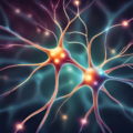
The mechanisms by which estradiol and other steroid hormones influence brain function are myriad and complicated. Beyond just reproduction, steroid hormones influence all aspects of neurological function, with estrogen, androgen, glucocorticoid (cortisol), and mineralocorticoid (electrolyte balance, blood pressure) receptors located throughout the brain. Steroid hormones produced in the body, because of their fat solubility, easily cross the blood-brain barrier where they bind to their receptors and regulate all sorts of processes. Perhaps even more remarkable, the brain has all of the machinery to synthesize its own steroid hormones and so when body concentrations fall, at least for a time, the brain can compensate. Eventually, however, brain synthesis declines and that is where we begin to have problems. Fortunately, natural reproductive senescence occurs later in life and the risk of neurodegenerative diseases is just that, a risk, not a foretold conclusion. This suggests that other variables are at play, ones that we may be able to modulate to improve health, offset and/or reduce the severity of the natural neurodegenerative processes. Again, however, we must ask, what happens when we induce reproductive senescence in young women? By all accounts, the effects are often devastating, leaving many to wonder if they will ever recover.

Estradiol and Mitochondrial Energy

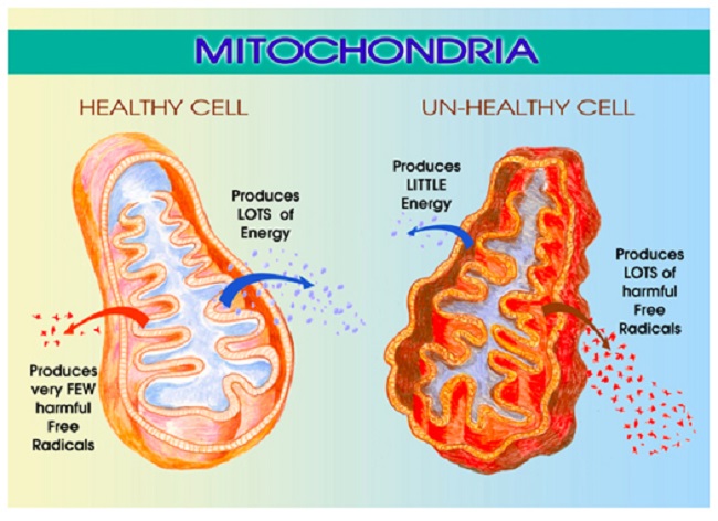
Among the myriad of functions mitochondria control, perhaps the most important is energy production. That is, mitochondria take the nutrients supplied by diet and convert them into adenosine triphosphate (ATP), the energy currency that cells use to perform all of the functions that keep us alive. The loss or diminishment of ATP is deleterious to health and can ultimately be deadly, by invoking a series of complicated processes

Estradiol is a critical component of that process and directly impacts mitochondrial energy production. That’s right, estradiol is part of the mitochondrial bioenergetic machinery such that when estradiol wanes, so too does energy production or ATP. As one might suspect, waning ATP is deleterious to brain health. In previous posts, I detailed the research showing how the loss of estradiol deforms mitochondrial morphology essentially disabling mitochondrial membrane potential while turning the mitochondria into misshapen donuts and blobs ripe for a slow, messy necrotic death; a process that evokes all sorts of deleterious reactions.

The Lupron Brain and Ketosis

Just recently, I stumbled upon research showing yet another mechanism of damage. In the absence of estradiol, brain glucose transport diminishes significantly. This effectively starves the brain for energy inducing severe bioenergetic deficiencies with all of the concordant neuronal damage one might expect. The reduction in glucose affects the mitochondria severely. Recall that glucose is one of the major fuel substrates of the brain, particularly where the Western diet predominates. The decline of glucose transport, therefore, is significant, and alone, without any other changes to the mitochondria, elicits a cascade of deleterious reactions. Oxidative phosphorylation and associated enzymes are downregulated, ATP production wanes, and ultimately may initiate the deformation of the very shape of the mitochondria, as observed in the research cited above. The ensuing reduction of ATP starves the brain of critical energy but also induces a state of hypoxia with the mitochondria incapable of utilizing molecular oxygen. With that hypoxia, inflammatory pathways are initiated further cementing mitochondrial death spirals and associated neuronal damage.

Interestingly, this reduction in aerobic activity coincides with the emergence of a ketogenic phenotype. That is, with the loss of one fuel substrate, ketones become the dominant source of fuel and the associated enzyme machinery is upregulated. Unfortunately, the Western diet is highly dependent upon carbohydrates and so a woman experiencing this loss of estradiol is not likely to consume sufficient fats and proteins to effectively weather this shift. Nevertheless, it does provide an opportunity for recovery. What if women who have lost the ability to produce sufficient estradiol either because of surgically (oophorectomy) or chemically (Lupron and other GnRH analogs) induced menopause adopt a ketogenic diet? Could we maximize the preferred energy source of the post-menopausal brain and reduce the neurological symptoms? I do not know the answer to that question, but given the severity of the suffering with surgical and chemical menopause, it seems worth the try.

We Need Your Help

More people than ever are reading Hormones Matter, a testament to the need for independent voices in health and medicine. We are not funded and accept limited advertising. Unlike many health sites, we don’t force you to purchase a subscription. We believe health information should be open to all. If you read Hormones Matter, and like it, please help support it. Contribute now.

Yes, I would like to support Hormones Matter.

Image by Angelo Esslinger from Pixabay.

This article was originally published on November 15, 2018.

Share

The Long Term Repercussions of an Unneeded Total Hysterectomy

11189 views

I have always preferred natural processes. I had four natural births using Lamaze technique. I breastfed. I was a registered nurse. I knew my ob-gyn doctors for years. I never expected my life to take a sharp and unnecessary turn for the worse, but it did, after I had, what was an essentially unneeded hysterectomy with oophorectomy.

A Total Hysterectomy for a Benign Ovarian Cyst

In 1990, I was 39 years old. I was physically active, I worked out daily. My blood pressure always in the 110/70 range. I scheduled my annual exam for mid March. My cycles were regular. I never had a suspicious pap smear. A couple years previously, however, I noticed some pain on ovulation. I asked the doctor if I might have endometriosis. She said she couldn’t find any evidence on exam.

My appointment was a shock. She found a mass near my right ovary. I was scheduled for an ultrasound. The ultrasound showed a 4cm mass.

I returned to the doctor with my husband. She said the mass was likely benign, but it should be removed. She asked if we planned any more children, we said no. She then recommended that I should have a total hysterectomy and my ovaries would also be removed (TAH-BSO). Her reason was that I might have future masses. She said I was in great shape, that I shouldn’t have any weight problems. She would do a Pfannenstiel incision (bikini cut) and it would fade to a small light scar. I was shocked, my mother was going through chemotherapy for cancer. What if I had another mass later that would be cancerous!

We scheduled surgery in two weeks. She gave us the ACOG hysterectomy brochure. I talked to a couple of friends that had the surgery. Not much to it, just rest a lot after surgery. They seemed fine, and hoped I would too. I called my doctor with a few questions, how will I feel after surgery and how might it effect sex. She said the Premarin would keep me feeling just as my natural estrogen has done.

Now the date was closing in, and my doubts were rising. I was busy with my four children, age 9 to 17. I read the ACOG brochure, it seemed too simple. The day before surgery, I asked my husband if we could go to the bookstore to see if we are missing something. He felt we had ample information from the doctor and friends, and yes, the ACOG brochure. We were both nervous. My husband didn’t want to lose me should I not have surgery. I was nervous about my future. My gut feeling was not to do it, but my brain told me that I don’t want a worse situation, if I don’t have the surgery. It haunts me to this day, I had that last chance to learn the truth and I did not take it.

A Total Abdominal Hysterectomy and Bilateral Salpingo Oophorectomy

The surgery was scheduled for April 5, 1990 at 1pm. I was terrified. My husband was so scared, he barely spoke to me. When I woke up in the recovery room, I felt a large dressing on my abdomen. I told the nurse “I just want to go home.” The doctor came in Saturday morning. She told me she found a lot of endometriosis. She detailed how much of it she cleared out. She said I would never have to deal with it again.

I went home Monday, I felt pretty good. I had a lot of abdominal swelling, nut that night I got up to go to the bathroom. It was then when I broke down and cried. Two weeks later, I took a good look at my abdomen and saw the horrible swelling over the incision. I showed it to my husband and he reminded me that it should take about six weeks to heal. He was very quiet and distant since the surgery.

I went to the doctor and she was startled when she saw my abdomen. She said it should be better in the coming months.

Learning the Truth

I was frustrated, I went to a bookstore to search what happened. I thought it was a hysterectomy issue. I found Hysterectomy: Before and After by Winnifred Cutler. As I glanced through the pages, it was a sharp contrast to the ACOG (sales) brochure. This was the information I needed BEFORE surgery! I began to tear up, I bought the book and went home. I could not understand why the doctor and ACOG would not inform women of these after effects. Now my abdomen problem was the least of my problems.

I asked my husband to read the book. He did but maintained the doctor gave us all the information we needed. I felt betrayed by everyone I had believed and trusted. No one had been honest with me. At that point, I was prescribed Prozac.

My post-op appointment was tense. I told the doctor that I had numbness in my right thigh, no response. I had insomnia, breast pain from the high dose of Premarin. There was no improvement with my abdomen. She told me “Just don’t look in the mirror”. By that time, my husband knew just how these doctors work. He had no doubt that the surgery was not necessary.

Hysterectomy Consequences

In March 1991, I went to another state and had a mini-abdominoplasty for scar revision. When the cosmetic surgeon saw the scar he said “That incision was closed unevenly!”.  The three cosmetic surgeons in my area never hinted that my gynecologist was at fault. So, after my surgery I submitted my records from him with before and after photos. Anthem reimbursed me for the abdominoplasty.

I felt better about my appearance, but the after effects of hysterectomy and ovary loss were beginning to pile up. I could not have the internal orgasms I had before. Still had insomnia, thigh numbness, and problems with Premarin. I wasn’t myself, I had anger and rage. I was tired, physically exhausted.

I was on Zoloft for a year and gained nearly 60 pounds. I finally ‘quit’ doctors. In 2001, I became a vegetarian. I lost most of my weight, but I still am 30 pounds over my ‘intact’ weight. I eventually could not wear contact lenses due to eye dryness. I still have episodes of rage and anger, panic attacks and on going depression. I have low libido, and few slight orgasms, along with bad back pain, knee pain, sciatica, and hip and joint pain.

I was 5’4 and now measure 5’2. I can only get one finger (normal is 2 to 3 fingers) between my ribs and pelvic bones due to post hysterectomy skeletal changes. In February 2017, I was diagnosed with bladder cancer. Fortunately, it was low grade and manageable. It bothers me to ‘need a doctor’.  There are studies linking hysterectomy and ovary removal to both bladder cancer and renal cell carcinoma.

Where I Am Now

I am now 67. I still grieve my organ loss and the negative impact on my life. I am trapped in a body that is not mine. I recently looked at a picture of me at one year old. I wondered if that little girl would have known her life and happiness would end in 38 years.

  • I could have gone to a bookstore to research the surgery the same day I scheduled it.
  • I would have had a second, third or fourth opinion.
  • I should have listened to my gut instinct.

Research, research and research. You can’t undo the damage! Don’t let the opinions of others cost you your well-being. You are the one that has to live with the consequences! They do not!

Share Your Hysterectomy Story

If you have a hysterectomy story, publish it here on Hormones Matter. Every story counts. Send us a note for more details.

We Need Your Help

More people than ever are reading Hormones Matter, a testament to the need for independent voices in health and medicine. We are not funded and accept limited advertising. Unlike many health sites, we don’t force you to purchase a subscription. We believe health information should be open to all. If you read Hormones Matter, like it, please help support it. Contribute now.

Yes, I would like to support Hormones Matter. 

Image by 412designs from Pixabay.

This story was published originally on May 16, 2018. 

Share

A Rant about Lupron and Oophorectomy, Some Mechanisms and Clues to Recovery

10337 views

A Rant

Women who have been given Lupron (leuprolide) and the other GnRH agonists and antagonists and women who have had their ovaries removed are thrust violently into menopause. Overnight. There is no gradual decline of ovarian hormones that allows for molecular adaptations to the new state of chemical senescence. No, none of that. Just the chemical trauma of the loss of hormones.

The experience is akin to castration; something that should never be considered as a viable treatment option for any benign disease process, especially in young people. Yet millions of young women and children (Lupron for precocious puberty, gender dysphoria, and now autism) are prescribed these drugs and undergo these procedures annually without as much as the slightest recognition that there might be negative sequelae. Indeed, women are told routinely that what they experience with Lupron, its analogs, or upon oophorectomy is not real, that it does not exist, and that their symptoms are no more than some form of psychosomatic stress – the long vestige of Freudian acquiescence called hysteria. It is not. The symptoms these women experience is real and directly related to how the loss of estradiol and other ovarian hormones damage the mitochondria.

Some Mechanisms

Estradiol regulates mitochondrial energetics via multiple mechanisms both directly and indirectly. I have written about the mitochondrial damage provoked by the loss of estradiol previously, see here, here and here. In brief, the loss of estradiol fundamentally changes the shape and functionality of the mitochondria, effectively disabling not only their ability to produce ATP (cellular energy), but also, reducing their capacity to perform the myriad of vital functions for which the mitochondria are responsible. The reduction of ATP is alone sufficiently damaging and I would argue the root of all disease. Without sufficient ATP all sorts of vital functions grind to a halt, but when one considers that the mitochondria are responsible for managing immune and inflammatory signals, the production of steroids, the sequestration of Ca+ (cell excitability), the removal of toxicants, both endogenous and exogenous, and even cell life/death cycles, the logic behind using a form medically induced castration as a treatment for any disease becomes suspect.

For all the young women who suffer through the loss of estradiol, either via Lupron, its analogs or via oophorectomy, the damage is believed to be irreparable, mostly because there is neither recognition of the ill-effects nor any research into possible recovery options. Some of the damage may be permanent, unfortunately. It is impossible to tell at this point. However, and this is a big however, the human organism is remarkable in its ability to heal and sustain life despite our best efforts to the contrary. I believe strongly that the body can recover from just about anything, save except death. One just has to give it what it needs to heal. That is, if we provide the core substrates, the appropriate fuel sources and nutrients and if we remove the toxins, healing can occur. It will not be quick and it may not be complete, but it will happen. This is where basic research comes into play. Though not ideal, it can guide us. I will explain, but first, let us review some components of mitochondrial illness.

Understanding Mitochondrial Damage

Mitochondria are central to cell survival, and thus, our survival. Mitochondria take the foods we eat and through a series of enzymatic reactions convert the food into chemical energy called ATP. ATP fuels everything. Without ATP, cells struggle to function, become hypoxic, protein synthesis and repair processes falter producing aberrantly folded proteins, until the damage becomes too great, overwhelming their capacity to function. Cells die, tissues and organs die, and eventually, we die. Before we die, however, a whole host of seemingly random and complex illnesses and symptoms emerge as a direct result of the diminished ATP. That is, in an effort to keep us alive, the mitochondria and the cells in which they reside initiate survival cascades, key among them, inflammation and immune reactivity. These cascades, if left unchecked, evoke even more damage, illness, and eventually death. Yes, mitochondrial damage can precipitate death or a life so painful that death may seem preferable. The pain and suffering these women experience is real.

What Causes Mitochondrial Illness?

Mitochondrial illness can be initiated via a whole host of interacting variables. Genetics play a role, but so too do epigenetics – environmental stressors that activate or deactivate genes. Diet is a huge contributor. Too much sugar, processed food, alcohol and not enough nutrients are key variables determining mitochondrial illness or health. Nearly every, if not every, pharmaceutical damages mitochondria via one mechanism or another. All environmental and industrial chemicals damage the mitochondria. Hormones too influence mitochondria. Estradiol is top among them, but likely not the only hormone influencing mitochondria, only the most frequently studied.

Presentation of Mitochondrial Illness

Mitochondrial illness breaks all the rules of modern medicine. The symptoms are highly varied and individual. They do not fit into our discretely compartmentalized view of illness, and how could they? Mitochondria are the engines of every cell in the body, powering all life sustaining functions, from the brain and nervous system to the heart, the GI system to the musculature and everything in between. So when the mitochondria struggle, we have symptoms everywhere, in every compartment of the body, but exactly how those symptoms present is as individual as we are.

When the mitochondria are struggling, the systems that require the most energy are taxed most, but depending upon the individual’s genetic/epigenetic and nutritional liabilities, all sorts of weird symptoms may emerge due to the lack of ATP, the survival cascades initiated, and the other mitochondrial functions that now struggle. In that regard, even folks with the same constellation of genetic variables, express their symptoms differently. The wild variability in symptom expression makes mitochondrial disorders simultaneously the most difficult and the easiest to diagnose; most difficult if one subscribes to a compartmentalized, organ specific form of medicine and easiest if one looks to root causes. At the root of every disease process, whether cause or consequence, are struggling mitochondria. All disease begins and ends in the mitochondria. As such, unless and until the needs of the mitochondria are addressed, healing cannot occur. The flipside, of course, is if we support mitochondria, healing becomes possible.

Mitochondria, Estradiol and the Problem with Medically Induced Menopause

As mentioned previously, when the mitochondria are deprived of estradiol, the membranes surrounding the mitochondria become deformed. The increased permeability of these membrane causes all sorts problems, but top among them, the transfer of nutrients is less efficient. Mitochondria require at least 24 vitamins and minerals to efficiently convert the food we eat into ATP.  Though it is not clear what happens first, whether the enzyme machinery responsible for processing ATP diminishes heralding the increased permeability and deformation of the mitochondrial membranes or whether the loss of estradiol deforms the membranes first and that then results in a loss of ATP, it is clear that the loss of estradiol severely constricts mitochondrial functioning. In both cases, the loss of functionality initiates the survival cascades, and what many call the death spiral, begins.

Clues to Recovery

The easy answer would be to replace the lost estradiol. The problem with that, however, is that the ovaries synthesize a whole host of hormones, not just estradiol, and our ability to replicate those hormones in the appropriate concentrations is just not there yet. Nevertheless, working with a physician or pharmacist experienced in bioidentical hormone replacement may be helpful, at least in the transition period while the body is adapting to the new state of chemistry. The use of synthetics, however, are likely to do more damage than they are worth.

I believe a more prudent approach would be to tackle the health and efficiency of the mitochondria themselves; an approach that had it been undertaken prior to the use of Lupron or oophorectomy may have been able to reduce the impetus for these procedures in the first place. In fact, these recommendations are not specific to Lupron or oophorectomy, but carryover to any type of mitochondrial illness.

Vitamins, Minerals, and Diet

Considering the mitochondrion’s primary purpose is to convert the food we eat into life sustaining ATP, the simplest and most often ignored component of recovery, is to feed the mitochondria what they need to function and avoid the stuff they do not. In other words, eat well and avoid chemical toxicants. In practice, however, this seems to be exceedingly difficult for most of us. Decades of processed food marketing has skewed our preferences away from ‘real food’, and in many ways, disconnected us from the purpose of eating – to fuel our bodies.

Mitochondrial Nutrients to Rescue Mitochondria

There are 24 of vitamins and minerals required to power the mitochondrial machinery responsible for converting food into ATP. Ensuring an adequate supply of nutrients is critical to repairing mitochondrial damage, perhaps more important than any other aspect of recovery. Top among them are thiamine and magnesium. We have written about this extensively on Hormones Matter and in our book. Thiamine is the gatekeeper to the mitochondrial factory, involved in the initial enzymatic reactions required to convert consumed carbohydrates, fats and proteins into ATP and just about every enzyme reaction throughout the process. More so than any other nutrient, thiamine is critically important to mitochondrial functioning, but its deficiency is least likely to be recognized, even though it causes a host of serious disease processes including death. Magnesium is an essential cofactor for thiamine. Deficiencies in thiamine and magnesium may well account for the vast majority of modern illnesses.

The other B vitamins are also important, as are a variety of minerals. Which ones and in what doses are required for recovery is individual, but often, the dosages are far higher than what is proposed by the RDA and in combinations that are not typically found in standard, over-the-counter (OTC) supplements or even in marketed mitochondrial cocktails, though some of these products are considerably better than the OTCs. In the case of mitochondrial recovery, vitamins and minerals are used at pharmacological doses in order to kick start, and in many cases, compensate for the increased nutrient demands caused by the damage and/or by genetic or epigenetic variables. For individuals with severe deficiencies and/or poor absorption, repeated intravenous vitamins/minerals may be warranted. The details of dosing are covered in our book.

The Mitochondria Diet: Eat Real Food

The second component of healing mitochondria involves diet more broadly. That’s right, what we eat can harm or heal us. Processed foods must be eliminated, as should alcohol and other toxicants. Diets should be high in protein and good fats and low in carbohydrates, and to the extent feasible, organic. Why proteins and fats? Chemistry. Health demands that protein synthesis outpace protein breakdown. That requires a ready supply of protein from the diet and the appropriate nutrients listed above to process them. There is much debate about how much protein, but broadly, and depending upon level and type of illness, one must consider.08 – 1.5kg of protein per kg of weight per day. In research of critically ill patients, higher protein consumption (~1.5kg/kg/day) is associated with better outcomes. With our current predilection for excess carbohydrates, most folks are nowhere near even the lower end of these requirements and that creates a barrier to healing.

The integrity of mitochondrial and all cellular membranes require fatty acids. For women recovering from the loss of estradiol, and the subsequent deformation of mitochondrial membranes, a diet poor in essential fatty acids would be doubly debilitating. Indeed, for most people, an increase in fatty acids would improve health considerably. Fatty acids are also a key fuel substrate for mitochondrial ATP (which just so happens to be thiamine dependent as well).

In general, lower carbohydrate intake is warranted. High carbohydrate intake exacerbates thiamine deficiency and in fact, by itself, with no other risk variables, can induce thiamine deficiency. Researchers from USC have shown that immediately following the loss of estradiol, brain glucose transporters begin to downregulate forcing the brain into ketosis.  That is, at least in mice, the loss of estradiol forces a metabolic shift towards using ketones as the primary fuel source to produce ATP. This means that our traditionally carbohydrate dense diets may not meet the brain’s energetic demands when estradiol is absent. Granted, this research used mice and is preliminary, but it suggests an additional route to healing post Lupron and oophorectomy would be to increase dietary fat. Ketosis may be an option to repair mitochondrial damage.

Having said that, there are disorders of fatty acid metabolism which make lower carbohydrate diets very difficult and sometimes even dangerous. Full blown manifestations of these disorders usually occur in infancy or childhood, but more subtle manifestations brought on by single nucleotide polymorphisms (SNPs) in key genes may remain somewhat latent until triggered. Anecdotally, the inability to metabolize fats seems relatively common among the folks who interact with us, manifesting in what can only be described as energetic collapse post ingestion of fats and proteins. In the cases where SNP analyses are available, the difficulties are related to what have been traditionally considered ‘rare’ variances in key enzymes. Nevertheless, the parameters of these recommendations hold true for most people. Eat real, nutrient dense foods. The specifics of the diet, like the specifics of the nutrient replacements is all that varies.

Avoid Toxicants

Finally, with poorly functioning mitochondria, taxing these organelles further by ingesting the chemically laden foodstuffs produced by conventional agriculture and livestock practices makes healing that much more difficult. To the extent dietary toxicants can be avoided, they should. Similarly, since most, if not all medications damage mitochondria and deplete vital nutrients by one mechanism or another, medication use should be evaluated thoughtfully assessing potential benefits against likely risks.

Is it Really That Simple?

Yes and no. In many ways, it is very simple. Feed the mitochondria and they will do the rest, but in other ways, it is not so simple. Diet is absolutely critical, but mitochondrial recovery also includes using supplements at pharmacological doses, sometimes intravenously, to rescue damaged mitochondria and induce a more favorable mitochondrial replication process. This is difficult for some folks to access. Few physicians have any background in this and many simply do not believe that mitochondrial damage exists or evokes illness. Worse yet and despite evidence to the contrary, many physicians are steadfast in their belief that pharmaceuticals have no bearing on mitochondrial function, and thus cannot possibly be responsible for these illnesses. This means that to the extent pharmaceuticals do not treat and often make matters worse in these cases, fuel to the ‘it-must-be-hysteria-or-psychosomatic’ fire is added. In many ways, the biggest stumbling block is the notion that mitochondrially-mediated disease processes exist at all. A close second, is that nutrients can be used to recover mitochondria and in many cases, override even genetic defects.

Making matters even more complicated, there is no one-size-fits-all diet or supplement protocol. While it is true that thiamine is critical and must be addressed if there is any hope of recovery, everything else downstream must be considered on an individual basis. We each carry a host of unique genetic, epigenetic and environmental exposures that combine to make a complicated chemistry; one that no one wants to untangle and few have the skills to do so. As a result, it is much easier to dismiss the symptoms as psychosomatic and prescribe an antidepressant or other pharmaceutical. I would also mention, even among those of us who have a background in this stuff, we do not know everything and cannot know everything. Untangling these patterns takes time, effort from the patient, lots of research from both parties, and a fair degree of trial and error. Finally, recovery is neither a straight line nor an absolute. There are setbacks, sometimes serious ones, and health has to be actively managed from this point forward. Even with all of these complications, however, better health is possible if one tends to the mitochondria.

We Need Your Help

More people than ever are reading Hormones Matter, a testament to the need for independent voices in health and medicine. We are not funded and accept limited advertising. Unlike many health sites, we don’t force you to purchase a subscription. We believe health information should be open to all. If you read Hormones Matter, like it, please help support it. Contribute now.

Yes, I would like to support Hormones Matter. 

This article was published originally on August 6, 2018. 

Share

Conquering the Uterus – Trends in Hysterectomy

6806 views

Every 10 minutes, 12 American women lose their reproductive organs, every day of every year. Hysterectomy is second only to cesarean in common surgeries. Approximately 660 women die each year in the United States from complications related to hysterectomy. Thousands more suffer long term side effects associated with oophorectomy – removal of the ovaries. The most common reasons for hysterectomy include:  uterine fibroids or rather the menorrhagia, heavy bleeding associated with the fibroids and endometriosis, an incredibly painful condition where uterine tissue grows outside the uterus. Both conditions are hormonally modulated, plague millions of women and take years to develop.

One would think that with such extended period of disease progression, 5-10 years, researchers and clinicians would have ample opportunity to develop innovative treatment protocols, long before the surgical removal of the uterus was necessitated. One would be wrong. Despite the cost of long term care leading to, and as a result of the hysterectomy; despite the outcry from the hundreds of patient associations, some with high profile members; despite the billions of dollars spent annually on performing what should be last resort surgeries, there has been no innovation in diagnostic tools for these conditions and no new therapeutics for women’s reproductive health developed in over 50 years, unless you call the re-purposing of old meds innovation.

Instead, innovation in women’s healthcare, much like American healthcare in general only magnified exponentially, comes at the end of the disease progression – when no other choice but surgery exists. Let’s build a cool robotic tool to remove even more uteri. Sure it will cost significantly more and have a higher complication rate, but the technology is so impressive that does not matter. Forget about developing early diagnostics and less invasive, more effective therapeutics, just take it all out and look cool doing so. Who would not want to perform surgery remotely with a million dollar piece of medical technology? Women don’t need their uteri anyway – a win win for all involved.

Robotic Assisted Hysterectomy

The robotic, joystick controlled, remote surgical tool is an impressive piece of engineering. With a price tag of over a million dollars per, it provides the cutting edge stature that all top-notch hospitals strive for. An added bonus, it makes gynecology, the long derided medical profession, the cool kid on the block. But does it work?

Well, not really. Sure it removes a woman’s uterus more quickly and with less scarring; a single ½ inch belly button scare versus two or three ½ inch abdominal scars, but it costs more and doesn’t reduce complications – may even increase them a bit. Compared to the minimally invasive laparoscopic hysterectomy, the robotic assisted hysterectomy costs $2000 more per procedure. As of 2010, about a quarter of all hysterectomies were performed robotically. That’s about $300 million dollars per year more to perform a robotic hysterectomy with no added gain health.  When combined with the costs multiple hospital stays, ineffective therapeutics and possible other surgeries that often led up to the hysterectomy, it is clear why women’s healthcare is so expensive.

Perhaps we could use our health dollars a little more wisely. Maybe we should spend some of those many billions of dollars or even a fraction of the $300 million spent annually on robot surgery, on prevention, early diagnostics or more effective therapeutics.

Update

Since this article was originally published in 2013, additional reports of complication rates for robotic surgery have been published. In a study of 298 patients undergoing robotic hysterectomy published in 2015, the complication rate was 18%. In 2017, a study of complication rates of a single surgeon using the robot, was 5.5% suggesting that some surgeons are better with this tool than others. In comparison, a study looking at 4505 hysterectomies performed by the same team between 1990 and 2006 (3190 were performed by laparoscopy, 906 by the vaginal route and 409 by laparotomy) saw the complication rates below 1%, significantly lower than that of the robotic surgeries, but again demonstrating that the skill of the surgical team is paramount.

We Need Your Help

More people than ever are reading Hormones Matter, a testament to the need for independent voices in health and medicine. We are not funded and accept limited advertising. Unlike many health sites, we don’t force you to purchase a subscription. We believe health information should be open to all. If you read Hormones Matter, like it, please help support it. Contribute now.

Yes, I would like to support Hormones Matter. 

Photo by Sander Sammy on Unsplash.

This article was published originally on March 18, 2013.

Share

Tank Estradiol and Lose Metabolic Flexibility: Pitfalls of Lupron and Oophorectomy

13694 views

Over the last several weeks, I have been looking at the role of estradiol in mitochondrial health. In the first post Hormones, Hysterectomy and the Aging Brain, we learned that estradiol depletion wreaks havoc on brain mitochondria turning them into misshapen donuts and blobs. Digging a little deeper, the next post (Lupron, Estradiol and the Mitochondria) pondered the connection between estradiol-depleting drugs such as Lupron, other Lupron-like drugs, and the devastating side effects that often follow suit. Could Lupron-mediated mitochondrial damage be at the root of these side effects? Quite possibly?  A question that remains is how. In this post, I will be digging even deeper into the role of estradiol in mitochondrial functioning, especially its role in something called metabolic flexibility.

A note of caution, while I focus on estradiol, the mitochondria, and what happens to health when we remove estradiol pharmaceutically via Lupron or surgically via oophorectomy, it is important to remember that estradiol is not the only hormone synthesized in the ovaries nor are the ovaries the only hormone-producing tissues. Moreover, the chemical castration induced by Lupron and other medications or via ovary removal disrupts and diminishes the synthesis of a myriad of hormones. Estradiol is simply where most of the research is focused, and so, it is where I too must focus, at least for the time being.

Steroid Hormones and Metabolic Flexibility: A Critical Factor in Post Lupron and Post Oophorectomy Ill Health

Steroid hormones regulate metabolic flexibility at the level of the mitochondria. Estradiol, the most frequently studied among the steroid hormones, plays a pivotal role in determining how food fuel is converted into cellular fuel or ATP.  When we eliminate estradiol with medications such as Lupron and other GnRH agonists or antagonists, or when we remove a woman’s ovaries, depleting her primary source for estrogen synthesis, metabolic flexibility diminishes significantly.  With the lack of metabolic flexibility comes several health issues, some noticeable, like weight gain, and others less noticeable, at least initially, like cardiac and neurodegenerative diseases. A common component of each of these conditions is mitochondrial dysfunction. Mitochondrial dysfunction can be initiated and accumulated via a number of mechanisms and over time, so estradiol is not the only variable, but it is a key factor that is often ignored.

Mitochondria

Mitochondria are the cellular powerhouses that consume oxygen and transform the foods we eat into a currency that cells can use (ATP) to perform all of the intricate tasks needed for survival and health. Mitochondria are also the site of steroidogenesis (steroid synthesis), immune signaling, and all sorts of other functions that determine cellular life and death. When you think about it, how well the mitochondria perform these tasks affects health at every level of organismal physiology. Without the appropriate amount of mitochondrial energy/ ATP, cell function becomes deranged, and ultimately, grinds to a halt. When that happens, disease is imminent. Indeed, genetic perturbations of mitochondrial function are some of the most devastating diseases known to medicine.

One has to wonder, what happens when we perturb mitochondrial function from the outside in – via toxicant exposure or by eliminating critical hormones or other co-factors such as nutrients that are necessary to mitochondrial operations? Worse yet, what if an individual with unrecognized genetic defects in mitochondrial functioning faces additional mitotoxicant exposures; what then? Complex, multi-system disease – that’s what. I would argue that mitochondrial dysfunction represents the final common pathway, a convergence point, connecting an array of seemingly disparate disease processes. Mitochondrial metabolism, and specifically, metabolic flexibility, may be at the heart of the derangement, with estradiol, and likely other hormones, in the driver’s seat.

Metabolic Flexibility: Adapt and Survive

When we think of stress and flexibility in general terms, it is easy to recognize that the more flexible one is in his/her behaviors or coping mechanisms, the easier it is for one to respond to, and survive stressors. Flexibility means that options exist for when everything hits the fan. Imagine if there were no options or if you had to respond to each and every stressful event in your life using exactly the same behaviors or response patterns. You would not get very far. The same holds true for cell behavior, and more specifically, mitochondrial behavior. The mitochondria need options to respond to the differing needs of the cells that they supply with energy. If those options become limited in any way, the mitochondria become less effective. They produce less energy, scavenge fewer oxidants (toxicants), and when stressors present, cannot easily adapt. In fact, the more inflexible the mitochondria are forced to become, the less likely they, and the cells, tissues, organs, and organism within which they reside, will survive. Estradiol is integral to mitochondrial flexibility. Remove the estradiol and the mitochondria become less metabolically flexible and less able to respond to the demands of a changing environment.

Estradiol Equals Increased Mitochondrial Efficiency and Decreased ROS

Estradiol maintains metabolic flexibility via two important mechanisms: increased mitochondrial efficiency and ROS management. With the former, estradiol regulates metabolic flexibility by altering the expression of genes that control the enzymes within the fuel conversion pathways. It is a complex algorithm of responses, with some proteins upregulated and others downregulated. The net result, however, favors increased efficiency in ATP production by maximizing metabolic flexibility or adaptability to the environment.

With the latter, estradiol, along with progesterone, manage the clean-up tasks inherent to any energy production process. In effect, estradiol manages ROS both on the front end and the back end of mitochondrial ATP production. On the front end, increased metabolic efficiency and flexibility equals fewer ROS byproducts. On the backend, estradiol cleans up the byproducts of processing -ROS – and tempers the damage these byproducts can cause.

Estradiol, Pyruvate, and ATP

Of particular interest to our work here at Hormones Matter, estradiol upregulates a set of enzymes called the pyruvate dehydrogenase complex, PDC. The PDC, responsible for converting glucose into pyruvate, is the first step in the long process that nets multiple units of mitochondrial ATP. The PDC is key to carbohydrate metabolism and more recently has been linked to fatty acid metabolism, making this enzyme complex central to mitochondrial energy production. Diminished PDC derails mitochondrial functioning, producing serious diseases. Children born with genetic pyruvate dehydrogenase deficiency suffer serious neurological consequences and rarely live to adulthood.

Importantly, the PDC (like all of the enzymes within these cascades) is highly dependent upon nutrient co-factors to function properly. Thiamine and magnesium, are critical to the PDC complex. Since PDC function demands thiamine, children and adults with thiamine deficiency also suffer significant ill-health, ranging from fatigue and muscle pain, to disturbed cognitive function, disrupted autonomic function affecting multiple organs, psychosis, and even death if not identified. Thiamine deficiency is most well known as a disease associated with chronic alcoholism but has recently begun re-emerging in non-alcoholic populations relative to medication and vaccine reactions.  Many medications and environmental variables deplete thiamine and magnesium, diminishing mitochondrial function significantly, by way of pyruvate.

Along with nutrient co-factors, estradiol is critical for pyruvate. Estradiol upregulates the expression of the enzymes that make up the PDC (in the brain). If estradiol is reduced or blocked, mitochondrial ATP production will take a hit. If estradiol is blocked in an already nutrient-depleted woman, the first step in mitochondrial fuel conversion would take a double hit. One can imagine the consequences.

In light of the direct role that thiamine, magnesium, and other nutrients play in the cascade of reactions required to produce ATP, can we maximize mitochondrial functioning with nutrients to compensate for the mitochondrial damage or deficiencies likely to occur post oophorectomy or as a result of GnRH agonist or antagonist drugs, like Lupron? I can find no research on the subject, but it is certainly a topic to explore given the millions of women already suffering from the mitochondrial damage induced by Lupron and/or pre-menopausal ovary removal. Even without the necessary research, correcting nutrient deficiencies and dietary issues should be undertaken for general health.

Another question in need of exploration, if we maximize mitochondrial functioning, does that then increase steroidogenesis in other endocrine glands? A section of the adrenal glands called the zona reticularus, for example, produces a complement of hormones similar to those of the ovaries. In postmenopausal women androgens, precursors for estradiol, produced by the adrenals account for a large percentage of total estradiol production. Could we take advantage of that to help stabilize circulating hormones?

Finally, beyond the nutrient requirements for mitochondrial ATP production, enzymes throughout the body, even those involved in post-mitochondrial steroid metabolism, require nutrient co-factors to function properly. Could we maximize those enzymes for more efficient steroid metabolism to net sufficient estradiol to maintain mitochondrial function?

What about Natural Declines in Estradiol?

It is not clear how menstrual cycle changes in estradiol affect mitochondrial functioning or how the postpartum decline in pregnancy hormones affects mitochondria. One would suspect there are compensatory reactions to prevent damage, but this has not been investigated. In natural menopause, however, researchers have noted that some form of compensation occurs as estradiol declines and, at least for a time, and in rodents, mitochondria maintain efficient production of ATP. In contrast, no such changes are noted with premature menopause or oophorectomy.

Also not investigated sufficiently, is the impact of chronic synthetic estrogen exposure on mitochondrial functioning. In other words, what are the effects of oral contraceptives, HRT, and the growing list of environmental endocrine disruptors, on mitochondrial ATP production? Since these compounds bind to estrogen receptors and displace the endogenous estrogens like estradiol, some evidence suggests endogenous production of estradiol is reduced. Do the mitochondria respond also by downregulating estrogen receptors or by some other mechanism?  Short-term, animal research suggests that supplementing 17B estradiol post oophorectomy reduces mitochondrial damage. In research in humans, where synthetic estrogens are used, results are less clear and longer-term studies do not exist beyond the broad brush strokes of epidemiology.

Metabolic Flexibility and Tissue Type

One of the more interesting aspects of estradiol’s role in metabolic flexibility is that it is site or tissue-specific and may point to novel therapeutic opportunities. Since different cell types, in different parts of the body, prefer different fuels for power to survive, when we eliminate estradiol from the equation, mitochondria from different tissues or organs respond differently to the lack of flexibility. Perhaps, we can utilize the information about fuel requirements to design diets that compensate for diminished metabolic flexibility.

Heart Cells. I’ve written about this research previously, not fully understanding the implications. Estradiol allows cardiomyocytes (heart cells) to switch from their preferred fuel of fatty acids to glucose during stressors such as heart attacks (and theoretically during any stressor like exercise). That ability to switch fuel types is protective and allows the cells to survive and heal. It may explain why women are more susceptible to heart damage post-menopause when endogenous estradiol declines. This may also point to a pathway for post oophorectomy and post Lupron declines in normal heart function.

Brain Health. Declining estradiol affects brain mitochondria differently. As I noted in a previous post, without estradiol, brain mitochondria become progressively less functional and misshapen. These structural changes impair mitochondrial ATP production. Unlike the heart, however, the brain prefers glucose as its primary fuel source. Estradiol appears to enhance glucose uptake from the periphery and across the blood-brain barrier. When estradiol is absent, brain glucose uptake diminishes significantly (in rodent studies), leaving the brain perpetually starved for glucose.

We know from brain cancer research, that with declining brain glucose, secondary fuels can kick in, but only when the mitochondria have sufficient flexibility to switch. For example, mitochondrial fuel flexibility is critical to battling brain tumors. Under conditions of stress and when brain glucose concentrations are low, healthy mitochondria can readily transition to ketone bodies for energy, at least in vivo. The transition from glucose to ketone bodies is believed to be an evolutionary adaptation to food deprivation allowing the survival of healthy cells during severe shifts in the nutritional environment. Estradiol appears to be key in maintaining that flexibility.

Weight Gain and Fat Accumulation. Post-menopausal, post-hysterectomy, and oophorectomy weight gain are well established research findings. Anecdotal complaints of Lupron weight gain are also common. These findings may be related to derangements in metabolic flexibility mediated by the relationship between estradiol and mitochondrial functioning. The increased lipid or fat accumulation in skeletal muscle though associated with impaired insulin-stimulated glucose metabolism may be related to the reduced capacity to adjust to a changing fuel environment. More specifically, weight gain may represent a declining ability to utilize fats effectively as a mitochondrial fuel source, possibly via a derangement in a mitochondrial channel responsible for shuttling fats and cholesterol into the mitochondria for processing. When the mitochondria become less flexible, a channel called the TSPO, shuts down, disallowing fats that would normally be shuttled into the mitochondria and processed for ATP (and steroid hormones), from entering. Instead, they are stored peripherally in adipocytes. I wrote about this in detail here: It’s All about the Diet: Obesity and Mitochondrial Dysfunction. It is possible in estradiol-depleted women that TSPO downregulation is a compensatory reaction to diminished metabolic flexibility.

It is also conceivable that the lack of brain glucose, as discussed above, leads to overeating and, more specifically, cravings for sugary foods. This would be a logical compensatory reaction to bring more fuel to the brain; one likely meant only for the short term and that when held chronically begins the cascade of other metabolic reactions known as obesity, diabetes, and heart disease. Perhaps, just as fat storage becomes a survival mechanism when mitochondria can longer process it effectively, the craving for sugar in estradiol-deprived women is also a survival mechanism.

Finally, adipocytes can synthesize estradiol. It is conceivable that in response to declining estradiol concentrations, the body stores fat to produce more estradiol.

Final Thoughts

Central to mitochondrial dysfunction, whether by genetic predisposition or environmental influence, is the inability to efficiently produce ATP (the fuel that all cells need to survive) and to effectively manage the by-products of fuel production and other toxicants. Estradiol plays a huge role in both of these processes. Eliminate estradiol and mitochondrial functioning becomes less efficient and less flexible initiating cascades of chronic and life-altering conditions. This suggests the ready application of medications like Lupron that deplete estradiol or the prophylactic removal of women’s ovaries is misguided at best, and dangerous at worst.

We Need Your Help

More people than ever are reading Hormones Matter, a testament to the need for independent voices in health and medicine. We are not funded and accept limited advertising. Unlike many health sites, we don’t force you to purchase a subscription. We believe health information should be open to all. If you read Hormones Matter, and like it, please help support it. Contribute now.

Yes, I would like to support Hormones Matter. 

Image by Triggermouse from Pixabay.

This post was published originally on Hormones Matter on February 11, 2015. 

Share

Hysterectomy’s Best Kept Secret: Figure Changes

56492 views

There are many misconceptions about the after effects of hysterectomy. There are a number of reasons for this. First and foremost, gynecologists are not honest with women. They present hysterectomy as merely the end of our ability to have children. A bonus is no more periods. Secondly, their professional society, ACOG, has a lot of influence on government and the media. Therefore, much of the information women find also misrepresents hysterectomy as benign. And last, but not least, most hysterectomized women fail to share the after effects. So it is no wonder secrecy abounds.

Over the years, I have written about many of the deleterious after effects of hysterectomy here, here, and here. Read the comments on any of these articles and see the thousands of women who have suffered. Among the least well recognized of these effects, however, are the figure changes that develop post-hysterectomy; changes that are related to both the anatomical effects of the surgery itself and the hormonal decline that ensues. Figure changes are hysterectomy’s best kept secret.

How Hysterectomy Changes a Woman’s Figure

How does hysterectomy change a woman’s figure? The “bands” (medically known as “ligaments”) that suspend the uterus are also the support structures for our midsection. They keep the spine, hips, and rib cage where they belong. The severing of these ligaments causes our entire torso to collapse. The hips widen, the spine collapses, and the rib cage drops onto the hip bones. This causes a shortened and thickened midsection, protruding belly, and a loss of the curve in our lower backs.

These unnatural changes lead to back and hip problems, loss of mobility, poor circulation in extremities, and chronic pain. Nerve injuries are another source of pain and loss of mobility. “Hysterectomy cripple” is a term from an old gynecology textbook that reverberates in my head. Two of my articles and readers’ comments on this best kept secret can be found here and here. Some women also talk about these changes here.

Other Harms of Hysterectomy

The uterus is essential for a woman’s whole life to keep her healthy. So are the ovaries. And the Fallopian tubes. We were not made to be disassembled. Studies prove it. Yet gynecologists continue to treat the female sex organs as disposable.

Although hysterectomy’s best kept secret is figure changes, there are a number of other harms. Hysterectomy’s effects on the bladder and bowel are explained here. Many women report sexual dysfunction including loss of desire. Feelings of emotional emptiness are common. So is chronic fatigue. Even the ovaries (vital endocrine glands) don’t escape unscathed. Their impaired function causes a whole other set of problems related to the diminished supply of vital hormones. For many, these life altering changes cause break-ups of romantic relationships and families. The effects can also end careers leading to financial hardship and shattered lives. The societal effects are far-reaching.

It is one thing to have cancer and have to live with these trade-offs. But over 90% of these surgeries are unnecessary since less than 8% are done for cancer.

Why Do We Not Know About the Figure Changes?

How can we not know that hysterectomy causes figure changes? Shouldn’t we have noticed this in women who had hysterectomies? Yes and no. Women gain an average of 25 lbs. in the first year after hysterectomy according to the HERS Foundation. That can certainly mask figure changes. Not only that, the torso collapses gradually so is not immediately discernible. And women tend to dress differently in an attempt to hide their altered figures. For women we didn’t know before their hysterectomies, we have no “before” view. Conversely, how much does any woman really critique other women’s bodies anyway? Not so much. Nor can we count on women to divulge these changes just as they fail to share other effects. Proof of this association does not require studies as it is evident from diagrams of the female anatomy. Hence, the reason hysterectomy’s best kept secret is figure changes.

So Much Despair

I had a hysterectomy 13 years ago at age 49. The effects were immediate and severe – physically, mentally, and emotionally. I never could have imagined that a person could age so quickly or feel that their very heart and soul were ripped out! You can read my story here. I quickly realized that my gynecologist of 20 years was dishonest about the consequences. And my medical records show that he also lied about my diagnosis and treatment options.

The changes to my figure amplify the despair that has plagued me since that fateful day. Like the author of the book Misinformed Consent, I cannot bear to look at myself in the mirror. And I shudder to think how much more height I will lose from my already small frame. Even more unsettling is the recent onset of hip and leg pain and midsection discomfort. I fear that reduced blood flow is causing my hip joint to deteriorate (known as “avascular necrosis” or “osteonecrosis”). I know some hysterectomized women who had hip replacements in their 40s or 50s for this reason. Great… more worries about my future health. The thought of any medical treatment, especially surgery, terrifies me!

The Harm of Female Organ Removal

There is long-standing evidence of the harms of female organ removal. Yet, nothing is being done to stop the abuse. It affects almost half of U.S. women. The states’ medical boards don’t care, and neither do legislators. Even women’s health organizations don’t care. Their platform is “reproductive choice.” I guess I was naive to think any of them would care. Then along came the #MeToo movement. I thought this was our opening to make our voices heard. But no. People don’t seem to view this as a form of sexual abuse or harassment. Evidently, perpetrators of surgical crimes against women get a free pass.

The ACOG works hard lobbying Congress and the media to keep it that way. One only need look at the Advocacy menu on their website. Hysterectomy is a big money maker. So maximizing these surgeries and denying the harm is in gynecologists’ best interest. The recent increase in resident minimum requirements from 70 hysterectomies to 85 is evidence of this. There is no training for myomectomy, or removal of fibroids, despite fibroids being a common reason for a hysterectomy. A gynecologist petitioned the ACOG to mandate myomectomy training, to make this uterine-sparing option more accessible. The ACOG rejected his petition. Clearly, the Ob/Gyn specialty puts profits before women’s health.

One has to question why insurance companies continue to authorize and pay for so many unwarranted hysterectomies. What documentation are gynecologists submitting to get these authorizations? My insurance company refused to divulge what my gynecologist submitted to get authorization. I had an ovarian cyst yet my medical records show authorization for a “hysterectomy.” There was absolutely nothing wrong with my uterus or other ovary as proven by pre-op imaging and post-op pathology. He should have removed only the cyst.

Protect Yourself

Don’t allow yourself to be deceived or bullied by a gynecologist. If you do go into an operating room, protect yourself. Modify the consent form to explicitly state what can and cannot be done and removed. Have the surgeon(s) sign off on all revisions.

You certainly don’t want to endure a hysterectomy’s figure changes or any of the other negative effects. The HERS Foundation and Ovaries for Life are good resources for understanding the lifelong importance of the female organs.

We Need Your Help

More people than ever are reading Hormones Matter, a testament to the need for independent voices in health and medicine. We are not funded and accept limited advertising. Unlike many health sites, we don’t force you to purchase a subscription. We believe health information should be open to all. If you read Hormones Matter, and like it, please help support it. Contribute now.

Yes, I would like to support Hormones Matter.

This article was published originally on June 13, 2019. 

Share

Hysterectomy: Bad for the Heart and Much More

26359 views

A Mayo Clinic study has shown that hysterectomy without removal of ovaries increases the risk of heart disease. Women who had a hysterectomy before age 35 are at a particularly high risk. Specifically, their risk was 4.6-fold for congestive heart failure and 2.5-fold for coronary artery disease. But this association is not new. This 1981 study showed a 3-fold increased risk of heart disease after a premenopausal hysterectomy. This would include most women who undergo hysterectomy. This 1982 study and this one from 1985 cite the uterine substance prostacyclin as the likely factor in women’s heart health. Since about 45% of women have a hysterectomy, it is no wonder heart disease is the #1 killer of women!

Heart Disease: Just the Tip of the Iceberg

Hysterectomy is bad for much more than the heart.

  • Hormone changes. Hysterectomy impairs the function of the ovaries which are part of the endocrine system. Multiple studies have shown this including this one and this one. This would logically predispose hysterectomized women to the same increased health risks and accelerated aging of ovary removal (castration). According to numerous studies such as this one and this Mayo Clinic one, the risks of ovary removal include heart disease, stroke, metabolic syndrome, osteoporosis, hip fracture, lung cancer, colorectal cancer, dementia, Parkinsonism, impaired cognition and memory, mood disorders, sleep disorders, adverse skin and body composition changes, adverse ocular changes including glaucoma, impaired sexual function, more severe hot flushes, and urogenital atrophy. Wow, what a list for such a common and rarely necessary surgery!
  • Figure changes. The uterus and its ligaments are key to the integrity of the pelvis. The cutting of those ligaments, the pelvis’ support structures, destroys pelvic integrity. As a result, a woman’s figure changes. The hips widen and the torso collapses until the rib cage sits directly on the hip bones. This causes a shortened and thickened midsection, protruding belly, and loss of the curve in the lower back making the derrière appear flat. These changes lead to back, hip, and leg problems, chronic pain, and impaired mobility. These effects are discussed here. Back pain after hysterectomy is one of the (many) “dirty little secrets” of Gynecology.
  • Organ dysfunction. The uterus sits between the bladder and bowel and keeps them where they belong. Hence, these organs drop and are adjacent to each other after hysterectomy. These changes can cause dysfunction as discussed here and here.
  • Sexual dysfunction. The uterus is a sex organ. Many hysterectomized women report a loss of sexual function – libido, arousal, and ability to orgasm – with or without ovary removal. Many also report feeling asexual and emotionally empty. This may explain why a renowned gynecologist referred to the uterus as a woman’s “heart center.” How ironic that the uterus is also essential to heart health!
  • Cancer risks. Last but not least, removal of the uterus increases the risk of some cancers. These include thyroid, renal cell (kidney), bladder, rectal and brain cancers.

The Devastating Toll of Hysterectomy

Women’s experiences are also compelling evidence of the devastating effects of hysterectomy. It can affect every relationship and aspect of life having far-reaching societal repercussions. Here, here and here are some heartbreaking stories of shattered lives. The Bleeding Edge documentary chronicles the stories of a few women who were harmed by Essure (tubal sterilization coils) and subsequently had hysterectomies. The HERS Foundation’s recently launched “In My Own Voice” project includes some women’s stories. Hopefully, more will come forward and share their experiences.

The uterus and ovaries are essential to a woman’s whole life. Female organ removal has been proven over and over again to be incredibly harmful as far back as 1912. Yet 45% of women end up having a hysterectomy. And over half are castrated at the same time which further increases the risk of heart disease. Additionally, more women have ovaries removed during separate surgeries. It is no wonder heart disease is the #1 killer of women.

Female organ removal is the biggest healthcare con as discussed here. Lack of informed consent is standard. And even worse, gynecologists commonly use unethical tactics such as instilling fear of cancer and intentionally misinforming women about the consequences. If women knew the facts, very few would consent to hysterectomy or oophorectomy.

In conclusion, the medical industry can no longer put its head in the sand or deny the horrific harm of these surgeries. Only 10% are done for a cancer diagnosis. Yet, it appears that they are gearing up to do even more. The Graduate Medical Education (GME) hysterectomy minimum was recently increased from 70 to 85. When will the ethical medical professionals or authorities address this intentional harm and sexual assault of almost half of U.S. women?

We Need Your Help

More people than ever are reading Hormones Matter, a testament to the need for independent voices in health and medicine. We are not funded and accept limited advertising. Unlike many health sites, we don’t force you to purchase a subscription. We believe health information should be open to all. If you read Hormones Matter, and like it, please help support it. Contribute now.

Yes, I would like to support Hormones Matter.

This article was published originally on February 12, 2019.

Share

Hormones, Hysterectomy, and the Aging Brain

11314 views

Everything slows down as we age. For some lucky folks, aging happens gracefully with nary a disease in sight. For others, the springs start popping off around 40 and by the time we reach ‘old age’ our bodies and brains are barely functioning. Arguably, diet and lifestyle have something to do with how well or how poorly we age, and of course, genetics contribute mightily, but beyond that, we really have no idea what’s happening with aging.

Sure, there are all sorts of physiological systems that become progressively less efficient over time. Wear and tear plays a huge role, but the relationships aren’t linear. There are always outliers. There are folks who, on a diet of smokes and scotch, live well into their nineties with all their faculties intact. Then there are the poor souls who are prodigiously healthy, who eat right and exercise, but yet, whose bodies seem set on wide-scale destruction, where the slightest change in lifestyle risks sending them into a morass of cascading illness. Somewhere in the middle, the rest of us live – sometimes healthy, sometimes not – aging in fits and spurts. What the heck?

From a physiological standpoint, aging is marked by two opposing factors: decreasing hormones and increasing inflammation. Where they intersect, age-related illnesses seem to accrue. Called endocrine senescence, researchers have long noted a relationship between declining hormones and declining immune function (marked by increased and inefficient inflammatory responses). Might there be some truth to the ever-young, hormone peddlers? Could hormones be the key to offsetting the age-induced inflammatory cascades? Possibly.

Hormones and Mitochondria

I just finished writing an extensive paper on acquired mitochondrial illness. Throughout the research, I stumbled upon a short essay linking mitochondrial structure and function to estradiol. More specifically, the rapid estradiol decline common post oophorectomy (ovary removal), fundamentally alters the shape, and ultimately, the function of mitochondria. Researchers found that a rapid decline in estradiol evokes significant damage in the brains (and presumably other organs) of female monkeys. Additional studies using estradiol starved mitochondria from female rodents showed similar shape alterations and consequent declines in brain bioenergetics. Interestingly though, with natural menopause, where estradiol declines more gradually, no such structural changes were observed. In fact, with the more gradual decline in estradiol, the mitochondria appear to increase their production of the lifesaving ATP as a compensatory reaction.

All Paths Lead to the Mitochondria

Recall, from previous posts, that mitochondria take dietary nutrients and oxygen, and change them into the chemical energy (ATP) that is used by every cell in the body. Without ATP, cell function grinds to a halt. So, anything that derails the mitochondria, imperils cell function and initiates cell death. Lack of nutrients, sedentary lifestyle, pharmaceutical, and environmental toxicants, all derail mitochondrial function. Cluster too much cell death together in one tissue or one organ and disease happens. Since mitochondria are in every cell of the body, mitochondrial damage induces disease broadly, but especially in regions with high energy demands like the brain, the heart, the muscles, and the GI system.

The cardinal symptoms of mitochondrial damage include fatigue, weakness, muscle pain, and depression. These are followed by dysregulated systems; a GI system, for example, that overreacts or under reacts or temperature dysregulation (hot flashes, cold insensitivity), insulin/sugar dysregulation, emotional volatility, migraines, seizures, syncope (fainting), and so on. It’s not a pretty picture.

In addition to providing the fuel for cellular respiration, e.g. life, mitochondria control a host of other functions, steroidogenesis is one of them. This means that if we fail to feed the mitochondria or hurl insults at them, hormone dysregulation is inevitable. Ditto for inflammation, as the mitochondria regulate inflammatory cascades. Every woman knows when her hormones are out of whack. Well, now we know that hormone dysregulation emerges from the mitochondria.

From a systems perspective, consider the mitochondria as central regulators of organismal health. Mitochondria both send and receive signals from all over the body and then adjust their functioning accordingly. With their role in hormone synthesis, we would expect there to be cross-talk between the mitochondria and circulating hormones. Indeed, there is. All steroid hormones have receptors on the mitochondrial membranes. When hormone concentrations increase or decrease, the mitochondria will initiate the synthesis of new hormones and send signals throughout the body to adjust other hormone-responsive systems as well.

No Estradiol Equals Misshapen Mitochondria: Donuts and Blobs

Removing the ovaries starves the mitochondria of one of its many feedback mechanisms and damages the brain mitochondria in the regions of the brain responsible for executive function and memory – the frontal cortex and the hippocampus. The mitochondria change shape, from spheres (healthy) to donuts and blobs, which represent early and late-stage mitochondrial damage, respectively. Misshapen mitochondria cannot provide the energy (ATP) needed to perform critical brain functions such as neural communication or the antioxidant tasks needed to clean up toxicants. Neurodegeneration ensues. In layman’s terms, and in the early stages, brain fog and memory loss. Researchers believe that it is this loss of functional mitochondria that contribute to the onset of neurodegenerative disorders like Alzheimer’s and other dementias. And, this loss of function is precipitated by an unnatural loss of estradiol.

Ovary Removal is Common with Hysterectomy – Now What?

For the millions of women who have had their ovaries removed with hysterectomy, this presents a problem. Amid the myriad of other side effects associated with ovary removal, and perhaps, the root cause of these effects, we can add mitochondrial damage and brain mitochondrial damage, specifically. The rapid decline of estradiol, and other hormones, places many women at risk for neurodegenerative disorders like Alzheimer’s. How could this be mitigated?

In animal research, hormone replacement with 17B – estradiol immediately after the ovaries are removed seems to temper the damage, at least in the short term. There are no long-term studies. Similarly, epidemiological studies in human women suggest hormone replacement immediately after open menopause and/or hysterectomy with oophorectomy reduces clinical symptoms associated with the diseases of aging – e.g. the cognitive decline of Alzheimer’s and other dementias. However, since the synthetic estrogens used pharmacologically are different compounds than those produced endogenously (and used in basic and animal research) and because there are no mitochondrial imaging or even mitochondrial function tests done with human females given hormone replacement, it is difficult to compare the two sets of literature.

Some data suggest that the use of synthetic estrogens damages mitochondria and further diminishes the synthesis of remaining endogenous estrogens (the adrenals continue to produce estradiol and other estrogens after the ovaries are removed). Women who have used synthetic estrogens such as those in oral contraceptives and hormone replacement therapies have lower concentrations of endogenous estradiol, estrone, androstenedione, testosterone, and sex hormone-binding globulin. Based upon the aforementioned research, the decline in endogenous hormones would suggest a commensurate derangement in mitochondrial structure and function, but there are no data either way. At the very least, caution is warranted when contemplating the use of synthetic estrogens, particularly in the current environment that is rife with estrogenic chemicals. There are no data on the use of ‘natural’ or ‘bioidentical’ hormones and human mitochondrial function. So, although the animal data are fairly clear, estradiol replacement begun early enough appears to offset the decline in endogenous estradiol, how this translates to human females is not known.

Other Hormones and Additional Pathways

A flaw common to most research in this field is the failure to address the other hormones involved in modulating health. Estradiol is but one of many estrogens produced endogenously. It is also one of many steroid hormones produced in the ovaries and regulated by mitochondrial function. How estradiol removal or add-back affects progesterone, the androgens, or even the glucocorticoids (cortisol) – is not known. Compensatory reactions are likely. Understanding how those reactions mediate mitochondrial function might determine a viable workaround for the depleted estradiol. The beauty of human physiology is a mind-blowing breadth and depth of compensatory reactions to maximize survival. So I would think, and this is purely speculative, that even if one has lost her ovaries, and even if estradiol treatment was not initiated immediately, or if synthetic estrogens were used instead, there should be other mechanisms to tap into and compensate for this loss. That is, there should be multiple pathways to help maintain mitochondrial function. What those are, I do not know, but they are worth exploring.

We Need Your Help

More people than ever are reading Hormones Matter, a testament to the need for independent voices in health and medicine. We are not funded and accept limited advertising. Unlike many health sites, we don’t force you to purchase a subscription. We believe health information should be open to all. If you read Hormones Matter, and like it, please help support it. Contribute now.

Yes, I would like to support Hormones Matter.

This post was published originally in January 2015.

Share
1 2 3 4