oxalate

Finding Your Inner Ox-alate

1917 views

 If you were seriously ill in ancient Babylon the priest-physician would have asked you to breathe into a sheep’s nose, whereupon the sheep was slaughtered to get a ‘reading’ of your illness and prognosis from your liver. It was believed the gods revealed their intentions through the master organ of the liver.  – paraphrased William Snively, The Sea Within

Oxalosis Is About Poor Liver Elimination and Not Solely Diet Related

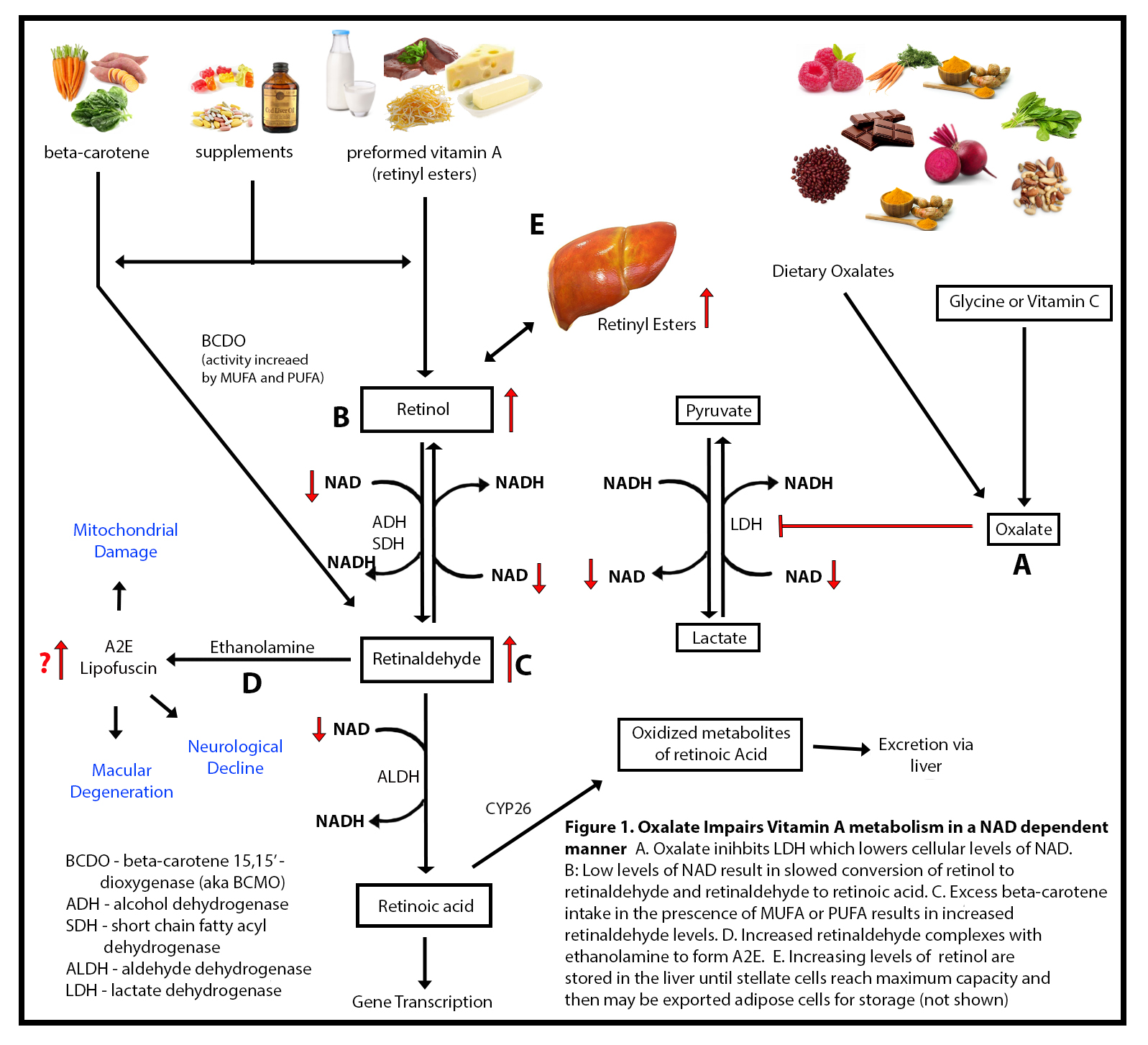
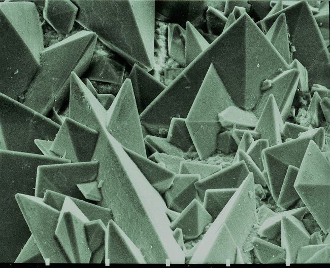
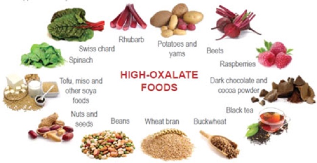
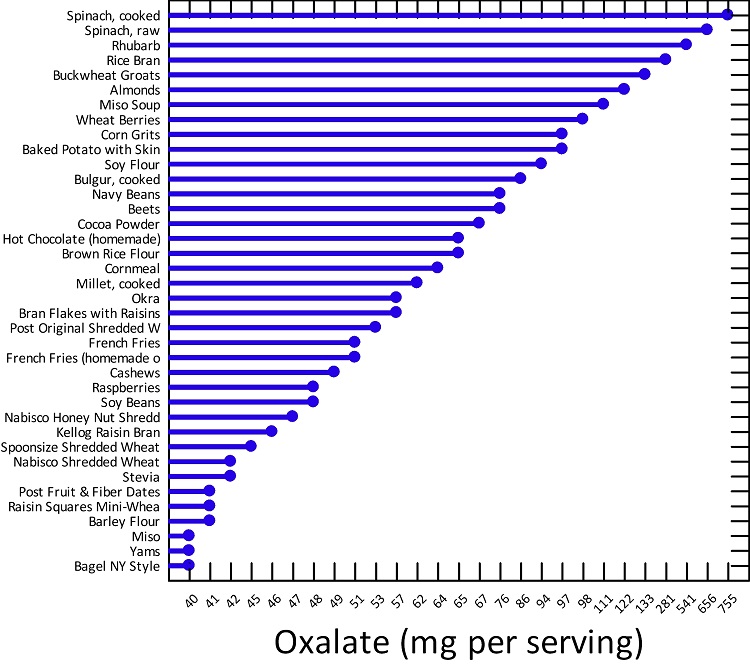
There is a truism in the sociology of occupational knowledge: “If all a doctor has is a hammer, then everything becomes a nail”.  All modern medicine relies on overkill dosages of drugs or surgery as the standard of care (Jennifer Daniels, MD, The Lethal Dose, 2013). There are no drugs or surgery that can alleviate the symptoms of poisoning from a natural pesticide found in plant foods but also produced in the liver. With this professional void, treating oxalosis has mainly been left to nutritionists. So, if you ask a nutritionist about oxalates, you will get a predictable answer to reduce oxalates in your diet. This is insufficient because most oxalates are produced by poor metabolism in the liver. Oxalate is a natural pesticide found in plant foods, but also is internally produced mainly from fungus and vitamin C (see How Oxalates Ruin Your Health). Oxalosis manifests as a syndrome of three main symptoms: oxalate crystals in tissues and kidney; histamine, mucous attack in nasal passages; and pseudo-gout mainly from acidity and eating cooked meat.

With Oxalosis, You’re Lost in an Ocean Without a Compass

I make no pretense to have expert knowledge or training on this issue beyond my personal knowledge of finding some solutions to my own oxalosis. Indeed, my own vitamin guru brother kept pushing on me mega doses of Vitamins C and D, avoiding calcium and iron, as a panacea for everything. Much later, I found that “health foods”, such as vitamins C and D, spinach and a plant diet, and the avoidance of calcium and iron were contributing to manufacturing oxalates in my liver.

Only after hitting a wall of brain fog, fatigue, pseudo-gout, nasal mucous attacks, extreme lack of thyroid hormone, pins and needles on the bottom of my feet, and crystals popping from my eyes, did I seek medical consultation for these symptoms. After several futile consultations, I found that doctors don’t know anything about oxalates. Finally, a naturopath doctor gave me an office blood blot test and stated I had oxalates. The only knowledgeable people I could find about what to do with oxalates was from a self-help group at TryingLowOxalates.io and the Hormones Matter.com website. I won’t bore you further with my medical system merry-go-round story that all those with oxalosis go through and instead will try and relate what I have learned to date, albeit all errors are my own.

Endogenous Oxalates Mainly Begin in the Liver

First, I learned that calcium is needed as a co-valent chemical binder to oxalate before meals to eliminate oxalate through the bowel or oxalic acid through with urine through the kidney. Secondly, I learned that plant foods like spinach, almonds, chocolate, and soy and were loaded with toxic levels of oxalates, a natural pesticide that protects plants from insects, worms, and herbivores. I also learned that high doses of synthetic Vitamin D (10,000 mg/day as a steroid) without Vitamin K2, could also lead to kidney stones and oxalosis. An iron deficiency may come into the picture as discussed below.

My learning curve with oxalosis ramped up when I read the 1950’s book Food is Your Best Medicine by Henry Beiler, MD. How could food be the best medicine when it was “healthy plant food” that was my pathway to chronic disease and eventually contributed to a minor heart attack (elevated troponin protein only)?

Beiler says all disease is a failure of the elimination systems (defecation, urination, expiration, perspiration, hydration, inflammation), not the immune system, in our body. The “master cylinder” elimination organ is the liver, which filters solid wastes. The kidney excretes wastes suspended in fluid such as oxalic acid. The lungs rid the body of C02 by coughing. Hydration dilutes oxalic acid in the gut making it less likely to form crystals. Perspiration expels toxins through sweating or skin rash, and inflammation is the depositing of toxins inside the body to quarantine them (e.g., cancer). Paraphrasing Beiler:

When the liver is congested it can no longer perform its eliminative function and waste matter is thrown into the bloodstream. Toxic blood must discharge its toxins, or the person dies (sepsis). So, nature uses ‘vicarious elimination’ of having the lung and kidney help-out in eliminating toxins and poisons in the liver. Fluid accumulates in the lung because the liver can’t filter food and toxins at the same time.

This is evidently how respiratory infection can begin without any airborne contagion from a virus.

He goes on to argue that toxic blood gets to the lung through the arteries and then permeates into the lung by cellular membrane diffusion in balloon-like alveoli sacks. For oxygen to get distributed throughout our body it first needs a transporter of red blood cells to carry oxygen-binding molecules called hemoglobin. Dietary iron is the building block of hemoglobin and is carried throughout the vascular system to bone marrow which is the farm for making red blood cells. Oxalates can bind to iron and subsequently lead to chronic anemia. So, oxygen production begins in the gut not the lung. Iron anemia is a predictor of poor outcome from lung infection. The oxygenated red blood cells then are carried throughout the body by the flow of blood.

He contends that cancer is the quarantining of toxins in tumors that switches cellular respiration from oxygen to sugar and oxalates can cause cancer in breast tissues. And finally, he says that heart disease occurs when there is a failure to get enough oxygen-rich blood to heart muscles (heart attack from irregular heartbeat arrhythmias) or to the brain (stroke).

Killer Proteins and Oxalosis

I shall try to show how oxalosis is related to excess unmetabolized protein in the human body. Beiler asserts “proteins can be body killers if we are not watchful of our diet”. Protein is necessary to grow and repair the body. However, poor metabolism with age often leads to making metabolites (non-nutrients) from fat, protein, or carbohydrate (not just from dietary oxalate consumption). Excess protein is one of the main sources of acidosis, which can be life threatening in the blood stream but not in the urine or saliva. Protein can change into fat or carbohydrate, but fat and carbs cannot morph into protein (Beiler).

One of the major symptoms of oxalosis is histamine or mucous in the nasal passages, for which there are no known explanations as to its mechanism of action with oxalates. “The more proteins are heated or cooked the more the colloidal form is changed”, the more mucous develops in the nasal passages. Unmetabolized proteins are expelled through a process called “vicarious elimination” through the nasal passages as mucous and or by diffusion into the spinal cord. Beiler adds:

When the adrenal glands are strong, they try to compensate for the liver’s failure by super-oxidation (requiring iron); this gives rise to increased kidney function. When both the kidneys and the liver become exhausted, the toxemia climbs to a higher level and often necessitates an attempt at vicarious elimination through organs that not normally excrete proteins.

For Beiler, oxalates are implicated in many cancers and heart disease where oxalate crystals in heart tissues or arteries potentially lead to fatal heart arrhythmias.

How This All Connects

My thesis is that internally produced oxalate is systemic and organic. It mainly manifests oxalosis (oxalate poisoning) and is produced in the liver, not solely from dietary oxalate, which can be managed with modest palliative measures. Oxalosis comes from poor elimination in the liver and not entirely from the food we consume. It is an interaction between the two in a feedback loop. I believe that the conventional approach to oxalosis, e.g. reducing the consumption of high oxalate foods, does not significantly reduce internal oxalate production, and thus, is merely palliative.

The motivation for this paper is that I could find no satisfactory explanation of the mechanism of action for endogenous oxalate other than vague references that it is produced in the liver, while dietary oxalosis happens in the kidney. Hereinabove I have hypothesized a plausible explanation of oxalosis from the liver from poor protein metabolism and oxidation. Since science is not just about finding evidence, but also about attempting to falsify a hypothesis, I am throwing this hypothesis out there for open refutation and clarification.

We Need Your Help

More people than ever are reading Hormones Matter, a testament to the need for independent voices in health and medicine. We are not funded and accept limited advertising. Unlike many health sites, we don’t force you to purchase a subscription. We believe health information should be open to all. If you read Hormones Matter, and like it, please help support it. Contribute now.

Yes, I would like to support Hormones Matter.   

Image by Ray Shrewsberry • from Pixabay.

Oxalate: A Potential Contributor to Hypervitaminosis A

4627 views

We have many backup pathways in the human body. When one pathway is broken, another pathway can pick up the slack. If that second pathway is broken, that is when disease occurs. Oxalate is a potential second pathway breaker in individuals with underlying mitochondrial disorders. As a dietitian, I have found many of my clients, especially those with Autism Spectrum Disorder (ASD) or other neurodevelopmental syndromes, are struggling with oxalate, which then impairs their ability to metabolize vitamin A. My clients have symptoms of vitamin A deficiency and have retinoic acid deficiency, but because of poor NADH/NAD recycling actually have retinol and retinaldehyde toxicity; a conundrum to be sure, until one understands the connections between oxalate, NAD, and vitamin A metabolism.

Briefly, nicotinamide adenine dinucleotide (NAD), derived from dietary niacin or vitamin B3, is a necessary cofactor in multiple enzymatic reactions involved in mitochondrial energy production. It is recycled endlessly back and forth between its oxidized and reduced forms NAD and NADH, respectively. The oxidized form NAD+ is required for the conversion of retinol to retinaldehyde and then to retinoic acid, the bioactive form of Vitamin A. With poor NADH/NAD recycling retinol and retinaldehyde are not converted to retinoic acid, and thus build up in the cell, presenting signs of both deficiency and toxicity simultaneously. The culprit behind this perplexing reaction, I believe, is oxalate damage to the NADH/NAD pathways via its interaction with an enzyme called lactate dehydrogenase (LDH). The chemistry in this pathway is a bit complicated, so bear with me. To help with the chemistry, I created a graphic (Figure 1.) to illustrate the pathways in question.

Oxalate Vitamin A Connection
Figure 1. Oxalate Vitamin A Connection

What is Oxalate?

The root of the dysfunction described above, I believe, begins with increased dietary oxalate and poor oxalate elimination. Oxalate is a component of plants that is impossible for the body to completely break down. It is a poison in large amounts. We absorb it at variable rates, but some of us make it in our bodies from vitamin C and glycine. Excess vitamin C becomes oxalate through direct degradation and without enzymes. Usually this occurs in vitamin C intake over 2000mg, but it can happen at lower doses as well. I never recommend vitamin C to “bowel tolerance” as this likely represents the death of the intestinal cells due to oxalate poisoning. Glycine is metabolized to oxalate in a B6 and thiamine deficient state, but when there is adequate B6 and thiamine, glycine does not become oxalate.

Oxalate Impairs Lactate Dehydrogenase

When oxalate is high, it impairs an enzyme LDH (Figure 1).We have to make some lactate to keep energy metabolism going, but when the mitochondrial respiratory chain is damaged, I believe this reaction becomes a more pivotal point for NADH to NAD recycling. While researching this possibility, Jenny Jones, a PhD in Human Molecular Genetics, shared this article to confirm my suspicions that the LDH reaction is a pivotal point of NADH to NAD recycling. The balance of NAD to NADH in cells is of the utmost importance to maintain normal cellular health.

When the body produces lactate, it also produces NAD+. This is what drives vitamin A (retinol and retinal) metabolism forward. What I found through researching literature, and through many of my clients experiencing hypervitaminosis A, is that oxalate does not directly inhibit alcohol dehydrogenase or retinol dehydrogenase or aldehyde dehydrogenase, which was what I thought originally, but rather, oxalate impairs lactate dehydrogenase (LDH), which then lower NAD+ levels. I hypothesized that oxalate takes away the “energy” needed to drive those reactions forward by impairing LDH.

Why Does Oxalate Inhibit LDH?

LDH is actually the last enzyme involved in the formation of oxalates. The benefit of oxalate being able to have a feedback inhibition on LDH is a safety mechanism to prevent oxalate poisoning. However, when dietary oxalate is too high this backfires and wreaks havoc on vitamin A metabolism and also energy metabolism. This means that high oxalate impairs LDH activity via NAD dependent pathway resulting in hypervitaminosis A in the form of retinol and retinaldehyde with a possible retinoic acid deficiency.

Oxalate Is Pathogenic in ASD

I mentioned in the introduction that oxalate has been implicated as a pathogenic substance in ASD. I propose that this is related both to a reduction in the NAD/NADH ratio which impairs overall energy metabolism, but also, due to the accumulation of retinaldehyde. High levels of retinaldehyde can form a complex with ethanolamine to form something called A2E in the skin and the eye. A2E is a lipofuscin (a pigmented by-product of failed intracellular catabolism) that has been found in the liver, kidney, heart muscle, retina, adrenals, nerve cells, and ganglion cells. It is studied predominantly in the eye as a symptom of advanced aging. In response to blue light, A2E increases reactive oxidant intermediates, but even in the absence of light, A2E has been shown to disrupt membrane integrity by acting as a detergent as well as by inhibiting key cellular function. A2E is just one possible way that poor vitamin A metabolism can contribute to altered mitochondrial function.

Retinoids and the various versions of vitamin A are mitochondrial toxicants when in excess. They cause the following problems due to alterations in cardiolipin (a stabilizer of the mitochondrial membrane) and displacement of cytochrome C oxidase. Cytochrome C oxidase, also known as complex IV, is the last enzyme of the mitochondrial electron transport chain before ATP is released. Damage to cytochrome C oxidase/complex IV causes all sorts of problems. The net effect of this mitochondrial membrane attack is:

  1. Increased free radical O2 production
  2. Increased nitric oxide (NO) which can then further impair cytochrome C oxidase
  3. Decreased ATP synthesis
  4. Decreased NADH to NAD recycling

I propose that the inhibition of these key cellular functions by aberrant vitamin A metabolism is causing the underlying inability to tolerate dietary oxalate in the absence of kidney stone formation, which then feeds back and causes more problems in the handling of vitamin A and dietary oxalate.

Since all of this is very technical, let me give you some real life examples of these patterns observed from family and my client base.

Case Evidence

My first clue to this pattern came from clients who rely on tube feeding where the food mixture contained high polyunsaturated fats and high oxalate foods such as carrots, sweet potatoes or almond meal of some sort. When the mixture contains high concentrations of beta-carotene (carrots, sweet potatoes) and is combined with polyunsaturated fatty acids, there is an upregulation of the beta-carotene monooxygenase (BCOM) enzyme leading to an increase in conversion of beta-carotene to retinaldehyde, as well as increases in cellular retinol binding protein 2 (RBP2) that facilitates uptake of vitamin A from the intestine.

Additionally, these foods are often also high in oxalate. The combination of a high oxalate tube feeding with an upregulation of BCOM and RBP2 leads to hypervitaminosis A of retinol and retinaldehyde with a deficiency of retinoic acid.

This pattern was not limited to just my tube fed clients. My daughter and four other clients also appear to have developed this pattern of high oxalate and hypervitaminosis A from their diets. In fact, regretfully, recommendations I gave to one of my clients, before I understood this pattern, pushed him into hypervitaminosis A with resulting liver failure and cognitive decline.

Client 1

For this client, I had prescribed a balance plate method for meal planning to help him with his cholesterol, triglycerides, and insulin resistance. I encouraged his family to cover half of his plate with vegetables at lunch and dinner to fill him up and provide more fiber. He loves carrots, so his lunch always had a large portion of carrots. Carrots are high in beta-carotene, but also oxalates. As I previously described, beta-carotene is a precursor to vitamin A but must be metabolized prior to being made into retinoic acid, the active form of vitamin A. It enters metabolism at the level of retinaldehyde, and in the presence of monounsaturated or polyunsaturated fat, is more readily converted to retinaldehyde. When combined with the oxalate induced downregulation of NAD/NADH pathway, this was a recipe for disaster.

With this diet, the client, who was already neurologically impaired, progressed quickly into dementia and now can no longer perform activities of daily living such as using a washing machine and changing his sheets. In addition, he now has elevated liver enzymes, metabolic acidosis, and insulin resistance. I had him checked for hypervitaminosis A by the only measure available, serum retinol levels. Indeed, his vitamin A levels were elevated. His intake of carrots in a NAD compromised state pushed him into worsening cognitive decline and liver failure caused by accumulation of vitamin A. Sadly, the balanced plate method is a standard diet prescribed to individuals with high cholesterol, triglycerides, and insulin resistance. It can be exceptionally high in oxalate and vitamin A creating a negative cycle of reactions that are damaging to health.

When we unpack his reactions, we can see that his liver toxicity can be explained by the fact that poorly metabolized vitamin A accumulated and contributed mitochondrial damage. This led to increased ROS which inhibited alpha ketoglutarate dehydrogenase. He was forced into a GABA shunt in the liver and also the brain leading to elevation in his AST and ALT enzymes as well as dysregulated behavior as his glutamate levels increased. However, because of an additional, what I believe to be, a functional B6 deficiency, he was unable to convert glutamate to GABA resulting in mood dysregulation from high glutamate and low GABA in the brain, as well as liver toxicity.  GABA is an antioxidant for the liver and an inhibitory neurotransmitter in the brain.

As to his declining cognitive dysfunction, his physicians have told his parents that he has early Alzheimer’s disease. The current plan for this particular client to slow the degenerative process, improve cognition, and reverse liver disease by:

  • Increasing dietary choline to restore cell membranes damaged by hypervitaminosis A
  • Improve vitamin B6 status
  • Reduce dietary carotenoids and insure Vitamin A intake is no higher than the RDA including any carotenoids.
  • Avoid dietary oxalates in excess of 50 mg per day. This will need to be lowered slowly over several weeks to prevent a retinoic acid surge and symptoms of retinoic acid toxicity such as rash, hair loss, fatigue, nausea, and headaches.

Other measures, specific to his case will also be employed.

Aldehyde Intolerance Due to Oxalate: My Daughter’s Experience

Over the past 11 years, my daughter Zoey has had moments throughout the day, and sometimes the entire day, in which she walked as if she were drunk. She also suffered from what doctors wanted to diagnose as “cyclic vomiting syndrome”. Looking back, the more oxalate that she was consuming, the more ataxia I would see, and the worse gastrointestinal symptoms she would have (reflux, constipation, nausea, vomiting). She would also have extreme mood swings and behaviors over the past two years that were so extreme that her neurologist recommended she be put on a “mood stabilizer”. These were only symptoms of underlying impaired ability to metabolize aldehydes and the A2E ethanolamine steal altering her autonomic nervous system function. These are also the same exact symptoms that my clients, adults with intellectual disability of various origins, are having.

I have since learned the mechanisms of this apparent drunkenness: impaired aldehyde metabolism. This could be genetic due to polymorphisms in genes related to alcohol and aldehyde metabolism or due to a decrease in the NAD:NADH ratio in cells. Alcohol dehydrogenase and aldehyde dehydrogenase both require NAD to work properly. If someone consumes a high oxalate diet, they are likely impairing LDH and causing low NAD levels. This slows aldehyde metabolism and results in aldehyde toxicity. Aldehyde toxicity can cause local cellular deficiencies of sulfur-containing antioxidants including glutathione as well as local deficiencies of thiamine, pyridoxine, folate, zinc, and magnesium which further impairs metabolism causing oxidative stress and membrane lipid oxidation. Aldehydes are also capable of forming adducts with DNA and causing DNA damage.

This means that oxalate was indirectly impairing her ability to metabolize alcohol and aldehydes. We actually do make these consistently during metabolism, and so any disruption in NAD will impair clearance of these byproducts of metabolism. My poor girl has been drunk on her own metabolites! I wasn’t giving her sips of beer! And now that I am no longer accidentally poisoning her with oxalate, she is not running into walls as much or falling as much. It must feel good to not be drunk. She also has no more nausea or vomiting, and no significant reflux.

Increasing Retinaldehyde Levels Have Far Reaching Implications?

I mentioned previously that individuals with ASD may have altered vitamin A metabolism to the point of having high retinaldehyde due to high oxalate intake. I am currently in search of a research laboratory willing to explore this mechanism and whether it is a widespread issue or something specific related to genetic alterations. It may be that individuals with underlying neurodevelopmental disorders are just more susceptible to alterations in vitamin A metabolism which leads them down a worsening pathway of neurological decline. I believe people with genetic syndromes should be closely monitored for impaired vitamin A metabolism.

However, the effect of higher levels of retinaldehyde due to poor metabolism may be widespread. Alzheimer’s disease is at least in part due to altered vitamin A metabolism due to dysregulation of a crucial enzyme in retinoic acid synthesis, ALDH1A1. In fact, altered retinoid signaling has been implicated in Alzheimer’s disease. Retinoic acid is very much needed for normal brain function. Anger and emotional dysregulation can be a serious issue in individuals with Alzheimer’s disease. The midbrain relies on ALDH1A1 to convert glutamate to GABA and if it is tied up in retinaldehyde metabolism it may lead to impulsive behaviors. In Parkinson’s disease, ALDH1A1 is crucial for dopaminergic neurons and dysfunction of this enzyme can lead to loss of fine motor control and impaired working memory. It would be interesting to evaluate if high retinaldehyde can contribute to the alterations seen in Parkinson’s disease. In addition, there is indirect evidence that psychiatric disorders such as schizophrenia, bipolar disorder, and major depressive disorder are symptoms of impaired Vitamin A metabolism. Overall, more research is needed to evaluate whether impaired vitamin A metabolism and elevation of retinaldehyde levels is contributing to neurological related disease.

Dietary Intervention for Poor Vitamin A Metabolism

At this time, I am working with my clients on various aspects of improving their production and recycling of NAD to help improve their vitamin A metabolism. Often, this means encouraging them to speak with their doctors about changes in medications. Some medications can alter the ability to metabolize vitamin A. These include PEG laxatives with molecular weights less than 4000 (3.7% metabolized to oxalate in the body), H2 receptor antagonists (inhibit vitamin A metabolism in a NAD dependent manner), Metformin (impairs respiratory complex one recycling of NAD), and high dose melatonin (which leads cells into a high NADH state).

And, of course, dietary oxalate reduction plays a major role in our therapeutic efforts to get vitamin A metabolism back on board. For these individuals with hypervitaminosis A induced mitochondrial impairment, reduction of dietary oxalate has become a key tool in solving their inability to metabolize vitamin A due to low levels of cellular NAD.

We have many backup pathways in the human body. When one pathway is broken, another pathway can pick up the slack. I have hope that once the mitochondrial damage is repaired, my clients will again be able to have their carrot cake and eat it too!

We Need Your Help

More people than ever are reading Hormones Matter, a testament to the need for independent voices in health and medicine. We are not funded and accept limited advertising. Unlike many health sites, we don’t force you to purchase a subscription. We believe health information should be open to all. If you read Hormones Matter, like it, please help support it. Contribute now.

Yes, I would like to support Hormones Matter. 

Image by Yerson Retamal from Pixabay.

What the Heck Are Oxalates and Why Should I Care?

21688 views

If you have searched the Internet looking for health solutions, chances are you have come across warnings about the dangers of oxalates in some foods. You may even have seen a reference to something called a low oxalate diet. What does that mean exactly? What the heck are “oxalates”? Well, let’s explore that a bit.

What Are Oxalates?

Oxalate is a relatively simple molecule in the grand scheme of things. Below is a diagram from Wikipedia.

oxalate

Oxalate is present to some degree or another in virtually all plant foods, but almost totally absent in animal foods. Why would that be? To begin with, oxalate is not a problem for a plant. Plants use oxalate to help regulate minerals like calcium, which is a metabolic process essential to the health of the plant.

Calcium is a 2+ cation; oxalate is a 2- anion. That means calcium and oxalate are attracted to each other; one might even say they are “preferred dance partners”. That’s an important point because oxalate helps to bind with calcium and allows the plant to draw this needed mineral up through the roots. It can also be used to help to obtain other needed minerals, including magnesium (another 2+ cation). So that makes oxalate very useful to a plant.

But this isn’t oxalate’s only purpose. In some plants, oxalate crystals participate in energy metabolism providing a source of carbon for respiration. In this case, oxalate may build up within the plant – but this is normal. Oxalate here may be protecting the plant from unfavorable environmental conditions, including drought and other stresses.

Plants have gone on to evolve other secondary functions for oxalate, including protection against both predation by insects and grazing by animals. One of these defenses can be the “Idioblast”. Idioblasts are specialized cells within the plant that may store plant chemical defenses, including oxalate crystals (as they do in the case of the Dieffenbachia plant). Oxalate may also be rich in the leaves of plants so that the mouths of insects may be damaged when trying to eat the plant.

Oxalates in Humans

The picture is vastly different for animals and humans alike. Oxalate has no useful purpose in the human body; instead, it can interfere both with our absorption and use of minerals, as well as with other important functions. The most commonly recognized issue that is directly related to oxalate is the dreaded kidney stone.

But that bias may be working against us; your general practitioner – and even your kidney specialist – may believe that the only issue with oxalate is the appearance of kidney stones. No stones? No problem!

For some, especially if their diet is not focused on the highest oxalate foods (see a list of the highest oxalate foods here), this may be true enough. For these folks, oxalate is a “mild” human toxin, as long as the intake is low enough, and the person is healthy. In this case, the body is able to clear it effectively, primarily through the kidneys. For other folks, however, consumption of even small amounts of higher oxalate foods presents significant problems.

Unfortunately, many of us are neither as healthy as we think, nor are our diets as low in oxalate as they were traditionally in the past.

Before the advent of modern food processing and shipping, many foods were seasonal and we simply couldn’t consume them in large amounts all year round. Good examples would be tender greens like spinach, which were eaten in season. Mind you, that might not have been enough to protect everyone from oxalate’s effects, and the fact that the historical record confirms the existence of kidney stones would be an indicator of that.

Another issue with the modern diet is that we often prefer foods raw; however, traditional cooking often saw certain plant foods boiled, which allowed oxalate to leach into the cooking water – which was then thrown away. With this modern preference for raw foods, we may unknowingly ingest extremely high oxalate in our diets. Research suggests that the daily oxalate consumption in western populations varies greatly, and can range as low as 44 mg/day and as high as 351 mg/day. Note that an average is smoothing out the highs and lows in intake, so your actual diet could range to extremely high levels. For instance, when extremely high oxalate foods like spinach, almonds, Swiss chard, beets, or rhubarb are consumed, your daily intake values may easily exceed 1000mg/day.

On one of the main support groups, Trying Low Oxalates, members report intake levels regularly in the area of 1000 mg a day. As a practitioner, I have personally seen oxalate intake as high as 3500 mg a day. If the individual is eating a Paleo low carb diet, and highly focused on leafy greens and nuts, reaching this level of intake is surprisingly easy. Keep in mind that hyperoxaluria – the condition of too much oxalate in the urine – is diagnosed at levels of oxalate in urine over 40-45 mg per day.

Where Do Oxalates Come From?

So where would the oxalate come from in your diet? Unlike other dietary exclusions, oxalate may be coming from a variety of sources, and each type of plant (and in some cases, even plant varieties) can have its own unique oxalate profile.


Category High Oxalate Foods
Beans/ Legumes Anasazi, Black/ Turtle, Cannellini, Great Northern, Navy, Pinto, Soy (whole bean not tofu), White
Carob/ Cocoa Dark chocolate, milk chocolate, chocolate substitutes using carob
Fruits Blackberries, Mission/ dried Figs, Guava, Kiwi, Pomegranate, Rhubarb, Star Fruit/ Carambola, Cactus/ Nopal
Grains Amaranth, Buckwheat, Quinoa, Teff, Wheat bran
Nuts Almond, Cashew, Brazil, Hazelnut, Peanuts, Pine
Seeds Caraway, Chia, Hemp, Poppy, Sesame
Spices/ Herbs Allspice, Celery seed, Cinnamon, Clove, Cumin, Curry powders, Fennel seed, Nutmeg, Turmeric
Vegetables Beets (root and greens), Bitter gourd, Burdock, Green beans (some varieties), Hearts of Palm, Jerusalem Artichoke, Okra, Plantain, Purslane, Potatoes (many), Sorrel, Sweet Potato, Swiss Chard, Spinach, Yam

As you can see from the list above, it’s not as simple as just giving up nuts or beans. It can take a review of your overall diet to discover what the sources of oxalate are, and how much they are impacting you.

Among the more common high oxalate diets are those that use fruit and vegetable smoothies either for ‘cleansing’ or weight loss. People who consume these smoothies may ingest upwards of 1000 mg of oxalates per day, a high concentration for anyone, but one that becomes especially problematic if the individual has underlying health issues. Chronic health conditions often reduce the capacity to process oxalates. In these cases, high oxalate ingestion can become deadly.

Let us review a few of those ‘worst-case scenarios.

Considering High Oxalate Diets: Worst-Case Scenarios

Oxalate metabolism presents problems with high intake, especially when the individual’s health is compromised in any way. High oxalate intake alone can kill insufficient dose – although we don’t hear about it often. Recent case studies show us how easily dietary intake can take us into this dangerous range.  We now have case studies of people – doing things that might be considered odd, but certainly within the realm of the ordinary – who then develop life-threatening issues.

The Green Smoothie Cleanse

Consider the case of a woman – well-intentioned – who began a green smoothie cleanse. Who hasn’t heard of such a thing in today’s world? Yet, after just 10 days, her kidneys failed. The green smoothie the woman was preparing contained over 1300 mg of oxalate per day, in spinach alone. Only her spinach intake is noted in the case study, although she may have had other oxalate sources, like nut milks or other vegetables. The material, in this case, indicates that the “normal” Western diet would have between 100 and 150 mg of oxalate per day.  This is 10x the amount that the average person is getting daily.

Imagine drinking only green smoothies for days on end and on top of that having pre-existing health issues. Over 10 days, she would have consumed a minimum of 13,000 mg of oxalate. It becomes easy to see how her health might be comprised!

In this case, the woman had other risk factors: she was 65 years old; had undergone gastric bypass (a known risk factor for oxalate absorption); and, she had also taken several rounds of antibiotic therapy, which is also a risk factor.

Age alone compromises metabolism and excretion. Compromised metabolism due to age means that we find it harder to absorb needed nutrients from our food. Compromised excretion due to age means it’s harder to get oxalate out of the body once it is in.

If to the issue of age we add gastric bypass and antibiotic therapy, we have significantly altered gut bacteria and diminished our ability to absorb and metabolize foods. Indeed, bypass prevents normal absorption altogether, by bypassing a portion of the stomach and the intestines. That is its purpose – to reduce nutrients to the body to help the patient to lose weight.

What we see is that the combination of gastric bypass, antibiotics, and high-dose oxalate consumption can be deadly.  It took only 10 days of what is a very common dietary protocol, to induce acute renal injury that could have progressed to full-fledged renal failure, had it not been caught.

Could a green-smoothie protocol induce such damage in other, healthier populations? Possibly. The variables that affect whether one has trouble dealing with oxalate are quite common.

It is well known that as we age, our ability to metabolize foods diminishes. One of the key things that we need to deal with the effects of oxalate is a good mineral level. Compromised digestion can easily compromise our level of many key nutrients, including minerals.

Antibiotics too, affect metabolism by disrupting the gut microbiota, and who among us has not been on multiple antibiotics over our lifetime? The likelihood that a person living in the Western world has had antibiotics is incredibly high. That means that our microbiome may or may not be ideal to handle dietary oxalate.

Then there is gastric bypass, which was performed on 179,000 people in 2013. This is a population whose metabolic function was already disturbed pre-surgery. Current statistics on the procedure are difficult to confirm, but given the US obesity rate was 38% as of 2015-2016, and appears to be rising, it is likely that the incidence of this procedure is rising.

Any one of the variables discussed (alone or in combination) would hamper an individual’s tolerance level for oxalate intake. If we then introduce an extremely high oxalate concoction like the green smoothie to the mix, it is entirely possible that oxalate issues will result, and that they will be serious.

Let us look at another case: a 51-year-old man on a low-carb diet, which also resulted in serious injury. Both “low carb” and “ketogenic” diets are very popular at the moment, although this particular vegan application was very strict and lacked protein. A low-carb protocol that included animal products would have meant less oxalate intake overall, as most animal products have no more than a trace of oxalate per serving.

This particular patient had done a protocol that included 6 meals of spinach, kale, berries, and nuts every day. With the current focus on eating more plant-based, it could seem healthy on the surface. Right? But here is the issue: not only was it excessively high in oxalate content, but the lack of animal protein and variety in the diet likely led to significant vitamin and mineral deficiencies, which in turn made the high oxalate content of his diet even more dangerous.

Finally, something as commonplace as star fruit consumption has been a cause of fatal injury in dialysis patients. Starfruit can contain as much as 300 mg of oxalate per 100 grams, putting a single fruit in the range of 270 mg. But that’s not as high as a single cup of raw spinach, which is over 300 mg for a single cup, eaten raw.

So what does this mean for us? While patients on dialysis have a clear risk factor, it may be surprising to note that there have been deaths in this population from consuming high oxalate foods. Note that as little as ½ a star fruit can be responsible for kidney injury for those who may have risk factors.

Not all risk factors are obvious either. How many people would adjust their diet because they had recently been on antibiotics? Once we are feeling well again, we would simply go back to our regular diet and all that is included.

Oxalate Consumption in Healthy Populations

This brings us to a discussion of those who are healthy. We might not be as “safe” as we think.

You may think that none of this concerns you because you have never had kidney stones and you do not have any of the pre-existing conditions noted in the case studies above. But as we saw from the green smoothie example, it is quite easy to reach excessively high oxalate consumption with common, everyday dietary practices.

Moreover, oxalate issues are not limited to kidney stones. Oxalate accumulation affects the entire body. In the second article in this series, we will explore oxalate consumption in ‘healthy’ populations and look at some lesser-known oxalate-related health issues.

We Need Your Help

More people than ever are reading Hormones Matter, a testament to the need for independent voices in health and medicine. We are not funded and accept limited advertising. Unlike many health sites, we don’t force you to purchase a subscription. We believe health information should be open to all. If you read Hormones Matter, and like it, please help support it. Contribute now.

Yes, I would like to support Hormones Matter.

Image by Robin Higgins from Pixabay.

This article was published originally on September 16, 2019. 

Fluoroquinolones, Beta Glucuronidase and Oxalosis: New Connections

3663 views

Have you ever taken antibiotics and felt they had possibly both beneficial and detrimental effects on your health? There are plenty of scientific journals that debate just this (here, here). In the functional medicine world, we hear many stories from people about moderate to severe adverse reactions to fluoroquinolones and other antibiotics. This website has many such stories about the fluoroquinolone reactions and has published several articles on the mechanisms. One way fluoroquinolones wreak havoc on health that has not be discussed here is their role in oxalosis. There are several straightforward ways that antibiotics may increase oxalate, but I would like to share some new ideas about what changes they may make that might impact oxalate and inflammation.

What Are Fluoroquinolones?

Fluoroquinolones are a family of antibiotics that have a powerful bactericidal effect by inhibiting bacterial type II DNA topoisomerase. Due to their potent antibacterial effect, fluoroquinolones are most commonly used to assist with a wide variety of infectious conditions. This class of drug contains a black box warning from the FDA, indicating that it can cause serious issues like tendinitis, tendon rupture, peripheral neuropathy, myasthenia gravis and central nervous system effects. The negative effects of these drugs have become so widespread that a nickname has been coined for those who have endured fluoroquinolone injuries, called Floxies.

In our functional medicine circle we have heard many of their stories. In some instances, the original problem for which the antibiotic was prescribed becomes worse after treatment. In other cases, Floxies seem to experience major hormonal shifts with changes to their skin, collagen, hair, and connective tissues. In the worst cases, patients experience tendon ruptures, often of the Achilles, which is one of the more likely areas to be affected. Additionally, some patients develop sudden onset of allergic reactions to things in their environment or diet that they were not previously allergic too. Angioedema, skin rashes, peripheral neuropathy, and air hunger are some of the many other common symptoms that develop after using fluoroquinolone antibiotics. Many of these may be related to newly developing oxalate issues driven by these antibiotics.

What Is Oxalate?

Oxalic acid is an organic acid that is highly corrosive and elevated levels in the body correlate to having high inflammation. Oxalic acid binds to minerals in the body forming insoluble oxalate crystals that can deposit in organs, tissues, joints and the vascular system.

We can ingest exogenous sources of oxalate in the foods we eat, with certain plant foods like spinach having the highest level of oxalate. The liver can produce oxalate endogenously if one has certain SNPs in oxalate metabolism or nutrient deficiencies. There are many other ways to increase oxalate, including certain antibiotics, like fluoroquinolones.

The Antibiotic-Oxalate Connection

Research shows that antibiotics are not selective for only the bad bacteria. They kill off good bacteria as well. Humans cannot metabolize oxalate. We need help from our microbiota, particularly the oxalate degraders. Antibiotics will kill off many beneficial oxalate degrading flora, like oxalobacter formigenes, allowing oxalate levels to increase.

The bacteria in the gut also absorb and produce many B vitamins. Most of the vitamins synthesized endogenously are used by the bacteria themselves, but a small percentage is released into circulation. Both poor absorption and reduced synthesis of B vitamins will have many deleterious effects on health. Among them, increased oxalate synthesis and decreased metabolism and elimination.

Thiamine (B1) and pyridoxine (B6), for example, in addition to all of their other functions in human health, are instrumental in preventing endogenous production of oxalate. The bacteria that produce these vitamins are affected negatively by antibiotics.

Menaquinone or vitamin K producing bacteria also take a hit when antibiotics are used. Reduced vitamin K impacts calcium handling. Calcium homeostasis is critical for cell function in general and oxalate management in particular. It is also important for something called matrix Gla protein expression. Matrix Gla is a protein produced in the bone that inhibits vascular calcification. In other words, matrix Gla prevents the accumulation of oxalate, when expressed. When it is not expressed sufficiently, because vitamin K concentrations are diminished either by diet, antibiotic use, or both, oxalate will accumulate (here, here). Vitamin K is also critical in balancing hormones by multiple mechanisms. It is important to androgen/estrogen balance as seen in this study about PCOS. Hormonal balance plays a significant role in oxalate metabolism, which will be discussed later. These are just a few of the ways in which antibiotics contribute to an increase in oxalate and oxalate-driven inflammation

Beta Glucuronidase: A New Player in Oxalosis

In addition to the antibiotic-induced changes to oxalate-degrading and vitamin-synthesizing flora, antibiotics also alter carbohydrate metabolism. Antibiotics inhibit an enzyme called beta-glucuronidase (BG). BG is a lysosomal enzyme needed for the breakdown of complex carbohydrates and for the proper degradation of other small molecules like glycosaminoglycans (GAGs) such as hyaluronic acid, heparin/heparin sulfate, chondroitin sulfate/dermatan sulfate, and keratin sulfate and glucuronides such as bilirubin.

We have both human (encoded by the GUSB gene) and bacterially produced BG enzymes. Some main bacterial producers of BG are Escherichia coli, Clostridium species, Bacteroides species and Staphylococcus species. As men age, BG will increase. For women though, BG declines over time.

In disease states, certain enzymes become elevated and can therefore reliably indicate various pathological conditions. Beta glucuronidase is one of those enzymes. With metabolic disease and diabetes, for example, BG is elevated and the mix of gut bacteria shifts considerably.

Elevated BG also interrupts an important part of phase II liver detoxification called glucuronidation. This is where a glucuronic acid molecule is added onto toxic substances to inactivate and make them water soluble and easier to excrete. Beta glucuronidase can break the glycosylic bonds freeing the carcinogen and allowing its reabsorption and enterohepatic recirculation, rather than the proper clearance. Elevated BG, because of its impact on glucuronidation/phase II liver detoxification, will decrease the clearance of sex steroid hormones, xenobiotics, pesticides, herbicides, and insecticides, BPA, mycotoxins, pharmaceutical medicines, and more.

Elevated BG can be a marker for, and can contribute to, various issues. The recirculation of these carcinogens can contribute to colon cancer. The poor clearance of estrogens is associated with hormonal cancers like breast and ovarian cancer and endometriosis. In prostate cancer, upregulated BG is related to disease progression. It is not just cancers where BG is problematic, in chronic periodontitis, an 8-fold increase in BG activity has been recorded along with 33-fold increase in BG in bacterial peritonitis. This study details some of these observations.

This is where the fluoroquinolones and other antibiotics enter. A key job that they perform is to inhibit BG enzyme activity and the BG producing bacteria, pushing these levels back down into range. BG inhibition helps limit the recirculation of toxins that contribute to a vast number of diseases. A quick look at studies shows that fluoroquinolones like ciprofloxacin significantly inhibit BG. This study reveals that a combination of other antibiotics such as penicillin and metronidazole can have an inhibitory effect of up to 50% on enzyme activity.

What Happens When Beta Glucuronidase Is Too Low?

While it is clear that elevated levels of BG contribute to serious disease states, there are many substrates in the body that have to be at just the right concentration; where either too much or too little can be harmful to health. BG is one of these substrates. If fluoroquinolones and other antibiotics inhibit BG, and BG manages oxalate accumulation, could repeated use of these drugs over-inhibit BG such that oxalosis develops? I believe so. Below are a few of the pathways that may lead to high oxalate.

Beta-Glucuronidase Effect on Flavonoids

There is a strong relationship between inflammation and deconjugation. Conjugation and deconjugation are terms used to describe the addition or separation of molecules during liver detoxification.

When tissues in the body are experiencing necrosis, inflammation and mitochondrial dysfunction one of the most helpful antioxidants is the flavonoid. Once ingested, flavonoids like quercetin, which is the most abundant flavonoid in our diet, are conjugated via glucuronidation in the liver to their inactive metabolites making them easier to transport through the blood to these problem areas. Once they arrive, they must be deconjugated back into their active aglycone form so that they can be used to suppress the expression of pro-inflammatory genes such as COX-2 and scavenger receptors.

When the inactive flavonoids arrive at the inflamed tissues, phagocytic cells surround the problem area and secrete large amounts of both lactic acid and BG. The lactate is required to create the acidic environment needed for the most optimal BG function. One study shows a 13-fold increase of BG activity from pH 7.4 to pH 5.4. Then, BG secretion is needed for the bioactivation of the inactive flavonoid metabolites back to their aglycone form. From here, the flavonoids can go to work quenching the inflammation. If BG has been inhibited by fluoroquinolones or other antibiotics, flavonoids like quercetin will not be re-activated and will be unable to temper the inflammation. Here are three fascinating studies that explain this in detail (here, here, here). It appears that elevations in BG might follow inflammation, in an attempt to help resolve it

In addition to suppressing pro-inflammatory genes, the diuretic, antioxidant, anti-inflammatory, and antibacterial properties of flavonoids help inhibit the formation of calcium oxalate stones. If BG has been over-inhibited, such that these flavonoids cannot be reactivated, do we not then lose the oxalate preventative benefit that they provide? Possibly, which means oxalate levels would increase.

Ironically, flavonoids are potent inhibitors of BG. Is it possible they are used to resolve the inflammation then inactivate the BG so that its continued accumulation is not able to progress hormonal and other issues? Perhaps BG is involved in a natural feedback cycle of sorts.

Beta-Glucuronidase Effect on Glycosaminoglycans

As mentioned previously, BG is a lysosomal enzyme, which catalyzes the degradation of glycosaminoglycans (GAGs), such as chondroitin sulfate, heparin sulfate, dermatan sulfate and hyaluronan. When there is a mutation in the GUSB gene resulting in a deficiency of BG the GAGs are improperly degraded with consequent intraliposomal and tissue accumulation, creating lysosomal storage issues in a disease called mucopolysaccharidoses.

Now, there is some major interplay between oxalate and GAGs. GAGs are protective against the growth and formation of calcium oxalate crystals by having a strong binding affinity to them (here, here). There appears to be a low excretion rate of sulfated GAGs in stone formers, possibly due to improper degradation. And like GAGs, oxalate also needs to be phagocytized or engulfed into the lysosome where it can be dissolved. If they tend to bind together, it would appear that the increase in BG level by phagocytes may also be important for proper handling of oxalate.

In tendon rupture, a well-recognized side-effect of fluoroquinolone antibiotics, there is an accumulation of non-degraded GAGs (here, here). We know, through observations of people with hyperoxaluria, that oxalate crystals can accumulate around the joints and within tendon sheaths. Could the over suppression of BG by fluoroquinolones and other antibiotics contribute to their accumulation? Possibly.

Beta-Glucuronidase, Oxalate, and Estradiol

We know a consequence of elevated BG is estrogen dominance (here, here), so low BG activity could translate to lower levels of estradiol. Could repeated use of fluoroquinolones diminish estradiol concentrations such that it too influences oxalate build up? Once again, possibly.

Low estradiol (E2) might contribute to elevated oxalate in a couple of ways. Estradiol inhibits the activity of glycolic acid oxidase (GAO), thereby decreasing the amount of glyoxylate converted to oxalate within the peroxisome. Studies show that E2 is significantly lower in kidney stone patients with a significantly higher frequency of calcium oxalate stones in the lowest E2 groups.

Estradiol is the body’s natural anti-androgen via its suppression of 5 alpha reductase, which converts testosterone to the substantially more potent androgen receptor ligand, dihydrotestosterone (DHT). So, it can also help to manage oxalate level by keeping DHT levels in check. A quick look at studies will show that androgens increase oxalate level and stone formation by increasing GAO activity, and by suppressing osteopontin levels while estrogens elevate it, respectively.

However, like BG, E2 is also one of those substrates that has to be kept in balance. There can be harmful effects if E2 becomes either elevated or deficient. This study shows that E2 might attenuate oxalate transport activity via the downregulation of SLC26A6 and therefore enhance cancer cell migration, creating a potential risk for nephrolithiasis and cancer.

Fluoroquinolones, Beta-Glucuronidase, Estradiol and Tendinopathies

Estradiol is very important to maintaining connective tissue in women by improving the tendon collagen synthesis rate and increasing the number of smaller versus larger fibrils. Conversely, this study shows that the natural ageing process, whereby estradiol declines, increases a woman’s risk of tendon rupture to a similar level as seen in men. They postulate that a reduction in tensile strength, decrease in collagen synthesis, fiber diameter, density, and increase degradation in tendon tissue, all play a part. This means that both elevated and diminished estradiol influence connective tissue. Consider, for example, pregnancy in which E2 is quite elevated. It is the E2 induced changes to various collagen synthesizing genes that allow the laxity and the stretching of tendons, ligaments and skin. But, going to the other extreme could low estradiol induce excessive rigidity, also contribute to injury? When fluoroquinolones are used, they inhibit beta glucuronidase. Since BG inhibits estradiol, it is possible that this is one of the mechanisms contributing to the rigidity seen in tendinopathies and ruptures after fluoroquinolone use.

It All Comes Back to the Gut

It is in the gut where the oxalate degrading flora live. It is in the gut where the microbes that absorb and synthesize vitamins and minerals live. It is in the gut where the microbes that produce short chain fatty acids and beta glucuronidase that help balance the estrobolome and other hormones live. All of these factors intersect to manage and determine our inflammation level. When we kill these microbes with antibiotics and other ‘kill tools’ we inevitably impact oxalate metabolism. Fluoroquinolone induced inhibition of beta glucuronidase may be a key player in the cascade leading to oxalosis. While the inhibition of BG may be useful when concentrations are too high, repeated or over-inhibition may be deleterious to health.

The good news is, the body wants to regain homeostasis when pushed out of balance. The cessation of any antibiotics that are depressing BG will allow it to recover over time. Perhaps by taking these other factors and associated biomarkers into consideration we might be able to gently push into some level of homeostasis more quickly to regain health.

We Need Your Help

More people than ever are reading Hormones Matter, a testament to the need for independent voices in health and medicine. We are not funded and accept limited advertising. Unlike many health sites, we don’t force you to purchase a subscription. We believe health information should be open to all. If you read Hormones Matter, like it, please help support it. Contribute now.

Yes, I would like to support Hormones Matter.

Photo by Vardan Papikyan on Unsplash.

Leaky Faucets Shouldn’t Be Ignored: A Story of Oxalate, Vitamin A, and Metabolic Decline

4357 views

Zoey. The name means life. She is beautiful. She is fearfully and wonderfully made. She has been labeled otherwise. She has been labeled broken and put into a “syndrome box”.  She drips out additional diagnoses like a leaky faucet.

She has accumulated these leaky faucets over the course of 11 years. She drips health problems out slowly with an audible, “DRIP….DRIP….DRIP”. Each year we find out something new, but most of the time the doctors won’t follow her on these new leaky faucets because they have put her in a “syndrome box”.  They say, “Oh, Zoey is just this way because she has 2q23.1 microdeletion syndrome, White Sutton Syndrome, and Non-classic congenital adrenal hyperplasia (NCCAH). That means she won’t sleep well, she won’t speak, and she won’t have a normal gate. She won’t, she won’t, she won’t”.

This year, a few of her best doctors stopped putting her in a syndrome box because the leaky faucet finally broke. This story centers around being placed in a kidney reflux and hydronephrosis box for 11 years to “watch and wait” to see if her hydronephrosis and reflux would spontaneously go away as kids typically just grow out of this. This watching and waiting was my biggest mistake. Now the pipes are broken.

Genetic Box Labels

Leaky Faucet Labels

  • Autism
  • Cerebral palsy
  • Diffuse cerebral function with epileptic discharges
  • Esophageal Dysmotility with pharyngeal backflow
  • Dysphagia requiring g-tube (she can eat solids, but not liquids)
  • Insomnia
  • Keratosis Pilaris
  • Onychotillomania
  • Impaired speech
  • Kidney Reflux
  • Hydronephrosis
  • Sleep Apnea
  • Ataxia
  • Fine motor delay
  • Gross motor delay
  • Constipation
  • GERD
  • Chronic ear infections
  • Chronic toe and finger infections
  • Submucosa cleft palate s/p repair
  • Tethered cord syndrome s/p repair and now fatty infiltration of thecal sac.
  • Severe astigmatism (diagnosed at age 11)
  • Growth hormone deficiency

The First Sign of a Leaky Faucet

Zoey has always had a weird urinary pattern. Ever since Zoey was a baby, she wouldn’t urinate at night. In the morning, she would only urinate a small amount, about 100 ml. After propping her up for a few hours she would urinate more and more. This continues to this day. The urologist praised me for my potty training efforts in a child with Autism because she is dry at night and during the day, but I didn’t do anything to potty train Zoey. She can’t pee. I have to rub her tummy to help her release urine. I asked about this and I was told, “She only has mild hydronephrosis. We don’t cath kids when it’s mild.” Honestly, though, Zoey would never let me do a straight cath on her. So that’s not even an option, but in case you have a similar issue, there is a suprapubic type of catheter that looks like a g-tube port. I was never offered that. Here is her weird pee pattern:

  • 8:00 AM 50 ml
  • 10:00 AM 300 ml
  • 12:00 PM 300 ml
  • 2:00 PM 400 ml
  • 5:00 PM 800 ml
  • 8:00 PM 400 ml

She seems to make more urine in the afternoon. I think this is related to gravity. As she is more upright, her kidneys drain into her bladder. At night she still sleeps with her knees tucked under her and her butt up in the air which worsens her hydronephrosis. She wakes up every morning feeling nauseated. It used to subside within a couple of hours, but by age 10 this was all day.

A Second Leak: High Oxalates

Zoey’s urinary issue has been going on for quite some time. When she was two years old, I ran a Great Plains Lab OAT test on her. She had many gut related issues, and evidence of metabolic issues for which I started many different supplements, but she also had severely high oxalate.

My husband also suffers from repetitive calcium oxalate stone formation, so there may be a genetic component. At two years old, Zoey had a nephrologist confirm two tiny little kidney stones. I was told to increase her fluids to 2400 ml per day. As a dietitian, I also changed her diet completely. I put her on a low oxalate diet, and increased her citrate levels through magnesium citrate (which also helped her constipation) so that we could dissolve the stones. It worked.

My husband had damage to his kidneys from multiple stone formations and ultrasound lithotripsy, so I wanted to spare her kidneys from damage. I mentioned this to our nephrologist and he seemed to be not impressed. He said we didn’t need to follow up and that urology can manage her kidney issues. I wasn’t the advocate I am now, so I took that as a good sign that she was going to be just fine, and just did yearly follow ups with urology.

The Third Leak: High Testosterone and Early Puberty but Poor Growth

At age six, Zoey started what seemed to be early puberty. I asked her PCP to check hormone labs on her because being short and starting puberty early would mean she would stop growing too soon and be extremely short her whole life. He initially refused, but eventually acquiesced and ran the labs. She had very high testosterone levels. He sent us back to the same endocrinologist that told me she wasn’t growth hormone deficient. That endocrinologist said, “Let’s watch these labs for a bit.”  We watched the labs for an entire year as the testosterone crept higher and higher.

I was so worried about testosterone hurting her body that I did a deep dive into journal articles and looked all along Zoey’s gene deletion to try to identify a gene deletion that was causing her hormone issues. I couldn’t find a single gene in her 7.29 MB deletion that could be a contributor. I was stumped. My then twelve year old daughter, after hearing me constantly talk about the damaging effects of testosterone and how Zoey will end up with a full beard, said, “Mama, if you are worried about Zoey, maybe you should be worried about me? I’ve been shaving my whole body and plucking thick hairs off of my chin.” And just like that, the older sister advocated for the younger sister.

We went to a new endocrinologist and Zoey and her big sister were diagnosed with NCCAH. Their particular version of this disease is caused by mutations on each allele for the 21-hydroxylase gene. So it was genetics, after all, but it wasn’t within Zoey’s large gene deletion or related to her mosaic POGZ mutation. They don’t make cortisol efficiently. Instead, they make too much testosterone. The solution is to take hydrocortisone to meet cortisol needs and to suppress the hormone that triggers the adrenal glands to make cortisol. This lowers testosterone levels.

Having NCCAH can also cause a person to stop growing sooner in life so the new endocrinologist checked Zoey’s bone age. It was close to normal, but she obviously saw Zoey’s short stature. She said, “Why hasn’t she started on growth hormone?” I said, “Well, she definitely has a risk of deficiency because the mouse model of MBD5 deletion causes growth hormone deficiency, but our last endo said she didn’t have that because her IGF was normal.” The new endo just stared at me for a few seconds. I said, “And….he missed her NCCAH…and he didn’t test her correctly, right?” She said, “Let’s schedule a growth hormone stimulation test to see what’s going on.” Then I said, “And he is wrong isn’t he? Growth hormone does cross the blood brain barrier, doesn’t it?”  She said, “Yes, but I can’t guarantee it will help her cognition.”   Well, if you want to see the results of just one month of growth hormone on a person’s cognitive abilities, please go to my YouTube channel. Growth Hormone Isn’t Just for Height Gain.

Once Zoey started on growth hormone in 2020 she grew quickly. She actually grew about four inches in one year! By September of 2021, we achieved the results I wanted to see on her kidney reflux. Her ultrasound showed resolution of kidney reflux completely and growth of each kidney! Woohoo!! Urology wanted to discharge us because she had finally “grown out” of her reflux. I was so happy, but being a more cautious advocate than previously, I asked to continue to follow up, due to my husband’s history. I requested a follow up. Again, the physician refused at first, but acquiesced with coaxing.

In September of 2022, Zoey went back for her follow up appointment. I had some concerns to talk about, because Zoey’s morning urine was cloudy, but cleared up as the day went on. She was also struggling with peeing and crying in pain. They tested her urine and she didn’t have a UTI. We actually had gone to urgent care a few times to test for UTIs because it was an ongoing issue. She had no infections at all, but was having serious urinary symptoms and nausea. The ultrasound revealed that Zoey’s kidney reflux was back and that she wasn’t emptying her bladder. The urologist thought it was probably neurological in origin though because she didn’t have any evidence of UTI or cloudy urine in his office.

A Few More Leaks: Elevated Vitamin A Concentrations, More Oxalate Issues, and the Return of Kidney Reflux

In September 2018, after having a g-tube placed, and being on a blenderized diet, her GI doctor checked Zoey’s vitamin A for the first time, and it was 40 mcg/dl (normal is 26-49). I was not supplementing with vitamin A due to the difficulty of dispensing it, and the tube feeding I made had natural sources of vitamin A such as egg yolks. Her doctor was pleased that she wasn’t malnourished from a blenderized diet that was a bit limited due to being low oxalate.

As Zoey was able to eat orally, she started back on a low oxalate diet with small amounts of plantain chips or random bits of plantain flour based on a handout I found that indicated this was a low oxalate food. Her vitamin A was creeping up slowly, but still in the normal range until March 2021 when it was 59 mcg/dl (normal range: 26-49). When I saw it, I put myself on a “watch and wait”, but asked the gastroenterologist about it. She said she wasn’t concerned and that she sees it go up sometimes. They only treat low vitamin A. I was comforted by the fact that in January 2022 it had come down just a few points to 51 mcg/dl.

Zoey  began to refuse our once a week snack of plantain chips. She would shake her head “No.” I should have realized then that there was a problem, but I thought it was because they were too hard to chew. She was struggling with painful baby teeth that she had ground to bits from bruxism. In August 2022, Zoey’s vitamin A had shot up again to 65 mcg/dl and I was quite alarmed. Looking back on this, it is easy to explain, as we had been giving her a full sandwich with two slices of bread that contained plantain flour, instead of just one half of a sandwich that had plantain flour.

With this change in diet, Zoey was extra tired all the time and she was having hypoglycemic episodes on and off. I asked her gastroenterologist about her increasing vitamin A and she recommended I ask the eye doctor to check Zoey’s eyes because I was concerned about the risk of a side effect of vitamin A toxicity, intracranial hypertension. Zoey had an eye checkup in August in September 2022 and all seemed fine except for a tiny change in her lenses.

From a Drip to a Flood

In January 2023, I decided to make banana muffins for Zoey only out of plantain flour and bananas. “So much better than white flour,” I thought. That week she asked for two at breakfast every morning, and because she had been so tired, I didn’t see a problem with it. Over the next five days she declined rapidly. She was walking as if she was drunk, and she couldn’t walk more than ten steps without having to sit down and rub her legs. She was telling us that she was sick by making sick sounds and asking for a thermometer, but she never had a temperature. She would tell us with her AAC, “headache, headache, vomit, vomit.”

After five days of plantain muffins, I went into her room to give morning meds and found her in her sleep safe bed lying in a puddle of vomit. When I opened the zipper compartment, she wretched and vomited some more. My heart immediately started to race because this is my child who aspirates liquids. She was going to aspirate her vomit! I turned her onto her side as she wretched some more. I wasn’t able to give her the hydrocortisone through her g-tube because she would just vomit it back up! I ran to get the inject-able hydrocortisone, solu-cortef injection, injected her, gave her Zofran, and rushed her to the emergency room. They started an IV and ran every test they could think of, but found nothing and diagnosed her with “vomiting”. Well, no one else in our family was sick or ever did get sick. While at the ER we left a urine sample and it was extremely cloudy. They checked it for a UTI, but said it was clean. They sent it off for cultures, but they came back normal later that week. After we left the hospital with a prescription for Zofran, the results were published to her medical portal. I saw what her urine and ultrasound of her bladder results said: Amorphous Crystals in Urine and Bladder Debris. Then it hit me. The only thing I had changed that week was plantain flour!!!

Hindsight has shown me that the cause of her returning kidney reflux the previous September was actually an increase in dietary oxalate from giving Zoey more plantain flour. I had been using the same handout for the past ten years from a kidney dietitian. The handout listed plantain flour as 1 mg of oxalate based on data from Harvard about 40 years ago. Well, plantain is now thought to have up to 524 mg per cup of flour. The oxalate in plantain flour actually was causing the kidney reflux as well as an alteration in Zoey’s ability to metabolize vitamin A leading to increasing serum vitamin A levels.  Zoey is not metabolizing dietary vitamin A into retinoic acid, which has multiple functions throughout the body, and is the only form of Vitamin A that can leave the body.

Now that we haven’t been giving Zoey plantain flour for the past three months, her kidney reflux has completely resolved. She still has cloudy urine, but that is related to hypercalciuria. Her serum vitamin A (retinol) levels remain high. I have learned that elevated retinol triggers calcium loss from bones due to alterations in osteoclast activity, although triggering of parathyroid hormone may also be involved. This causes hypercalciuria. My sweet girl has developed 10+ cavities in the past six months from rapidly depleting calcium stores. Her dentist was shocked. He joked that he didn’t know what to do…because he eats oxalate all the time. In addition, Zoey now has severe astigmatism and has to wear “coke bottle” glasses. Astigmatisms can be caused by retinoic toxicity. Zoey now be making retinoic acid to quickly. However, we are on a path of healing now and working out ways to help her metabolize vitamin A better in her body. She even can walk a straight line now and has energy!

History of Supplements

  • Birth -1 years old: No extra vitamin supplements. Was using Neocate Junior, 1 scoop per feeding of breast milk (8 scoops per day usually).
  • 1-2 years old: No supplements. During that time her diet was horribly high in oxalate because I didn’t know about them. Had GPL labs done at 2 years old showing hyperoxaluria and multiple vitamin issues.
  • Age 2 to 6 years: Calcium Citrate 300 mg at all meals. No multivitamin because she was allergic to cobalt. She would break out in hives. She refused to take the separate vitamin supplements orally, and when I did give them to her, she would vomit, so I gave up because the vomiting was horrible for her.
  • Age 6 to 6 years 8 months: She had a g-tube and was on a homemade blenderized diet where I added the vitamins listed  below (except only 25 mg of thiamine) to her g-tube including the calcium citrate. She tolerated them fine in microdoses over the day through her g-tube.
  • 6-11 years old: All vitamins were given via her enteral tube feeding bag with just water. It’s basically more of microdosing every 2 hours with 400 ml of vitamin infused water. If I push them all at once she vomits. During these 5 years, the routine was not consistent because I had to rely on a hired caregiver when I was at work. Again, during this time she only received 25mg of thiamine and not the amount on the list.
  • Last three months: We have been consistent with the vitamins and the higher dose of thiamine and she has come back to life and appears to be recovering. I currently have Niacin on hold due to I’m exploring the relationship between NCCAH and the Kynurenine Pathway.

Current Supplements

  • 500 mg Thiamine HCl
  • 400 mcg 5-methyl folate
  • 25 mg Pantothenic Acid
  • 17 mg of B6 (Pyridoxal -5- Phosphate)
  • 5 mg Niacin
  • 5 mcg Biotin
  • 25 mg Alpha Lipoic Acid
  • 250 mg Vitamin C
  • 500 mg Acetyl L-Carnitine
  • 500 mg Glutathione
  • 325 mg Calm Magnesium (Magnesium Malate)
  • 2 Capsules of Mega Multi Mineral (half of a dose) contains:
    • 500 mg of calcium amino acid chelate
    • 11 mg potassium phosphate
    • 5 mcg iodine (from potassium iodine and kelp)
    • 5 mg Zinc
    • 25 mg Copper
    • 25 mcg selenium
    • 5 mg Manganese
    • 25 mcg chromium
    • 25 mcg molybdenum
    • 59 mg Potassium
    • 5 mg Boric Acid
    • 5 mcg Vanadium
    • 25 mg Glutamic acid

Why My Kid?

Sometimes I wonder why it has to be my kid that suffers. Maybe it is only so that we can help other people. This experience has actually helped me with my own clients who have disabilities of various origin. Many of them are also suffering from this same inability to metabolize vitamin A. I was only able to spot this after I learned what the symptoms of this condition looks like by experiencing it with Zoey.

I tell Zoey every time that something I learned from her health circumstances helps another person. She will clasp her hands under her chin with one elbow up in the air and one elbow down and squeal in delight. It is the only thing that keeps us going. We love to help other people with nutrition. I definitely understood nutrition before Zoey was born, having been a dietitian for 10 years before she entered this world, but her health circumstances forced me to fine tune my nutrition knowledge. She made me dive deep into the research. She made me learn how to read metabolic tests such as plasma amino acid tests and urinary organic acid tests. I have been a dietitian for 22 years, but the past 11 years has turned me into a nutrition detective. I am thankful for all that my sweet Zoey Abigail has taught me.

We Need Your Help

More people than ever are reading Hormones Matter, a testament to the need for independent voices in health and medicine. We are not funded and accept limited advertising. Unlike many health sites, we don’t force you to purchase a subscription. We believe health information should be open to all. If you read Hormones Matter, like it, please help support it. Contribute now.

Yes, I would like to support Hormones Matter. 

PPARs and Oxalate Metabolism: Do They Intertwine?

2714 views

In a world where disease states characterized by high levels of inflammation are increasing, more and more scientists are taking a closer look at the connection between oxalate and these conditions.

Oxalate is an inflammatory molecule, that when bound up to minerals in the body, can crystallize and accumulate in organs and tissues. By binding minerals, oxalate can induce nutrient deficiencies impairing many important systems and processes in the body.

High levels of oxalate will also trigger an inflammatory cascade where the NLRP3 inflammasome, which is like our innate immune system’s alarm bell, becomes over-activated increasing NF-kB activity and increasing the release and expression of inflammatory mediators and cytokines like TNF-a, IL-1B and IL-6.

This over-activation can compromise our innate immune system’s ability to jump into action when the next “immune attack” comes along. Whether it is a pathogen-associated molecular pattern (PAMP) triggered by something like influenza A or Covid-19 or a damage-associated molecular pattern (DAMP) triggered by something non-infectious, like uric acid crystals or hyaluronan fragments or oxalate, chronic immune activation precipitated by oxalate will threaten the immune system’s ability to fight the next illness.

The other problem with constant over-activation of the NLRP3 inflammasome is that we now have elevated inflammatory cytokines, which will increase with the next attack, potentially driving us into a cytokine storm syndrome. So, keeping this type of inflammation low is the goal for having a strong immune response when it is needed.

Genes Influencing  Oxalate Metabolism

Inborn errors in three particular genes can contribute to faulty glyoxylate metabolism leading to hyperoxaluria – excessive oxalate storage. The glyoxylate pathway is where oxalate metabolism takes place.

  • A problem with alanine glyoxylate aminotransferase (AGT/AGXT) will cause someone to convert less glyoxylate to glycine, creating a buildup of glyoxylate which can convert to oxalate. This is characterized as primary hyperoxaluria type 1 (PH1).
  • An issue with glyoxylate reductase/hydroxypyruvate reductase (GRHPR) will lead to a lesser conversion of glyoxylate to glycolate and less push by hydroxypyruvate reductase of glyoxylate towards gluconeogenesis, again leaving an accumulation of glyoxylate available to convert. This is referred to as primary hyperoxaluria type 2 (PH2).
  • Primary hyperoxaluria type 3 (PH3) involves SNPs in 4-hydroxy-2-oxoglutarate aldolase 1 (HOGA1) and over activity can form excessive glyoxylate from hydroxyproline.

A loss of function mutation in any of AGT, GRHPR or HOGA1 will allow glyoxylate to pool and become available for lactate dehydrogenase (LDH) to convert to oxalate.

Non-genetic Pathways to Poor Oxalate Management

In the Trying Low Oxalates group we see a lot of people without defects in these genes still having issues with oxalate. Here, we have to consider other factors that contribute to elevated oxalate. Some main factors include, but are not limited to:

  • Endogenous production (can be caused by both genetic mutations and/or nutrient deficiencies in specific B vitamins)
  • Exogenous food sources, particularly in dietary extremes
  • Certain supplements that convert or can be degraded to oxalate
  • An unhealthy microbiome and estrobolome in general with fewer oxalate degrading microbes and potential hormonal imbalances, often spurred on by past antibiotic use but this is not the only cause

I believe the loss of peroxisome proliferator-activated receptor activity might also play a key role. This family of nuclear receptors are important in many different areas but particularly inflammation and immunity.

What Are Peroxisome Proliferator-Activated Receptors and Why Are They Important?

Peroxisome proliferator-activated receptors (PPARs) are ligand dependent transcription factors that regulate the expression of many genes involved in inflammation, fatty acid metabolism, energy homeostasis and metabolic function. There are a number of ways that PPARs might impact oxalate issues. The glyoxylate cycle itself occurs within the peroxisome. So from a purely mechanical perspective, any derangement of peroxisome function would potentially impact oxalate metabolism.

Additionally, a quick look at studies show that PPARs may alter activity levels of some key genes in oxalate metabolism (AGT, GRHPR). Third, PPARs play a key regulatory role on (intestinal) tight junctions (here, here). Tight junctions help prevent hyper-absorption of oxalate through the gut, while leaky junctions (leaky gut), allow oxalate to disperse into the bloodstream and travel to different tissues and organs where they cause damage.

Lastly, PPARs regulate macrophage polarization and therefore crystal phagocytosis (clearance of oxalate). They also increase antioxidant activity (here, here).

PPAR activity seems directly important to oxalate metabolism due to its control over the genes, tight junctions, macrophages and antioxidant status. A lack or loss of PPAR activity might also affect our glyoxylate cycle and levels, leaving plenty available for conversion to oxalate. Perhaps the lack of PPAR activity plays a role for those who do not have obvious SNPs in the important oxalate genes, yet still wind up in oxalate overload.

PPAR Activation and Inflammation

PPARs play a critical role in inflammation. They have a regulatory effect on both crystal-related enzymes and pro-inflammatory enzymes such as iNOS, metalloproteinase MMP-9 and COX-2, but their main function in inflammation is to promote the inactivation of NF-kB thereby decreasing the production of inflammatory cytokines (here, here). The exact mechanism behind how these are reduced is not clear though. I believe that the reduction of highly inflammatory oxalate in plasma contributes to this. The research seems to support this hypothesis. It is a bit technical, so bear with me.

  • Alanine glyoxylate aminotransferase (AGT) is positively regulated by PPAR-alpha (PPAR-a). This means that in absence of sufficient PPAR expression, AGT activity will be low. This allows glyoxylate to accumulate and potentially be converted to oxalate.
  • PPAR-a activation is crucial in inducing transcriptional activation of glyoxylate reductase hydroxypyruvate reductase (GRHPR) in mice but in humans GRHPR expression was shown to be PPAR-a independent due to promoter reorganization during primate evolution. (here, here). This means that without proper PPAR activation GRHPR activity will be lower, again allowing glyoxylate to accumulate. However, there is uncertainty if this works the same way in humans as it does mice due to the promoter reorganization that has occurred in man.
  • Mice deficient in PPAR-a present higher plasma levels of oxalate and as expected, administration of a PPAR-a ligand reduces plasma oxalate levels.
  • PPAR-gamma (PPAR-y) activation suppresses calcium oxalate crystal binding and oxalate-induced oxidative stress.
  • Yet oxalate itself impairs PPAR-y expression and phosphorylation creating a very important “negative feedback loop”.
  • PPAR-y agonists enhance barrier function through the upregulation of tight junction molecules claudin-1 and -4, occludin, and tricellulin at the transcriptional level, and will thereby be protective against hyper-absorption of oxalate through the “leaky gut”. PPAR expression also leads to a significant increase in tight junction strands, which will create a stronger barrier between the apical and basolateral membrane domains limiting the excess passage of oxalate and other proteins and lipids (here, here).
  • Classically activated M1 macrophages (which are inflammatory and cause tissue damage) facilitate renal crystal formation while alternatively activated M2 macrophages (that are anti-inflammatory and focused on tissue repair) suppress it (here, here). Macrophages have an important role in crystal phagocytosis, which is the process where phagocytes engulf and destroy foreign substances, like oxalate. PPARs play a key role in regulating this macrophage polarization to ensure that the anti-inflammatory macrophages are activated and assisting with tissue repair and crystal phagocytosis.
  • PPAR-y agonists can regulate TGF-β1 and HGF/c-Met to exert antioxidant effects against hyperoxaluria and alleviate crystal deposition and can exert an antioxidant effect through the PPAR-γ-AKT/STAT3/p38 MAPK-Snail signaling pathway.

Nutrients That Influence PPAR Activity

We know nutrient deficiencies play into oxalate issues so it is not surprising that B vitamins (B1, B2B3, B5, B6,  biotin, folic acid, and B12) activate PPAR-a and PPAR-y.

There are many natural ligands that can activate PPARs, like essential fatty acids and eicosanoids, but as indicated in this study they are required in extremely high concentrations of up to 100 uM for PPAR activation. A quick glance at ctdbase.org reveals that a wide variety of nutrients are capable of activating PPARs, but again, I would guess they would be required in huge pharmacological doses.

A couple notable ligands (but certainly not limited to) are free fatty acids with a preference for long-chain polyunsaturated fatty acids (PUFA’s) like arachidonic acid, alpha linolenic acid, EPA and DHA, linoleic acid, carnitine, alpha tocopherol, various carotenoids, endocannabinoids, PGJ2, leukotriene B4 and more. Ligands worth noting that have inhibitory effects are caffeine. There are other nutrients that have seem to have both activating and inhibitory effects, like zinc and copper.

Beta carotene appears to have both inhibitory and activating effects on PPAR’s depending on concentration, but BCMO1 is also transcriptionally regulated by PPAR-y, so might be worth digging into for anyone with SNP’s there (here, here).

Folic acid appears to have differential effects on PPAR-a and PPAR-y activity, so could be valuable to consider for those with MTHFR SNPs (here, here).

PEA and PPARs

I have wondered how much palmitoylethanolamide (PEA) comes into play. PEA is a fatty acid amide produced endogenously by neurons and glial cells in the central nervous system and also contained in egg yolks, peanuts and soybean. PEA is involved in the neuroprotective mechanisms that are activated following tissue damage and inflammation. In the cell, PEA is hydrolyzed (broken down) by fatty acid amide hydrolase (FAAH). Upregulation of this FAAH gene can cause us to rapidly break down PEA in the cell making it unavailable for its anti-inflammatory and analgesic effects.

In the body, it is produced on demand and acts locally, and its production is increased in areas of inflammation. PEA has been shown to activate both PPAR-a and PPAR-y. In doing so, it regulates inflammation and crystal-related genes and in this way helps to quench inflammation. So, elevated activity of FAAH may work against us with regards to inflammation by too rapidly breaking down this helpful endogenous fatty acid amide.

Genes involved in FAAH activity:

  • rs324420 A allele will cause lower and C will cause higher FAAH activity
  • rs2295633 G allele will cause likely higher activity
  • rs3766246 G allele will cause possibly higher activity

PEA is a ligand for the PPAR nuclear receptors, but it is not clear if it is PEA’s direct effect that is most beneficial or the “entourage effect” that improves PPAR activity. In the entourage effect, PEA competitively inhibits FAAH’s hydrolysis of and provides a sparing effect of anandamide (AEA). AEA is known as the “bliss molecule” as it can bind to cannabinoid receptors, providing an analgesic effect and creating feelings of happiness and wellbeing.  AEA is the more potent ligand for the PPAR’s so it is believed PEA’s prevention of AEA breakdown is also important for better PPAR activation.

It is also possible to have mutations in the 3 PPAR’s themselves which can lead to a loss of function, so they may be worth looking at too.

Conditions that Overlap with Oxalate and PPAR Activity

There are some key conditions that have traditionally been recognized as overlapping with oxalate overload. Notably, these include: COPD and respiratory disorders, cystic fibrosis, chronic kidney disease, thyroid disorders, vulvodynia, and autism.

We are also beginning to see that oxalate intersects with many other conditions. Studies show and we have learned through discussions on TLO that there is a clear tie between oxalate and conditions like metabolic syndrome, diabetes, obesity, mast cell activation syndrome, chronic fibromyalgia type pain, mood disorders, fatty acid oxidation issues and more. It seems that dysfunctional PPAR activity is central to most of these, as well.

Inadequate PPAR expression may affect overall oxalate concentrations and allow many of these conditions to develop alongside oxalate overload. This is one of the reasons that inflammation is finally being recognized as a clear contributor to neurological and mood disorders. We know oxalate can also be causative for some of these conditions, so there is a viscous cycle of inflammation that needs to be broken. I hope that factoring in PPAR activity and its effect on oxalate metabolism might be helpful to your own research and health journeys.

In the next article, I will take a closer look at some of the conditions discussed here and how oxalate and the PPARs might both play a role.

We Need Your Help

More people than ever are reading Hormones Matter, a testament to the need for independent voices in health and medicine. We are not funded and accept limited advertising. Unlike many health sites, we don’t force you to purchase a subscription. We believe health information should be open to all. If you read Hormones Matter, like it, please help support it. Contribute now.

Yes, I would like to support Hormones Matter. 

Marginally Insufficient Thiamine Intake and Oxalates

29821 views

Over the last few years, it has become increasingly apparent how important thiamine is to overall health. Thiamine (thiamin) or vitamin B1, sits atop the mitochondria at multiple entry points involved in the metabolism of foods into cellular energy (ATP). It is also critical for several enzymatic reactions within the mitochondria. We have illustrated repeatedly how thiamine deficiency leads to mitochondrial dysfunction, which in turn leads to complex multi-organ system illnesses characterized by chronic inflammation, disturbed immune function, altered steroidogenesis. Each of these is related to deficient mitochondrial energetics. When serious or chronic, thiamine deficiency leads to erratic autonomic function, now called dysautonomia, and a set of disease processes called beriberi.

Long before those symptoms emerge and absent severe deficiency, marginal thiamine status evokes subtle changes in metabolic function. Among these changes, enzymes that would normally metabolize certain foods fully and into useful substrates for other functions are downregulated, shifting the metabolic pathway towards more toxic end-products. The chemistry is complicated and we will go over it in a moment, but first I would like to propose a framework for understanding metabolism. For me, it is useful to imagine metabolism visually as giant maze of right and left turns; where wrong turns lead to dead ends and dead ends lead to the build up of endogenous toxins. Among the primary variables determining the route metabolism takes is enzyme nutrition.

Enzymes require nutrient cofactors to perform their metabolic tasks. When the appropriate nutrient co-factors are present in sufficient concentrations for the enzymes to operate fully, the food we eat is successfully metabolized into end-products that are useful for all manner of processes and cellular energy is produced. Even in the case of genetic aberrations that limit enzyme function endogenously, there is evidence that nutrient manipulation can overcome inadequate enzyme activity. When nutrient co-factors are in short-supply, however, resources are reallocated. Metabolism shifts directions, it takes a right turn when it should move left or vice versa. Different enzymes are activated and metabolism eventually reaches a dead end but not before potentially toxic, unused waste products build up. As these toxins build up, other systems become disrupted, inflammatory and immune responses are activated, demanding ever more energy to resolve. It is this energy spiral, I believe, that induces and maintains many of the illnesses we see today. This means that observing how one reacts to certain foods may point us to correctable nutrient deficiencies.

The Rise in Food Sensitivities

In recent years, I have become fascinated by the growing preponderance of food sensitivities and intolerances. It seems everyone has a problem with something. Given the current practices used in industrial food production, I suppose it is no wonder. We use a staggering number of chemicals to grow and process foods; chemicals that reduce the nutrient content of supposedly healthy foods, but also, present as toxicants that must be dealt with metabolically when ingested. The double hit of low nutrients/high toxicants is disastrous for metabolism. Throw in the generally high calorie content of the western diet and one has to wonder how our mitochondria function at all. And yet they do. Well, sort of. If we don’t count the exponential growth in chronic and seemingly intractable illnesses, but I digress. I believe that food, or lack of quality food, is top among the core contributors to modern illness and food sensitivities are among the key early warning signs of poor metabolism and by definition, faltering mitochondria.

Oxalate Problems

One of the more intriguing and troubling food intolerances that has become increasingly common is to the high oxalate foods. Oxalates are natural substances found in many healthy foods, especially dark leafy greens like spinach, that bind calcium and other minerals, and when left unmetabolized, can form crystals leading to kidney stones. Approximately 10% of men and 7% of women experience at least one episode of kidney stones across the lifetime. Beyond the kidney stone, oxalate intolerance is linked to wide range of chronic health conditions largely due to the build up oxalic acid which may or may not bind calcium, but causes problems nevertheless. Poor oxalate metabolism disrupts gut health, shifting the microbiome unfavorably causing dysbiosis, damages the mitochondria and induces system wide oxidative stress, inflammation and immune reactivity. Problems with oxalate metabolism have been found in individuals with autism, multiple sclerosis, arthritis, and fibromyalgia to name but a few. A common and usually somewhat successful remedy is to avoid the consumption of high oxalate foods. Below are some of the more common high oxalates.

Figure 1. High Oxalate Foods


Absent genetic aberrations leading to poor oxalate handling, I cannot help but wondering if the avoidance diet is the correct response, especially permanently. Certainly, it would help short term, and there may be foods that result in oxalate accumulation that could or should be avoided long term, but an across-the-board and permanent avoidance of most oxalate producing foods seems problematic nutritionally. If we consider that many who suffer from oxalate issues may also be sensitive to other foods, the avoidance approach could limit dietary options considerably. What if we are approaching this issue incorrectly? My gut tells me, and research seems to back it up, that barring genetic issues with oxalate metabolism, the dietary component is not simply one of excess oxalate consumption. It is a problem with inadequate nutrient consumption in the face of excessive non-nutrient foods – e.g. it is a problem with the modern western diet in its entirety.

Other Dietary Contributors to Oxalate Buildup: Processed Foods

If we dig into the oxalate issue a little more, we see that foods resulting in excess oxalate storage are not necessarily limited to whole foods listed above in Figure 1. A number of foods classified as high oxalate, are simply processed food products, high in carbohydrates, trans fats and low in nutrients. Below is a graph of some of the higher oxalate foods as compiled by the University of Chicago via Harvard’s School of Public Health. Notice, how processed foods make this list. Sure, their oxalate status is significantly lower than other foods, but consider what portion of the average western diet these products comprise. Click the links above to see a more complete listing foods that result in high oxalate accumulation. When you search through those lists (especially, the one from the University of Chicago), it becomes apparent that virtually all processed foods can result in oxalate problems.

Figure 2. Oxalate Content in Common Foods

food-oxalate-graph

One could argue that oxalate buildup involves shifts in the metabolic pathway that are directly related to nutrient deficiencies and those nutrient deficiencies emerge from the consumption of the modern diet. Processed carbohydrates, for example, in the presence of thiamine deficiency, are metabolized quite differently than when thiamine is present, with the former resulting in oxalate build up. Since a diet high in processed carbohydrates is one of the leading causes of thiamine deficiency in the first place, this begs the question, is the issue really oxalates or a sort of high calorie malnutrition resulting in thiamine deficiency, where oxalate accumulation is just a side-effect. Similarly, when thiamine is absent, fatty acid metabolism can go awry, making highly processed, high carbohydrate, high fat foods damaging on two fronts.

Finally, there are many other foods that can lead to high oxalate production in the presence of low thiamine including: beer, wine, tea, coffee, yogurt, bread, rice, soybean paste, soy sauce, and oil, along with foods that have been fermented, roasted, baked, or fried. And just like high carbohydrate diets can lead to thiamine deficiency, as nature would have it, all alcoholic drinks, coffee, and tea decrease thiamine uptake thereby both creating and exacerbating the thiamine deficiency that leads to oxalate accumulation. It could be that problems with oxalates is simply the early sign of thiamine deficiency and it may very well be a protective mechanism of sorts, a metabolic diversion, albeit an unhealthy one, to forestall the other issues associated with insufficient thiamine intake.

I should also mention that oxalate problems may not be solely related to diet. Inasmuch as all pharmaceuticals damage the mitochondria and either decrease thiamine directly or increase the demand for the need for thiamine indirectly, regular use of pharmaceuticals may also contribute to the problem. Similarly, a number of environmental exposures increase glyoxal (a precursor to oxalate build up in the face of low thiamine), including: cigarette smoke, smoke from residential log fires, vehicle exhaust, smog, fog, and some household cleaning products.

Is Thiamine Really the Problem?

It may be. The chemistry is complicated and detailed below, but basically, marginal thiamine status, prevents the proper metabolism of certain foods leading to the build up of toxins while simultaneously crippling the natural detox pathways. The combination of increased toxins and decreased detox ability leads to all sorts of damage and illness, high oxidative stress, and as illustrated by the graphic below, can lead to cancer (to be discussed in a subsequent article). Thiamine prevents this. A paper published in 2005, (from which Figure 3., is taken) details just how many mechanisms that lead to oxalate accumulation are initiated by low thiamine.

Figure 3. How Low Thiamine Leads to Elevated Glyoxal and Cancer

glyoxal pathways

The specifics involve a metabolic diversion that leads ‘food’ metabolites down what is called the glyoxal pathway, the pathway responsible for oxalates. Each of the red ‘X’s’ indicates an impaired thiamine dependent enzyme.

With Marginal Thiamine

  • Elevated glyoxal and methylglyoxal
    • Diminished activity of thiamine dependent enzymes (transketolase, pyruvate dehydrogenase, branched chain ketoacid dehydrogenase, and a-ketoglutarate)
      • Low transketolase = low NADPH
      • Low NADPH = low glutathione (the primary detoxification agent in the body; glutathione also requires vitamin C)
        • Low glutathione = poor detoxification of glyoxal and methylglyoxal = increased carcinogenic protein adducts
    • High Oxalate Foods
  • Diminished pyridoxal kinase (PK) activity*
    • *This is not discussed in the aforementioned paper, but should be included. PK is the enzyme that converts the inactive form of vitamin B6 (pyridoxine 5-phosphate) into its active form, pyridoxal 5-phosphate (P5P). Low P5P prevents glyoxalate from being converted back into glycine, leading to high oxalates. Many mistakenly assume that low vitamin B6 is responsible for high oxalates. While that is possible, it is also possible, and often more likely, that low thiamine is responsible.

With Sufficient Thiamine

  • Thiamine dependent enzymes work appropriately
    • Sufficient transketolase activity = sufficient NADPH
  • Glyoxal and methylglyoxal are metabolized into other substrates and/or excess is detoxified
    • Sufficient NADPH = sufficient glutathione
  • Glyoxalate is converted to a-hydroxy-b-ketoadipate or glycine and not oxalate
    • Alanine glyoxylate amino transferase (AGT), the enzyme required to convert glyoxalate into to glycine instead of oxalate has sufficient activated vitamin B6.

This is not to say that there are not other vitamin and mineral deficiencies also associated with hyperoxaluria, there are. Research has shown that low magnesium (a requisite co-factor in many of same enzymes as thiamine), along with low vitamin A, in addition to the low vitamin B6, mentioned above play a role. Vitamin E may also be involved.

Take Home

The majority of modern illnesses are the result of poor diet and environmental exposures directly, cumulatively, and generationally. Over the span of a few short generations, we have forgotten that food is fuel and that good, clean, unprocessed food is required for health. The allure of processed foods and cheap agriculture through chemistry, has left much of the population starved of nutrients, while simultaneously bearing a high toxicant load. The result is all sorts of metabolic disturbances which may manifest as food insensitivities and intolerances. It is interesting to note that the metabolic changes involved in oxalate buildup do not require what we would consider a full-scale thiamine deficiency, but rather, a sort of thiamine insufficiency initiated by marginal thiamine intake, something that is likely common across populations.

A less complicated overview of the low thiamine > high oxalate connection can be viewed below.

Is Your Body Producing too much Oxalate?

We Need Your Help

More people than ever are reading Hormones Matter, a testament to the need for independent voices in health and medicine. We are not funded and accept limited advertising. Unlike many health sites, we don’t force you to purchase a subscription. We believe health information should be open to all. If you read Hormones Matter, like it, please help support it. Contribute now.

Yes, I would like to support Hormones Matter.

Image: Scanning electron micrograph of the surface of a kidney stone showing tetragonal crystals of weddellite (calcium oxalate dihydrate) emerging from the amorphous central part of the stone; the horizontal length of the picture represents 0.5 mm of the figured original. Image credit: Kempf EK – Own work, CC BY-SA 3.0.

This article was published originally on August 15, 2019. 

Oxalate Degrading Microbes: Reconsidering Pathogenesis

27905 views

An exciting new paper was published recently which should turn the oxalate world upside down. The authors hunted public databases and compiled a list of microbes that possess variations of a gene called oxalyl CoA decarboxylase. That gene makes a protein that degrades oxalate.

Oxalate is an ion used by microbes for communication between fungus and bacteria, but it is found at varying levels in plant food. Our bodies also manufacture oxalate during a stress response. Everybody makes some amount of oxalate in their bodies and eats sometimes huge amounts of oxalate in foods like spinach, beets, nuts and seeds, and also (alas!) chocolate. When oxalate in our bodies gets too high, it causes dysbiosis and becomes dangerous, tangling with mineral metabolism, and delivering harsh changes to mitochondrial function, adding in distressing levels of oxidative stress.

Humans lack any oxalate-degrading gene in our own genome. That means we cannot degrade oxalate using our own talents. Instead, we are reliant upon our microbes in the gut to degrade oxalate. From studying isolated human beings who have never seen an antibiotic, scientists have realized that like them, our ancestors had plenty of oxalate degrading microbes in the gut (termed oxalotrophic microbes). The job of these microbes was to keep oxalate from food from getting into the inside of their bodies. That system worked well even when the diet was high in oxalate, but things changed when our microbiome began to be damaged by antibiotics and antifungals, and other hits.

In this discussion, I would like to examine findings from this recently published study from China:  Abundance, Functional, and Evolutionary Analysis of Oxalyl-Coenzyme A Decarboxylase in Human Microbiota where they identified the microbial species that possess one of the two genes that we know are used to degrade oxalate in our intestines. We will also examine some other issues that this new knowledge unwraps.

Oxalate Degradation Is Dependent upon Thiamine and Is Impaired by Antibiotics

The protein oxalyl coenzyme A decarboxylase is dependent upon thiamine. It requires thiamine that is in other forms to be converted into thiamine diphosphate (TPP). Scientists have told us that this is the primary form of thiamine that microbes will make and use.

Some of us have inborn errors in our thiamine chemistry. Those errors might limit our delivery of thiamine to the inside of our gut so that our oxalate degrading microbes can use it. Another issue is that some antibiotics kill bacteria by attacking those microbes with a direct hit to their thiamine chemistry.

I had my own tangle with these issues very many years ago. Back in 1967, I was given two rounds of chloramphenicol, an antibiotic that attacks the thiamine chemistry in bacteria. In March of the following year, four months after I stopped taking the antibiotic, I developed bone marrow failure and almost died. The same blood disease, called aplastic anemia, has been found on occasion in primary hyperoxaluria, which is a genetic disease where the body makes fatal levels of oxalate coming mostly from producing oxalate in the liver.

Chloramphenicol was taken off the market in the US in 1968 because so many people died from blood problems that came on slowly like mine. Another antibiotic that attacks thiamine chemistry is Alinia and it is used broadly by functional medicine doctors. There may be even more antibiotics that would also cause this problem, but no one has done a systematic review of vitamin deficiencies caused by antibiotics. If someone would do this type of analysis of all licensed antibiotics, then doctors would have a list of antibiotics to avoid if their patients had a genetic weakness or a deficiency in thiamine or other vitamins. Also, some viral infections may purposefully impair thiamine chemistry to weaken the host. Polio is an example. That is why after certain infections, someone may actually develop a new oxalate problem.

Oxalate Degrading Species May Not Be Pathogenic

In the paper mentioned above, the researchers found 1739 Oxalate degrading species in humans. All of the different species they identified were equipped with oxalyl-Coenzyme A decarboxylase that allows microbes like oxalobacter formigenes to degrade oxalate. You may be surprised to learn that you have probably never heard of most of these species. Because I was curious about their identity, I looked for how many oxalate degrading bacteria show up on the GI Microbial Assay Plus (GI-MAP) test from Diagnostic Solutions Laboratory, or were found on the comprehensive-digestive-stool-analysis-(CDSA) from Genova Diagnostics. These are tests frequently ordered by functional medicine doctors or naturopaths. I have noticed how often these tests are ordered when a patient has some sort of GI distress because I routinely review that sort of lab work during consults and find out from the patient what his doctor prescribed after seeing the results of the test. Most often, the patient was given antibiotics. Of course, which antibiotic the doctor chose varied significantly.

Included in the list of bacteria that degrade oxalates, were many species that are believed to be pathogens because they were elevated on tests that were ordered at a time of increased symptoms. It is natural to assume these microbes were the primary cause of the symptoms, however, I cannot help but wonder if the relationship between these bacteria is what we thought it was. If oxalate is a communication method between microbes, how many of those symptoms could have been caused by the elevated oxalate or the effect of that oxalate on intestinal microbes? If oxalate is a favored food for these types of microbes, might they expand their population whenever oxalate is increased? This might be similar to how ants or flies may show up in numbers when you uncover food at a picnic. Flagging the increased count of these species on lab tests might have persuaded the doctors to treat with antibiotics, and there was no other game plan. Would reducing oxalate have helped solve the problem without antibiotics?

Oxalobacter Formigenes: An Oxalate Obligate

Within the oxalate field, a great deal of attention has been given to the microbe oxalobacter formigenes.  The man who discovered this microbe, Dr. Milt Allison, had a lot to do with inspiring me to start looking more carefully at oxalate in autism and other conditions, and that grew to include pain conditions, autoimmunity, and gastrointestinal distress.

The uniqueness of oxalobacter formigenes, as far as we know, has been that it is the only microbe that requires oxalate to survive. Its dependency on oxalate is why scientists call it an oxalate obligate. This trait is why this microbe has received the most attention from scientists and became the launching point of this Chinese paper.

In a different study that I have reviewed before in the TLO Research Corner on the Trying Low Oxalates Facebook group, scientists looked at the differences in the diversity of microbes that survive in a very high oxalate condition (which in this case was chronic kidney stones) and compared that to normal controls. These scientists found out that oxalobacter doesn’t tolerate a high oxalate environment very well. Please note that their title implies that oxalate causes dysbiosis and not the reverse.

In the last two decades, a company called Oxthera and its predecessor have spent millions of dollars trying to develop oxalobacter as a drug for primary hyperoxaluria.  Sadly, they still have no product on the market. Oxalobacter formigenes may have been the wrong microbe to pursue because the paper on dysbiosis found that this microbe really doesn’t like extremely elevated oxalate.  This may be like humans having a hard time eating a hundred hamburgers in one sitting. This Chinese paper shows that scientists now have many more choices of oxalate-degrading microbes to study for research.

What Might Cystic Fibrosis Teach Us About Oxalates?

I have talked to our TLO group about this before, but cystic fibrosis is a genetic condition very important for oxalate research. This condition involves a broken intracellular regulator which governs the secretion of oxalate and sulfate among its other duties. This is why people with cystic fibrosis are elevated in oxalate. If someone has this gene defect, the mucus becomes very thick in the lungs and it is prone to infection. People with CF often live from cradle to grave with antibiotics. Pseudomonas aeruginosa often becomes their most common infection, and yes, this microbe showed up on the Chinese list of microbes that degrade oxalate. Might pseudomonas aeruginosa be growing too high levels and turning pathogenic just because it is responding to oxalate as its favorite food?

We are used to watching with distress as flies and ants discover our food at a picnic. Does oxalate become a picnic for certain microbes?

Have we made other mistakes assuming the worst about microbes when they were actually providing a benefit to us? I recently reviewed a paper in the TLO Research Corner that showed that intestinal infections with candida protected mice from systemic infections, including systemic infections with candida.  Using antifungals destroyed that protection. Have we been confused about what was going on in microbial communities, putting black hats on microbes that might be trying to protect us from something worse?

Counting Microbial Species In Cystic Fibrosis

I used PubMed to discover that many of the oxalate degrading microbes identified in the Chinese paper have been commonly reported as infections in cystic fibrosis. This is what I found:

  • Pseudomonas – 7838
  • Burkholderia – 1624
  • Mycobacteria – 708
  • Achromobacter –  206
  • Klebsiella – 118
  • Pandoraae – 59

Is there a chance that excess oxalate in cystic fibrosis patients (which is known to occur) could be attracting and feeding these microbes in the lungs? Might the antibiotics used to kill these microbes be accomplishing something equivalent to killing the policeman or fireman who is trying to get rid of the flames to save your house? Could we have been making similar kinds of mistakes by not knowing which issues (like oxalate) were encouraging particular microbes to prosper?

Because of this Chinese paper, scientists may now have a very new direction to pursue.  Unfortunately, this direction may be politically risky for them because antibiotics have been the main thrust of treatment for decades and are considered to be lifesaving in cystic fibrosis.

Is it too late in this game for a shift of focus to happen?

Pathogenic Bacteria in Stool Tests: Maybe Not

I went through the list from this Chinese paper and identified microbes that showed up on the standard stool-sample-based test that a lot of doctors are now ordering rather routinely. Here is the count of bacterial species that are covered on these tests but which the Chinese paper identified as being microbes capable of degrading oxalate. The number of species is coming from the oxalate paper and not from the lab tests.

  • Escherichia – 252
  • Mycobacterium – 221
  • Lactobacillus – 70
  • Shigella – 46
  • Bifidobacterium – 38
  • Proteobacteria – 6
  • Salmonella – 6
  • Klebsiella – 4
  • Enterobacter – 3
  • Pseudomonas – 3
  • Yersinia – 2
  • Bacillus – 1
  • Bacteroides – 1
  • Citrobacter – 1
  • Clostridium – 1
  • Prevotella – 1

I discovered that this list of microbes from stool tests covered 48% of the species that the Chinese study identified. Other species they found that degrade oxalate will be less familiar to everyone.

Probiotics and Oxalate Degradation

The Chinese study found that 78 species of lactobacillus and 38 of bifidobacteria possess the oxalyl-coA carboxylase that degrades oxalate. These two types of bacteria are included in most probiotics, and now we know why this sort of probiotic has been so helpful maybe for centuries. Of course, our ancestors who began to use yogurts and kefirs certainly had no idea that a chief mode of their action was degrading oxalate. Were people with this habit the people who routinely ate potatoes or beets or Swiss chard? The following article on kefir also helped to identify the bacteria from kefir that the Chinese article found could degrade oxalate: acetobacter and pseudomonas as well as lactobacillus and bifobacteria.

Rethinking Our Relationship with Bacterial Oxalate Degraders

What do we know about other species they mentioned and when those species might show up? Did this list of species expand in the intestines in people after those people became high in oxalate? Might the bacteria also have increased when oxalate was leaving tissues where it had been stored during a phenomenon that our oxalate project calls dumping? This involves a sudden increase of blood and urine oxalate when previously stored oxalate comes out of tissues in a kind of rush.  Scientists have described this happening but never named it.

Could a mobilization of stored oxalate also have happened when someone was fasting while getting ready for surgery, or maybe fasting for their health? How do these bacterial populations shift when someone goes carnivore, and do we know if and when and how such a change may induce dumping?

Many previously unnoticed populations of microbes could have expanded because someone recently took an antibiotic that either killed the competitors of these microbes, or perhaps killed other oxalate-degrading microbes. Do we have any idea how these microbes would share an oxalate burden? Do we know under which circumstances one of them, versus another, would increase their population to meet that challenge?  Scientists suddenly have so many questions they need to answer.

The most glaring question is whether the symptoms that prompted a doctor to order a lab test, instead of being a response to “overgrowth”, were instead caused by the disturbances made by the way elevated oxalate affected both our microbes and our intestinal cells. Could the symptoms have arisen due to the conversations taking place between our microbes and our intestinal cells about a distressing level of raised oxalate?

Urinary Tract Infections: E. Coli

It didn’t take long for me to recognize that the genus the Chinese paper reported as the most largely represented among the oxalate degraders was E. coli, with a record number of 252 species identified. Did you know that E. coli is the most frequent microbe identified in urinary tract infections? Of course, the urinary system is where oxalate can reach a critical concentration that may provoke kidney stones. Is the E. coli showing up there in order to protect us from the oxalate in urine?

Many doctors routinely do urine tests to identify bacteria in urine during well woman visits. If they find bacteria present like E. coli, they may prescribe an antibiotic. Most frequently, this will be Cipro, a fluoroquinolone that may especially target oxalate-degraders, but it also likes to damage tendons. Previously, I have reported in the TLO Research Corner that scientists found that when doctors prescribe antibiotics for non-symptomatic urinary tract infections, it actually leads to a worsened patient outcome. That becomes glaringly obvious after a future symptomatic infection takes place after the microbes that were targeted by the antibiotic became antibiotic resistant. There is much here to think about.

Oxalate and Dysbiosis

I am listing next the species that were found to be present at higher levels in those with kidney stones versus controls in a paper I reviewed. That paper boldly stated that oxalate causes dysbiosis, rather than the reverse. Recently I looked again in their supplementary materials and found their list of species that were much more prevalent in those with kidney stones than in their control population. Those kidney stone patients had greatly elevated oxalate compared to controls. I looked for which of the microbes from that list had been identified in the Chinese paper as oxalate degraders, and these microbes made the cut:

  • Bacteroidales
  • Bacteroides
  • Bifidobacterium
  • Burkholderiales
  • Clostridia
  • Enterobacteriaceae
  • Gammaproteobacteria
  • Prevotella

Please note that many of these oxalate-degrading microbes are also on the tests for microbial overgrowth.

Are you, like me, gnawed by the question of whether these microbial populations increase merely because they found excesses of oxalate to degrade? When your doctor or practitioner orders a stool test, if these species seem to be in overgrowth compared to their reference population, will your doctor think about first suggesting that you try a low oxalate diet or identify other sources of oxalate in you BEFORE he considers the use of antibiotics?  Might addressing oxalate first be safer for your long-term intestinal health? We have learned that antibiotics might make your situation worse by perpetuating your issues with a longer term dysbiosis. Unfortunately, no one knows how to restore the anaerobic bacteria you lose with antibiotics. Probiotics won’t help you there since probiotics are cultured where air is present.

Rethinking the Role of Thiamine

We have all been in the habit of thinking that vitamins were in our diet just for our own benefit. It is a bit odd to think of vitamins also being there to nourish and equip our microbes. A new paper recently made it more certain that microbes in our colon actually make vitamins that can nourish our own colon cells and I am talking about the cells called colonocytes.

Other scientists have identified yet another thiamine requiring gene in a type of bacteria that generates acetic acid, which is a substance most of us know better as vinegar. This other protein is called oxalate oxidoreductase. They explain that the protein called oxalate oxidoreductase (OOR) metabolizes oxalate using thiamine pyrophosphate (TPP). The reaction generates two molecules of CO2 and two low-potential electrons. The gene is there to help the bacteria make acetic acid from oxalate.

This simple but elegant mechanism explains how oxalate, a molecule that humans and most animals cannot break down, can be used for growth by acetogenic bacteria.

So oxalate is good for those particular microbes, but only because they have this special gene that is only found in this type of bacteria.

A Giant Rethink Is In Order

If we have misunderstood the purpose of so many microbes, perhaps it is time that we change our thinking!

In much of the world this last year some of us learned that there were prejudices we were taught that gave us different points of view about many groups that we thought we understood. We learned that many of us needed to listen to people from other groups to find a different perspective. Groups we had belonged to had taught us to define ourselves by membership within their ranks, but those groups also perpetuated our having a narrow point of view.

Similar human influences have shaped what scientists and the public and even doctors were able to notice within scientific findings. Instead of realizing that microbes were a beneficial part of our bodies, we instead assumed they were dangerous. Why? We didn’t understand what exactly the microbes were doing with their superset of genes that outnumbered our genes by at least 140 to one. We had no tools to recognize ways that they were doing good things for us.

Now we are learning how they degrade oxalate and we are learning that their job of ridding us of oxalate is apparently a lot more important to human life than anyone ever knew before. We also learned that their task was accomplished by a much more diversified team of players than we thought. Scientists are diligently working to understand relationships that were unknown to us before.  These relationships are being revealed as we rid ourselves of some major assumptions.

So much of what we learned through these scientists deserves a giant rethink…like so many things that have happened to us this past year.

We Need Your Help

More people than ever are reading Hormones Matter, a testament to the need for independent voices in health and medicine. We are not funded and accept limited advertising. Unlike many health sites, we don’t force you to purchase a subscription. We believe health information should be open to all. If you read Hormones Matter, like it, please help support it. Contribute now.

Yes, I would like to support Hormones Matter. 

Photo by CDC on Unsplash.

This article was published originally on June 7, 2021.