thiamine deficiency - Page 12

Post Gardasil Heart Failure, Ragged Red Fibers and Thiamine

8455 views

I recently became aware of a case of a boy who had died from a myocarditis and subsequent heart failure after he had received the Gardasil vaccination. He was reportedly completely healthy before the vaccination and the death had been directly attributed to it. The autopsy report was very striking. Examination of heart muscle had revealed a “long narrow band of dark reddish discoloration, somewhat darker than the rest of the myocardium”. Although this was not described more fully, it strongly suggested that the description was that known in medical jargon as “ragged red fibers”.

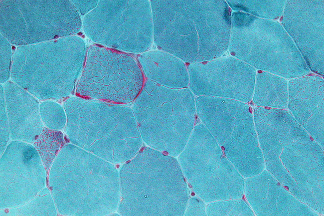
Ragged red fibers are commonly seen with mitochondrial dysfunction in muscle tissue. The ragged red fibers indicate an accumulation of abnormally functioning mitochondria in the muscle tissue. Though ragged red fibers have been studied extensively in individuals with inherited mitochondrial disease, it is not clear whether this type of damage can be acquired or induced in individuals with or even without mtDNA mutations. Studies in rats, however, have shown that thiamine deficiency does result in the finding of ragged red fibers in muscle tissue, suggesting that this damage can be induced. In view of the many posts on this website discussing the association of thiamine deficiency with Gardasil vaccination, I became very interested and began to search the literature for what was known about the relationship of thiamine with ragged red fibers.

Mitochondria

Before I offer an explanation, let me remind the reader about mitochondria. We have between 70 and 100 trillion cells that make up the human body. Each cell has a prescribed function and each of the body organs consist of highly specialized cells. What we tend to forget is that our food provides the fuel from which energy is generated. To use a simple analogy, a car without an engine would not be capable of functioning. Mitochondria are the “engines” that exist in each one of those cells. They provide the energy for which function is dependent. We inherit from our parents thousands of genes that control how we look, behave and perform as personalities. These are known as cellular genes, but mitochondria have entirely separate genes inherited only from the mother. They control the mechanisms of the “engines” (mitochondria) in generating energy. Disease can be caused by genetic mutations in cellular genes but in the past decade it has been recognized that mitochondrial genes can be responsible for disease and more than 50 different mutations have been described. The majority of these mutations are single base changes in the DNA strand. They do not necessarily produce disease by themselves. Other factors related to nutrition, lifestyle, and as I suspect, medications and vaccines, may have to come into play.

To express this fully, a deficiency of thiamine (or other vitamins) can be so mild that the symptoms, if any, are regarded as inconsequential or ascribed to other causes. This obviously becomes more important if there is an associated unknown genetic risk that affects the metabolism of the vitamin. Because thiamine is so vital to energy synthesis, the imposition of a stress factor such as a mild infection or an inoculation can precipitate more severe symptoms. Meeting stress requires adaptive energy. To provide an analogy, a car with an inefficient engine may be adequate on the level but be inadequate to meet the stress of climbing a hill.

Mitochondrial Disorders are Multi-Systemic

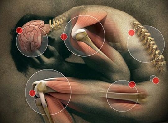
I should note that mitochondrial disorders are often multi-systemic due to impaired oxidation that results in defective mitochondrial energy production. That means there can be symptoms from damage to multiple organ systems simultaneously. Mitochondrial disorders are also phenotypically different amongst family members with the same mutation and amongst individuals with acquired mitochondrial dysfunction. In other words, how mitochondrial disorders or damage can present symptomatically varies radically from person to person. This variation is what makes diagnosing mitochondrial dysfunction difficult for many practitioners. The symptoms don’t always fit into nice, neat, discrete diagnostic categories that so many of us are accustomed too. This variability in mitochondrial dysfunction makes it difficult to attribute the action of a vaccine or medication as the cause of a subsequent illness, or in some cases, death. How is it possible for a medication or vaccine to induce so many seemingly disparate symptoms? To answer that question, we need to understand a few mechanisms.

Thiamine Deficiency Post Gardasil

Over the last several years, we have identified several cases of laboratory confirmed thiamine deficiency post Gardasil. Additionally, when lab testing was unavailable (few labs offer the appropriate assays for thiamine testing), clinical response to thiamine treatment has been confirmatory. In more recent research, we have identified thiamine transporter gene mutations (SLC19A2) in a group of young women who experienced severe reactions to the Gardasil vaccine (reported within this article). Combined, this suggests that thiamine deficiency is involved in some of the adverse reactions observed and that the potential danger from the use of a vaccine requires more information from the patient and his/her family. How can something as simple as thiamine cause so many adverse reactions and even death? And can a medication or vaccine induce thiamine deficiency?

Thiamine is Critical for Mitochondrial Functioning

Thiamine is a critical co-factor in multiple pathways involved in mitochondrial energy production (ATP). It is necessary for carbohydrate processing via the pyruvate pathway and it is necessary for fatty acid processing because of its involvement with the HACL1 enzyme.  In other words, the mitochondria depend upon thiamine to function. Diminish thiamine and all sorts of compensatory reactions are initiated which, if not stopped, can cause death. Thiamine deficiency in adults, particularly those with chronic alcoholism, is considered a medical emergency. It has not, however, been readily recognized in reference to other causes of malnutrition where there is an imbalance between the ingested calories and the necessary vitamins – high calorie malnutrition. This particularly applies to thiamine.

The Gardasil Thiamine Relationship

There are multiple mechanisms by which a vaccine or medication can induce thiamine deficiency or push an existing or subclinical deficiency into a danger zone. Beginning with the later first, the modern western diet is replete with highly processed foods that are dense in calories but lack non-caloric nutrients. It is entirely likely that many individuals, even those that appear healthy, are borderline thiamine deficient or intermittently thiamine deficient when stressors or illnesses arise. Vaccines are toxicological stressors to the immune system and broadly speaking, any stressor, but particularly one that demands an immune reaction like a vaccine, is capable of inducing a thiamine deficient state. In individuals with latent errors in thiamine absorption (GI disorders), distribution or metabolism (like those with thiamine transporter mutations), or anything that evokes even a slight degradation in thiamine nutrient availability, thiamine deficiency will be exacerbated exponentially.

The Gardasil vaccine was developed using a yeast type base*. The yeast produces an enzyme called thiaminase that inactivates thiamine. Again, against the backdrop of poor diet or diet high in foods that also produce thiaminase (coffee, tea, certain fish), but especially, against the backdrop of a genetic or acquired mitochondrial issue recognized or latent, the reaction to the vaccine (or medication, as many medications can block thiamine directly or indirectly), can be devastating.

Finally, vaccines, because of the adjuvant carriers like aluminum, damage to mitochondrial functioning more broadly, with both structural and functional changes are noted. Damaged mitochondria are not only less capable of producing appropriate amounts of cellular energy but are also incapable of performing the myriad of other functions with which the mitochondria are tasked.

Ragged Red Fibers and Cardiomyopathy

Let us continue with this case and the ragged red fibers observed in the myocardium, the heart muscle, of the deceased boy. For those who study mitochondrial disorders, one of the more common histological hallmarks of the disease process in mitochondrial disorders are ragged red fibers.  These are muscle fibers with abnormal focal accumulations of mitochondria. According to the coroner’s report:

“a long narrow band of dark reddish discoloration which is somewhat darker than the rest of the myocardium, extends over a length of 6 cm and has a width of 0.4 cm extending from the anterior base of the heart almost to the apex. ..this lesion is limited to the anterior free wall”

was observed. The coroner concluded that the boy developed asymptomatic myocarditis in weeks preceding his death. The myocarditis evoked a heart attack which was the determined cause of death. A subsequent review by a medical expert hired by the attorneys presenting the case against the vaccine manufacturer, went a little deeper, attributing the dark fibers to a vaccine-induced inflammatory reaction resulting from the first dose of Gardasil. He argued that the first dose of the vaccine initiated a heart attack that was somehow not noticed by the child, as he continued to play football. Upon receiving the second dose, however, the damage initiated by the first dose was exacerbated, slowing heart function until it failed entirely. In either case, the heart muscle was irreparably damaged such that the child died in his sleep with the Gardasil as the causal agent.

Given my background in thiamine research, and thiamine’s role in heart function (as well as in brain function), I immediately wondered if the observed “band of darkish reddish discolorations” could be the ragged red fibers so common in mitochondrial dysfunction and if there presence indicated thiamine deficiency. Furthermore, I suspected that the fact that he died in his sleep strongly suggested that the automatic respiratory mechanism governed by the brain stem was implicated. This too, is a strong support for thiamine deficiency. I should note, I did not have access to the full report; only that which was published online.

Thiamine Deficiency and Ragged Red Fibers: Experimental Evidence

As I have argued previously and elaborated above, the HPV vaccines can induce and/or exacerbate thiamine deficiency. The question is whether thiamine deficiency can induce ragged red fibers in muscle.

To that end, I discovered a manuscript in the Archives of Neurology: Neuropathic and mitochondrial changes induced in rat muscle, showing that experimentally in rodents this was possible. Thiamine deficiency could induce ragged red fibers in muscle tissue. In this particular study, two groups of rats were compared. One group was fed a normal diet and the other group was fed a diet deficient in thiamine. The rats with thiamine deficient diets developed ragged red fibers in the muscles. Other abnormalities were described not found in the muscles of the control rats.  The authors concluded that thiamine deficiency was responsible for  the observed ragged red fibers and may be involved in what are now called the “ragged-red diseases”.

Case Studies: Ragged Red Fibers, Thiamine and Mitochondrial Disease

Japanese investigators studied two siblings with muscle disease due to mitochondrial dysfunction, a mutation in the mitochondrial DNA, and familial thiamine deficiency. Ragged red fibers were found in muscle biopsies. The older brother had presented at the age of 20 years when he developed muscle disease and beriberi heart disease. Thiamine deficiency was present in the siblings and parents and ragged red fibers were noted in muscle biopsies from the siblings. The development of symptoms at the age of 20 years certainly indicates that it was not a purely genetically determined disease.

Another article in a Japanese journal reported a nine year-old boy with muscle and brain disease in whom thiamine administration gave temporary improvement. A muscle biopsy had revealed numerous ragged-red fibers.

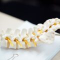
Mitochondrial diseases have a special predilection to involve the brain in view of its high metabolic demand. Patients with a form of disease known as myoclonic epilepsy have ragged red fibers in muscle tissue thus identifying the underlying mitochondrial cause.

Mitochondrial Dysfunction in Myocardial Infarction and Sudden Death

In a recent review of mitochondrial cardiomyopathies we see some striking similarities between this case and what has been recently recognized. Accordingly:

The presentation of mitochondrial cardiomyopathy includes hypertrophic, dilated, and left ventricular (LV) noncompaction, and the severity can range from no symptoms to devastating multisystemic disease. Severe cardiac manifestations include heart failure and ventricular tachyarrhythmia—which can worsen acutely during a metabolic crisis —and sudden cardiac death. Mitochondrial crisis is often precipitated by physiologic stressors such as febrile illness or surgery [a vaccine] and can be accompanied by acute heart failure.

Bioenergetic derangements are increasingly recognized as major culprits in the development of cardiac hypertrophy and in the progression to heart failure, in both acquired and inherited disease. The mitochondria are a crucial platform for energy transduction, signaling, and cell-death pathways that are broadly relevant to heart failure, even in the absence of an underlying mitochondrial myopathy. Oxidative stress and mitochondrial dysfunction are key factors in the development of most heart failure.

Connecting the Dots

The question remains, how could this boy’s death from a vaccination have been predicted and thus avoided? It is clear that there was temporal relationship between the vaccine, the damage to his heart, and his subsequent death. Mechanistically, the evidence is collaborative with this association. From an evidentiary standpoint, the vaccine appears capable of inducing mitochondrial dysfunction via its ability to diminish thiamine, and likely, via other, yet to be identified, mechanisms. Of key importance, however, is that thiamine depletion on its own, can induce ragged red fibers in muscle tissue, probably including the heart muscle. When the vaccine is given to an individual with genetic or other risk factors (like comorbid health issues, poor diet, and/or the high metabolic demands of sports training), the results can be devastating. Given that this combination of variables includes most teenagers, it is difficult not to see the dangers of this vaccine. In conclusion, if the long band of dark reddish muscle tissue described in the heart muscle of the boy had been shown to be ragged red fibers, it would have supported mitochondrial dysfunction as the cause of death.

*It should be noted that the Cervarix HPV vaccine was not developed using a yeast base, and thus, it is not clear by what mechanism(s) it might diminish thiamine concentrations.

We Need Your Help

More people than ever are reading Hormones Matter, a testament to the need for independent voices in health and medicine. We are not funded and accept limited advertising. Unlike many health sites, we don’t force you to purchase a subscription. We believe health information should be open to all. If you read Hormones Matter, like it, please help support it. Contribute now.

Yes, I would like to support Hormones Matter.

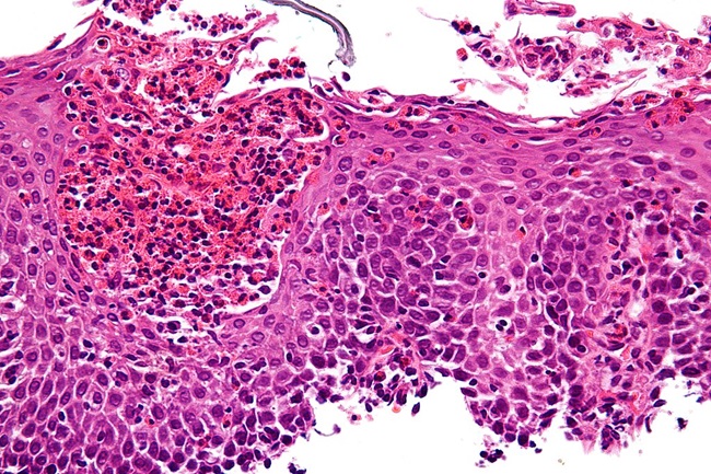
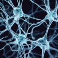
Image: Very high magnification micrograph showing ragged red fibres (also ragged red fibers), commonly abbreviated RRF, in a mitochondrial myopathy. Gomori trichrome stain.

Nephron, CC BY-SA 3.0, via Wikimedia Commons

This article was published originally on Hormones Matter on January 5, 2016. 

Connecting the Symptom Dots: Discovering My Thiamine Deficiency

21571 views

As a registered dietitian nutritionist (RDN), I was surprised to find out that I had a thiamine deficiency in December 2015. My diet wasn’t perfect, but it was close. I never imagined I’d spend so much time trying to treat my own deficiency, but it’s been over a year the first lab work showed the deficiency and I’m still struggling with it. I’ve been asked to share my symptoms and experiences, so I’ll start back around the initial diagnosis.

Let me preface my story by sharing some information about myself. I’m a 46 year old female and I’ve always considered myself fairly healthy. I’m active, and I complete a minimum of 12,000 steps/day and often much more. That includes some form of aerobic activity daily. I’ve dealt with some annoying health problems, but nothing I considered major. I’ve had issues with insomnia, depression, nerve problems, migraines, hypoglycemia and GI distress (mostly diarrhea) for years or decades. I’ve also had some discomfort on the left side of my chest, on and off, which goes unexplained. I’ve seen many different types of doctors, including cardiologists, neurologists, gastroenterologists, psychiatrists, sleep specialists, endocrinologists, allergists, etc. Also, I have very early visual symptoms of glaucoma, but my doctor said there aren’t any signs of disease in my eye. No familial history of glaucoma, and I’ve never been diagnosed with diabetes. Separately, all of these symptoms seemed minor. Only within the last few years or so, did I begin to wonder if there was some sort of connection.

In the fall of 2014, I started a post-bachelors program in dietetics. I had returned to school almost two decades after completing my bachelors, and the road to this program was a long one. My insomnia seemed to be severe the night before exams. Sleep eluded me, even with the prescription sleeping pills. Anxiety, right? It never occurred to me that it was something else. After all, I’ve had insomnia issues for at least a decade. Sometime during the semester, I had seen a neurologist for some nerve testing. I had numbness and tingling in my feet, hands and arms. It would wake me up at night. I began seeing a doctor of osteopathy for manipulations to help with the nerve problems, too. Also, I had noticed some garbled speech and numbness in my tongue, but thought I was imagining it.

During finals week in December, my insomnia became severe. My physician prescribed Xanax, but I hated the way it made me feel. I felt my anxiety actually increased.  Even after finals were over, sleep eluded me. I was piecing 3-5 hours of sleep together, if that. I had trouble eating a full meal and was losing weight. In addition, I was having discomfort on the left side of my chest, something that I had experienced in the past but was yet unexplained. All of this was attributed to anxiety. By the end of December, my physician prescribed a daily anti-anxiety medication. This medication made me nauseous and I had diarrhea. Of course, these symptoms didn’t help the weight loss. At no time did my physician do any lab work while this was happening. I was so miserable that I emailed my advisor to inquire about dropping out of the dietetics program. Fortunately, she wouldn’t entertain the idea and encouraged me to continue, noting that I could take an Incomplete if necessary.

By February of 2015, I was down to 103 pounds, (I’m 5’ 4” and 130 pounds currently). I was dragging myself to school. I had lost a lot of muscle mass, and couldn’t sit for long in class because of the lack of muscle. My face looked quite thin and my temples were hollowed out. In March 2015, I was weaned off the medications and began taking 7.5 mg Remeron, and Ambien as needed. The Remeron helped my appetite and I began regaining weight and strength. With the support of my professors, I was able to complete the semester, and even maintained a high grade point average!

Early in the fall semester, I listened to a lecture by an RDN who is an integrative and functional medicine certified practitioner (IFMCP). Based on her lecture, I knew my instincts about an underlying connection to all of my symptoms was correct. In November 2015, I had an appointment with that RDN. She recommended some blood work, which my primary care physician (PCP) reluctantly agreed to do. It was a lot of blood work, and fortunately my insurance covered it. There were many positive or problematic results, but among them was low thiamine (whole blood) at 29ug/L, a positive ANA test, TPO 693, as well as magnesium and ferritin were in the low normal range. After further autoimmune testing, it was determined that I have Hashimoto’s disease, too.

The low thiamine level could explain many of my symptoms, including, insomnia, nerve issues, migraines, precordial pain, weight loss and problems processing carbohydrate. The question is why was my thiamine level low? I had always thought my diet was relatively healthful. For years, I watched my added sugar intake because of trouble with hypoglycemia. My fiber, protein and water intake seemed adequate. I’m very careful with my fat intake because I had a cholecystectomy in 2009 and still have problems with lipid digestion. I rarely drank alcohol because of the hypoglycemia and insomnia. The only other beverage I consumed was tea, usually 1-3 cups per day. Furthermore, because of my hypoglycemia, I ate mostly whole grains and very little gluten, if any.

In January 2016, I began taking a B vitamin complex, magnesium, lipothiamine and some other supplements, including Ortho-Digestzyme to aid in lipid digestion. I made changes to my diet, including dairy free and gluten free. I began seeing some health improvements. Eventually, I added yogurt and cheese back into my diet, but remained gluten free. I was having fewer migraines and began sleeping without Ambien. That spring I was taken off the lipothiamine, but continued the B vitamin complex and magnesium. I graduated from the dietetics program in May 2016, something I feared wouldn’t happen only one year earlier.

At the end of October 2016, I had an infection (perhaps, due to an insect bite) on my outer ear which wouldn’t go away. My PCP prescribed a cephalosporin antibiotic for 10 days. Towards the end of November and into December, I was having increased nerve issues, occasional insomnia, mild apathy and anxiety, which was strange given I had nothing to be anxious about. Also, I had the same chest discomfort again. My thiamine level was tested and it was low at 32 ug/L. I was taking the B vitamin complex and magnesium all along, so my PCP was unsure what to do. I’ve since learned that some antibiotics, like the one I took, can deplete thiamine.  I saw the RDN again and began taking lipothiamine again on 12/23/2016. I was taking 50 mg, twice a do with magnesium, in addition to the B vitamin complex.

My PCP planned to retest in a month to see if it was working. However, on January 20, 2017, I had an emergency appendectomy. During the surgery, I was given a cephalosporin antibiotic, but it was only during the surgery, not afterwards. It should be noted that I only missed one day of supplements because of the surgery. By the end of the first week, I strongly suspected my thiamine level had bottomed out, because my symptoms of anxiety, insomnia, nerve pain, etc., reminded me of what happened two years earlier. During that week, I was taking 50 mg lipothiamine twice a day, 200 mg magnesium and a potent multivitamin. Personally, I think the antibiotic, surgical procedure and recovery, and resulting diarrhea contributed to the low thiamine despite supplementation. I almost went to the ER in hopes that they’d give me a thiamine injection or IV, but decided to wait until Monday to see my PCP. Her suggestion was that I continue my supplements, then we’d retest in a month. One month later, my thiamine level was low still at 32 ug/L. My PCP said she isn’t comfortable giving intramuscular thiamine injections and suggested I see a gastroenterologist. I mentioned information I found on Hormones Matter, but I don’t believe my PCP was interested in reading the material.  I feel like I’m being bounced around from one doctor to another. I’m going to see the gastroenterologist, whom I’ve seen before but I’m not hopeful that she’ll be able to help. I saw a neurologist recently, who was very kind and listened intently, but could only suggest an MRI and a DO, who “might” be able to help me, but that DO’s office is 1.5 hours away. Next week, I’ll go back to the cardiologist for a check-up because of the ongoing discomfort on the left side of my chest.

For now, I’m sleeping at least 6 hours a night, which feels like a lot to someone who’s experienced severe insomnia. My hypoglycemia is under control. I’m not sure if that’s because of the thiamine supplementation, the gluten free diet or both. The last time I had gluten, I experienced both mild insomnia and hypoglycemia, but again, my thiamine was likely low too. I feel I still have occasional memory issues, but maybe that’s age related. Also, the numbness and tingling in my extremities continues. Migraines occur much less and are less severe, usually. The mild vision problems linger, as well.

The RDN I’m seeing is uncomfortable with me taking more than 100 mg lipothiamine per day. At this time, she is recommending supplements to treat continued GI inflammation too.  Here is my current regimen: 100 mg lipothiamine/day, 200 mg magnesium/day, multivitamin 1/day (RDN wants me to take 2/day), 28 mg iron w/vitamin C, sodium butyrate 600mg 4/day, NAC 600mg 2/day, Ortho-Digestzyme 2 capsules before each meal to help with lipid absorption, and about 4000 IU vit D3.

Unfortunately, I feel I’m just one missed dose of my supplements away from problems all the time now. I’m not sure how to find a physician who can help me solve this ongoing thiamine problem and don’t know where to turn next. Again, I’m going to see a gastroenterologist and cardiologist this month, but feel it may be more of the same. My father died at 45 years old of cardiovascular disease. I know thiamine deficiency can lead to cardiovascular problems too, which is why I’m going back to the cardiologist.

Any suggestions are welcomed!

We Need Your Help

Hormones Matter needs funding now. Our research funding was cut recently and because of our commitment to independent health research and journalism unbiased by commercial interests we allow minimal advertising on the site. That means all funding must come from you, our readers. Don’t let Hormones Matter die.

Yes, I’d like to support Hormones Matter.


Thiamine, Fibromyalgia and Chronic Fatigue

11107 views

Fibromyalgia affects roughly six million Americans, mostly women. Its symptoms include all-over muscle and tendon pain, increased pain sensitivity, chronic fatigue, sleep disturbances and brain fog. Fibromyalgia is often reported as feeling like a flu that never leaves. Similarly, symptoms of Chronic Fatigue Syndrome (CFS) overlap with many of those of fibromyalgia and the two conditions are often co-morbid. With chronic fatigue, however, the predominant symptom is a fatigue that never lets up versus all-over muscle and tendon pain.

Both fibromyalgia and chronic fatigue are co-diagnosed frequently in women with endometriosis, especially those who have had Lupron treatments. Similarly, we are finding a high incidence of chronic fatigue and fibromyalgia post Gardasil/Cervarix and post fluoroquinolone. All over muscle and tendon pain, coupled with never-ending tiredness seem to be common symptoms post medication or vaccine reaction. Could they be linked to a broader problem, specifically, thiamine deficiency?

What is Thiamine?

Thiamine or vitamin B1 is necessary for cellular energy. It is a required co-factor in several enzymatic processes, including glucose metabolism and interestingly enough, myelin production. We can get thiamine only from diet. When diet suffers, as in the case of chronic alcoholism where most of the research on this topic is focused, when nutritional uptake is impaired (leaky gut and other GI disturbances), or when other factors inhibit the enzymes necessary to carry out intracellular reactions, thiamine deficiency ensues. And thiamine deficiency can elicit a whole host of problems that are consistent with the current definitions of chronic fatigue and fibromyalgia.

Thiamine and Fibromyalgia – A Few Hints

A recent case study suggests that what is currently diagnosed as fibromyalgia and/or chronic fatigue may be attributable to thiamine deficiency. A very small case study (n =3) from Italian physicians found a significant reduction in fibromyalgia symptoms in patients given high dose thiamine. Researchers found:

  • Patient 1:  71.3% reduction in fatigue; 80% reduction in pain.
  • Patient 2:  37% reduction in fatigue; 50% reduction in pain.
  • Patient 3:  60.7% reduction in fatigue; 60% reduction in pain.

Thiamine and Chronic Fatigue

In a little bit larger study – 17 patients with Chronic Fatigue, researchers found a functional reduction of the enzymes involved in vitamin B metabolism (aspartate aminotransferase -pyridoxine, glutathione reductase and transketolase) compared to healthy controls, suggesting thiamine deficiency.

What This Means

It’s way too early to tell if thiamine deficiency is at root of fibromyalgia and/or chronic fatigue symptoms, or if adverse reactions to medications and vaccines can elicit the symptoms of fibromyalgia and chronic fatigue, but there are hints pointing in that direction. Much more research should be done. In the meantime, if you suffer from fibromylagia or chronic fatigue or undiagnosed neuromuscular pain, why not consider testing for thiamine.  And while you’re at it, since many of these symptoms overlap with those of hypothyroidism, particularly of the autoimmune Hashimoto’s sort, why not get tested for that too. If you test positive for either of these, tell us about it, it will help other patients find solutions. To learn more about thiamine deficiency and other topics, search our growing library of research and patient stories here on Hormones Matter.

We need your help.

Hormones Matter needs funding now. Our research funding was cut recently and because of our commitment to independent health research and journalism unbiased by commercial interests, we allow minimal advertising on the site. That means all funding must come from you, our readers.  Don’t let Hormones Matter die.

Yes, I’d like to support Hormones Matter.

This article was first published on Hormones Matter in October, 2013.

Treating Sensory Processing Disorder in Children

9326 views

On Tuesday, August 16, 2016, I read an article in the Wall Street Journal with this title: Treating Children for Sensory Processing Disorder.  Since I have treated hundreds of these children, I am posting here some of the facts that I have learned. First of all let me provide some extracts from this article that is all about a diagnosis of “Sensory Processing Disorder” (SPD). The article says that SPD is believed to affect 5% to 16% of children in the United States.

I want to make it clear what we are talking about. This article describes a three-year-old child who, when accompanying his mother to the grocery store would have meltdowns. His mother was quoted as saying “he would literally bite me throughout the grocery store”. An occupational therapist determined that he had SPD: “a condition in which the body and brain have difficulty processing and responding to sensory stimuli in the environment”. The article goes on to say that “some people with SPD are hypersensitive to loud noises or different textured foods. Others may be agitated by the touch of a clothing tag”. The Director of Occupational Therapy (DOT) research at Cincinnati Children’s Hospital Medical Center was reported as saying

“occupational therapists treat dozens of SPD patients every week. It can affect just one sense, such as hearing, touch or taste, or multiple senses. Sensory processing problems can also affect the body’s vestibular system, creating difficulties with balance, or the proprioceptive system, leading to problems with clumsiness and body positioning”.

The DOT “has worked with some children with SPD who are academically gifted and don’t have autism or ADHD. It is clear from the article that the method of treatment, entitled “sensory integration”, looks upon SPD as abnormal psychological behavior. This is  in spite of the fact that a professor of radiology and bioengineering at University of California, San Francisco stated that studies showed that children with SPD had less developed white matter mostly in the back of their brain, compared with typically developing children. This posterior region of the brain is where a lot of sensory processing takes place. This is a major clue as we shall see shortly.

Beyond the Bad Parenting Theory of Sensory Processing Disorder

Now I ask you dear reader, is it common sense to claim that this kind of disorder in 5% to 16% of our children is purely psychological from bad parenting, acceptably normal in a young child, or caused by genetic changes? Since the Wall Street Journal article claims that “adults can have SPD”, it is clearly not confined to children. To believe that any of these facts, or all of them together, can result in so much willful behavioral deviation is a reduction to absurdity. It is absolutely certain that Mother Nature never makes that kind of genetically determined mistake in so many individuals.

The article in the Wall Street Journal reports that “a common treatment at Cincinnati Children’s is called sensory integration, involving three sessions a week for about six weeks”. The founder of the STAR Institute for sensory processing disorder in Denver involves an intensive treatment program of some 31-hour sessions nearly every day for several weeks. The cost is about $175 per session.

When I was a consultant pediatrician at Cleveland Clinic Foundation, I saw many children who were referred because of “emotional problems”. The accepted cause at that time (and still is by many pediatricians) was lack of good parenting. In discussions with parents, I found that bad parenting was rare, but lousy diet was common, particularly because of the enormous overload of sugar, often started in infancy. In fact, sugar was used as an inducement to good behavior, not recognizing the fact that the sugar was the cause of the bad behavior in the first place. By doing a blood test on these children I repeatedly found evidence of thiamine deficiency. To me, the extension of the absurdity is that there is no mention at all in this article about the role of nutrition. I have posted a number of articles on this website concerning vitamins, particularly  vitamin B1 (thiamine). I have pointed out many times that overloading the diet with empty calories, particularly from sugar, automatically induces thiamine deficiency relative to the excessive calories. The scientific evidence for this has been known since 1936. Any attempt to depict thiamine deficiency by measuring its blood level in a person eating “empty calories” will be doomed to failure. The concentration of thiamine in the blood is only normal in relation to a normal calorie content of the diet. It is the calorie/thiamine ratio that counts.

Sugar, Thiamine and SPD

By pointing out to the parents that they had to get rid of the sugar and providing the child with a supplement of thiamine and magnesium, all the symptoms of “psychological misbehavior”, no matter what pretty name was given to it, quickly resolved. For literally a few dollars and cents, this form of treatment is overwhelmingly simple and effective. The “posterior region of the brain where a lot of sensory processing takes place” is peculiarly sensitive to thiamine deficiency. It will affect balance and in its extreme form, can affect brainstem mechanisms where the control of heart rate and breathing is automatically conducted. This is why an excess of sugar is incredibly dangerous, not because the sugar is a poison in its own right, but because of the secondary effect on energy metabolism in that part of the brain that is essential to life itself.

What seems to be poorly understood is that thiamine deficiency produces the same effect in the brain as lack of oxygen and sensory perception becomes exaggerated. Pain is felt more intensely and may give rise to a phenomenon known as “hyperalgesia”(acute pain perception). Sound and light may be so much more perceived that the sufferer puts hands over his ears or closes his eyes, because the perception is offensive. Touch is grossly exaggerated and may even give rise to screaming by the child when being physically examined by a physician. Because of this poor understanding, the behavior of the child is regarded as “psychological”. Under such circumstances a mild injury to an ankle may give rise to severe pain in the leg. It used to be known as “acute sympathetic dystrophy”. The name has been changed to “acute regional pain syndrome” or “complex regional pain syndrome“. Let it be clearly understood that no matter what kind of injury, obvious inflammatory reaction or source of discomfort occurs in the body, the pain is perceived by the brain. If the mechanism of sensory perception is exaggerated, the pain will be more intense.

Conclusion

It is becoming abundantly clear that a diagnosis of sensory integration, ADD, ADHD, OCD and many other diagnostic refinements are not separate diseases at all. Like variations on a symphonic theme in music, the biochemical changes in the brain are responsible for creating the symptomatic expressions on a completely variable basis. It also explains in practical terms why many of the so-called SPD children in the Wall Street Journal article “were unusually gifted”. Like different models of cars with different horsepower, surely the more intelligent brain requires efficient energy metabolism to meet its “gifted” requirements. For those interested in further details of this concept, turn to the post on “Eosinophilic Esophagitis” on this website. There you will find that the unfortunate patient described with this disease was misdiagnosed for many years as psychosomatic. I will go further than this and say that if the symptoms that are commonly represented by changes in brain processing are neglected, and the malnutrition continues, we can expect damaging changes to take place. I would expect this to lead to a whole series of diseases that also go by different diagnostic nomenclatures, Parkinson’s disease, Alzheimer disease and various forms of dementia that represent the end point damage that has accrued over years. Are we collectively insane?

We Need Your Help

More people than ever are reading Hormones Matter, a testament to the need for independent voices in health and medicine. We are not funded and accept limited advertising. Unlike many health sites, we don’t force you to purchase a subscription. We believe health information should be open to all. If you read Hormones Matter, like it, please help support it. Contribute now.

Yes, I would like to support Hormones Matter. 

Photo by Alireza Attari on Unsplash.

The Sugar – Thiamine Connection in Adverse Reactions

8862 views

As published on this web site previously, we have scientific evidence that two girls and one boy were shown to be thiamine deficient (TD) after Gardasil vaccination. On the other hand, a girl who had similar symptoms to these three had not received the vaccine and her laboratory test proved that she also had TD.  On the face of this information, it suggests that the vaccination has nothing to do with the illness of these individuals.  In a previous post, however, I have suggested that the vaccine is a “stress factor”, given to an individual in a state of marginal, or even asymptomatic thiamine deficiency, thus exacerbating the first appearance of symptoms. In this light, medications and other vaccines may also be considered stress factors and evoke or exacerbate a previously asymptomatic thiamine deficiency. There are a number of facts that need to be seen collectively in order to understand the hypothesis that follows.  In order to make this clear I am going to present the material under subheadings.

What Does Thiamine do in the Body?

All simple sugars that we take in our diet are broken down to glucose, the primary fuel of the brain.  This oversimplified fact has long been used to suggest that taking sugar is the way to meet energy demands in the body.  It is, in fact, an extremely complex chemical process which is well beyond the scope of an article like this.  It can, however, be simplified by comparing glucose, as a fuel, to gasoline in a car.

Gasoline + Oxygen + Spark Plug = Energy  + (ash/oxides)

Glucose + Oxygen + Thiamine = Energy + (ash/oxides)

Each one of these equations represents combustion, a combination of fuel with oxygen.  Because combustion is always incomplete, waste products (oxides) are formed and must be got rid of as waste.  It is obvious that combustion of gasoline without oxygen and spark plug, or glucose without oxygen and thiamine, will not occur.  What is not quite so obvious is the fact that an excess of gasoline causes choking of the engine, black smoke from the exhaust pipe (unburned hydrocarbons) and loss of engine efficiency.  This could be referred to as “oxygen/spark plug deficiency” since each of the three components must be present in proper concentration to produce efficient combustion (oxidation).  The three component parts, glucose, oxygen and thiamine are the equivalents in the body.  An excess of glucose “chokes” the “engines” (mitochondria) that create energy in all of our cells. This particularly applies to the brain because of its high rate of metabolism (energy consumption), thus providing a potential explanation for why the vaccine seems to pick off the brightest and the best students.

The Reptilian Brain and the Limbic Nervous System

All animal brains are built on the same basic principle, a lower, more primitive part and a higher, increasingly complex part. The lower part of the human brain, the limbic system, also known as “reptilian”, computes all the reflex mechanisms by which we automatically adapt to our environment.  For example, we sweat when it is hot and shiver when it is cold, both adaptations to the ambient temperature.  It also controls our emotional reflexes, represented by body language that we recognize easily.  It uses two mechanisms, the autonomic nervous system and the endocrine system.

Autonomic and Endocrine Systems

We have two nervous systems. The one that we use to will our actions is controlled by the upper brain, here described as cognitive. The autonomic nervous system (ANS) automatically controls all the actions required by body organs to meet day- to- day adaptation.  It consists of two major branches, known as the sympathetic and parasympathetic components.  The sympathetic branch prepares us for mental and physical action while the parasympathetic switches us to a period of rest.  As one goes into action, the other one is withdrawn. The endocrine system is represented by a group of glands, each of which produces one or more hormones.  These are really messengers that induce actions in the cells to which they are aimed.  When either or both of these systems are not functioning in their ordained manner in the brain/body of an individual, we can refer to him/her as maladapted.

Explanation of Symptoms in Reference to Thiamine Deficiency

As explained in previous posts on this web site, the disease known as beriberi occurs as a result of TD.  The mother of a Gardasil affected girl had done her own research and had come to the unlikely conclusion that her daughter suffered from beriberiRed cell transketolase, a blood test used to depict TD, showed that she was correct in her conclusion. Her daughter did in fact have beriberi and has responded, at least partially, to thiamine supplementation.  We know, from historical data, that long term beriberi responds slowly to treatment and sometimes not at all, depending on chronicity.  Since she has had her symptoms for approximately four years, I think that it would be fair to call this chronic. When the ANS is not functioning properly, it is called dysautonomia (dys, meaning abnormal: autonomia refers to the ANS).  Beriberi in its early stages is the prototype for dysautonomia, the commonest effect being dominance of the sympathetic branch of the ANS.

Published Effects of Gardasil Vaccination

Although many symptoms have been reported related to this vaccination, two resultant conditions have been nominated: POTS (Postural Orthostatic Tachycardia Syndrome) and Cerebellar Ataxia.  POTS is one of the many conditions that are described under the heading of dysautonomia and I have already reported in a post that the first case of thiamine dependency was in a six year old boy who had intermittent episodes of cerebellar ataxia, each of which was triggered by a stress episode that included mild infection, mild head injury or inoculation.  A critical enzyme that depends on thiamine for its energy producing action was able to function until some form of physical or mental stress was imposed.  The existing mechanism was insufficient to meet the energy requirement imposed by the stress.

Sugar, the Autonomic Nervous System and the Liver

New research provides one more clue to our emerging theory of thiamine deficiency in post vaccine and medication adverse reactions.  The study: The Autonomic Nervous System Regulates Postprandial Hepatic Lipid Metabolism by Bruinstroop et al. demonstrates the influence carbohydrate intake has on autonomic control of liver lipid metabolism. Triglycerides are measured in a medical laboratory as part of what is known as the “lipid profile”, that includes the various components of cholesterol. The Bruinstroop study found that when the parasympathetic system was deactivated and carbohydrates were ingested, triglyceride levels rose significantly, inducing metabolic dysregulation. Other studies have found stress, combined with diets high in refined carbohydrates can increase blood triglyceride concentrations also inducing metabolic syndrome. Indeed, stress and the concurrent increased sympathetic system activity seem key to metabolic functioning with sugar intake triggering the ill-health.

Interpretation of Technical Language

The work by Bruinstroop and associates was done in rats.  To understand what they found, it is necessary to remind the reader that the two branches of the ANS, sympathetic and parasympathetic, work synchronously.  As one branch becomes active the other one is withdrawn. This is automatically controlled by the “reptilian” brain, thus enabling us to adapt to the physical and mental changes we encounter on a day -to-day basis.  These authors were able to show that abolishing the parasympathetic input to the liver resulted in marked elevation of triglycerides in the blood. This would induce continuation of sympathetic dominance in any “stress reaction” in the animal if it was in a free living state. The effect was modulated by sugar intake. That is, when the animals were fed more, the effects were larger.

Hypothesis: High Sugar Diets Lead to Thiamine Deficiency, A Risk Factor for Adverse Reactions

I am proposing that an excess of carbohydrates in the diet, particularly fructose, results in a mild degree of thiamine deficiency.  We know, from studies done as early as 1943 (Williams R D, et al. Arch Int Med 1943;71:38-53), that this results in what is typically called psychosomatic disease, in which a large component is reflected in emotional lability (instability), so common in the modern child and adolescent.  Physical symptoms, such as unexplained “pins and needles”, in the hands or feet, may be so slight as to be ignored.  The stress of the vaccination or a medication reaction triggers an energy crisis in the “reptilian” brain, specifically evoking autonomic dysregulation, typically with sympathetic system dominance and resulting in beriberi, POTS, or cerebellar  ataxia and potentially other syndromes.  Perhaps a rise in blood triglycerides as suggested by the Bruinstroop study, indicates the partial crippling of the parasympathetic branch of the ANS and sympathetic dominance.  High blood triglycerides might well be a  mark of the early stages of underlying autonomic dysregulation and thiamine deficiency and a potential risk factor for adverse reactions to certain vaccines or medications.

We Need Your Help

More people than ever are reading Hormones Matter, a testament to the need for independent voices in health and medicine. We are not funded and accept limited advertising. Unlike many health sites, we don’t force you to purchase a subscription. We believe health information should be open to all. If you read Hormones Matter, like it, please help support it. Contribute now.

Yes, I would like to support Hormones Matter. 

Addendum

A 5th case of post Gardasil thiamine deficiency has been identified; a young woman who developed severe idiopathic hypersomnia, a variant of narcolepsy, post vaccination. The patient is undergoing treatment with success. A full case study will be presented soon.

Image by 🌸♡💙♡🌸 Julita 🌸♡💙♡🌸 from Pixabay.

This article was published on Hormones Matter previously in January 2014.

More About Eosinophilic Esophagitis

8512 views

Seeing some of the comments following the appearance of my post Eosinophilic Esophagitis May Be a Sugar Sensitive Disease, it seemed that it was necessary to provide a little more explanation for how the conclusions were reached. Hopefully this may produce less misunderstanding.

Compartmentalized Medicine

The present model for disease is being rapidly outdated, so let me first of all review how a diagnosis is made in modern medicine. When a patient pays a visit to a physician, a medical history is recorded. The history begins by the patient describing symptoms, the sensory afflictions experienced since the loss of health began. This is followed by a physical examination when the physician is looking for evidence of malfunction. For example, this may include finding enlargement of a given organ, point tenderness when pain is elicited or a neurological deficit. Family history and the history of previous illnesses are both taken into account. The physician may or may not have a working idea of the nature of the disease process at this stage and a series of laboratory tests are requested. All of this is put together and the physician then has to consider what is generally referred to as a differential diagnosis. Which part of the physical examination, combined with the tests, all point conclusively to a diagnostic category?

This method of making a diagnosis was derived from the Flexner report initiated by Rockefeller in 1910. It was adopted from the German method in which laboratory confirmation was emphasized. This gave rise to the methodology that we now call “scientific medicine”. The symptoms, signs and laboratory reports are then put together and a given disease is named as the most likely fit.

So let us examine for a moment how this confuses us. All sensations are perceived in the brain and symptoms are merely a method by which the brain/body provides a warning that something is wrong. The “wrongness” has to be interpreted. In the present model, each constellation of symptoms, signs and laboratory reports are then given a name. For example, because somebody by the name of Parkinson was the first to describe a given constellation, it is called Parkinson’s disease, even though the underlying cause is completely unknown. Research has been aimed at finding a cure for that disease without giving full recognition to the fact that the constellation of findings overlaps with the constellations exhibited in other brain diseases, each being named separately. Furthermore, if the constellation points to an organ as the seat of a given problem (such as the intestine), the patient is referred to a specialist (a gastroenterologist) whose practice is confined to diseases of that organ (organic disease). An attempt to improve the symptoms by prescribing drugs is the chosen method, without considering the complex connection of the sick organ with the brain. An “anti-inflammatory” drug is prescribed, without asking why or what caused the organ to become sick.

In the case that I wrote about previously, the disease process called eosinophilic esophagitis or EoE, results from ingesting food. The presently accepted cause is “food allergy”.

Understanding Disease Differently: A Connected System

Let me provide an example to illustrate the change in perspective that occurs if the whole person is considered. On one of these posts a mother reported that her daughter had eosinophilic esophagitis, “associated with idiopathic gastroparesis” (partial or complete paralysis of the intestine). The word idiopathic stands for the simple sentence “the cause is unknown”. Evidently, no attempt had been made to connect the two conditions together. Is it likely that two unusual conditions will exist at the same time in one individual? By recognizing that the brain is always involved with body disease and brain disease is always involved with the body, it is possible to provide a solution for a connection between eosinophilic esophagitis and gastroparesis. It depends completely on an understanding of the profound genius of the brain/body interconnection.

The post that led to all of these comments asks the question, is this disease caused by the ingestion of sugar? We know that ingestion of sugar can easily induce thiamine deficiency because we have the ancient model of beriberi where white rice (without its surrounding cusp) ingestion, consumed as a staple, was found to be the cause. (Rice grain is starch and is broken down in the body to glucose. The cusp around the grain contains the vitamins. When the cusp of the rice is removed, as it is in white rice, the vitamins are removed leaving only the starch, which is converted to glucose.)

Digestion: Where Mechanical Meets Chemical

The vagus nerve is the 10th cranial nerve. Its action, initiated in the lower part of the brain, is to send outgoing messages to the spleen, an important organ that is used for controlling inflammation. The vagus nerve uses a neurotransmitter called acetylcholine and it also deploys messages to the esophagus and the entire intestinal tract. The wave pattern in the respective parts of the intestine that is induced by this nerve is called peristalsis. It pushes the contents along while the complex process of digestion occurs. Without going into details, the synthesis of acetylcholine depends on vitamin B complex, dominated by thiamine. Without thiamine, there is less acetylcholine and without this vital neurotransmitter, the control of inflammation and peristalsis in the esophagus, the intestinal tract, or both, are all compromised.

Eosinophilic Esophagitis and Food Allergy

In EoE, food sensitivity, occurring for whatever reason and known as food allergy, is causing inflammation that might occur in either the esophagus or any other part of the intestinal tract. When it occurs in the intestine it is called eosinophilic enteritis. Although the mechanism is the same, the locality differs but the esophagus is more commonly the affected part. The inflammatory response gets out of control because the vagus nerve, lacking acetylcholine to transmit the necessary information, is failing to suppress esophageal inflammation by sending a proper message to the spleen. The association of eosinophilic penetration into the intestinal tissue is part of the inflammation and it is interesting that a similar event has been associated with asthma in bronchial tubes. Asthma was a recurrent problem in the history of my patient.

Like the famous poem:

“for the want of a nail a shoe was lost; for the want of a shoe a horse was lost; for the want of a horse a battle was lost; for the want of a battle a kingdom was lost”.

To paraphrase this in biochemical terms “for the want of thiamine (vitamin B1), action of the citric acid cycle (engine of the cell) was lost; for the want of the citric acid cycle, acetylcholine (neurotransmitter) was lost; for the want of acetylcholine, suppression of inflammation was lost; for the want of acetylcholine, normal peristalsis (wavelike action) in the esophagus and intestinal tract was lost.

The loss of the peristaltic wave in the intestine was given the name “idiopathic gastroparesis”, a clear indication by the diagnostician that “its cause is unknown”. Like the blind men and the elephant the present medical model looks at a segment of the problem and fails to see the big picture. The trouble with this failure to understand the full nature of the problem is because we have divided brain disease from body disease. If it is suspected that the brain is the cause of the problem and all laboratory studies are negative, it is assumed that the symptoms are psychosomatic in nature and have been “imagined by the patient”. When the patient is told that it is “psychological”, it naturally induces anger.

My patient’s symptoms, recurring through infancy to the age of 8 years, were thought to be psychosomatic until endoscopy revealed the esophagitis. The “psychosomatic symptoms” were resulting from thiamine deficiency affecting the brain. His dramatic growth spurt during treatment strongly suggested that the autonomic (automatic) nervous system was at the seat of the complex problem. That conclusion can be supported by the medical literature concerning a well known genetically determined disease called Familial Dysautonomia, a disease whose clinical course results in growth failure. In the case of my patient, the dysautonomia was reversible and the result of thiamine deficiency, hence the growth spurt.

Nobody is looking for evidence of a vitamin deficiency because it has been assumed that that kind of disease is of only historical interest. This idea is so impregnated in the modern medical psyche that we can actually miss such a diagnosis when it is staring us in the face! That was the case here and may be the case in many other instances of eosinophilic esophagitis or enteritis.

We Need Your Help

More people than ever are reading Hormones Matter, a testament to the need for independent voices in health and medicine. We are not funded and accept limited advertising. Unlike many health sites, we don’t force you to purchase a subscription. We believe health information should be open to all. If you read Hormones Matter, like it, please help support it. Contribute now.

Yes, I would like to support Hormones Matter. 

Very high magnification micrograph of eosinophilic esophagitis.

Nephron, CC BY-SA 3.0, via Wikimedia Commons.

Diabetes and Thiamine: A Novel Treatment Opportunity

14926 views

Underlying all diabetic conditions is poor sugar control or hyperglycemia. Hyperglycemia can be due to a lack of insulin as in Type 1 diabetes or insulin resistance as in Type 2 diabetes. In either case, the corresponding diabetic complications that evolve over time in many diabetics, the cardiovascular disease, retinopathy, peripheral nerve and vascular damage, represent the effects of sustained hyperglycemia. Until recently, the mechanisms by which diabetic vascular damage developed eluded researchers. Although multiple, seemingly discrete biomarkers had been identified, no single, unifying mechanism was understood. It turns out that diabetics, both Type 1 and Type 2, are severely deficient in thiamine or vitamin B1 and that thiamine is required for glucose control at the cell level. Why is thiamine deficient in diabetics and how does thiamine manage glucose control? The answers to those questions highlight the importance of micronutrients in basic cellular functioning, particularly mitochondrial functioning, and the role of excessive sugar in disease.

Thiamine

Thiamine (thiamin) or vitamin B1 is an essential nutrient for all living organisms. The body cannot synthesize thiamine by itself and so it must be obtained from diet. Thiamine is present in yeast, pork, fish, various nuts, peas, asparagus, squash and grains (unprocessed) and because of the severity of the illnesses that thiamine deficiency evokes, many processed foods have been fortified with thiamine. Nevertheless, thiamine deficiencies thought resolved by modern nutritional technologies, are emerging once again. Modern thiamine deficits appear to be caused by diets of highly processed, carbohydrate and fat laden foods, exposures to thiamine blocking factors such as alcohol and those found in many medications (fluoroquinolones, possibly others) and vaccines (Gardasil, possibly others), environmental toxicants and some foods. Thiamine deficiency is also common after bariatric surgery and in disease processes like AIDS and cancer. Over the course of our research, thiamine deficiency has been observed in previously healthy, young, non-alcoholic patients, post medication or vaccine, along with symptoms of dysautonomia.

Thiamine Deficiency Symptoms

Thiamine deficiency at its worst is linked to severe decrements neurological functioning, like Wernicke’s Encephalopathy that include noticeable ataxic and gait disturbances (loss of voluntary control of muscle movements, balance and walking difficulties), aphasias (language comprehension and/or production difficulties), and if it persists, Korsakoff’s Syndrome (severe memory deficits, confabulations and psychosis). Early on though and as the deficiency is evolving, thiamine deficiency presents much like the mitochondrial disease that it is – with the myriad of seemingly unrelated symptoms, that are not typically attributed to thiamine deficiency, such as fatigue and excessive sleeping, hair losscardiac dysregulationGI disturbances such as gastroparesis and others, autonomic instability, demyelinating syndromes and hormone irregularities, especially thyroid, but also reproductive hormones. In diabetics, thiamine deficiency may present as ketoacidosis, lactic acidosis, hyperglycemia and persistent encephalopathy. Thiamine deficiency attacks the mitochondria. Mitochondrial dysfunction presents diversely. In fact, with mitochondrial dysfunction, symptoms are as varied as the individuals who experience them. Diabetes, may be just one more phenotype of among many.

Thiamine Deficits in Diabetes

With diabetes, thiamine deficiencies are common, though likely under-recognized. Diabetics are susceptible to thiamine deficiencies mediated by diet and exposures like most of the Western world, but also have added risk factors associated with the disease itself. In diabetics, kidney function is altered which decreases thiamine reabsorption while increasing thiamine excretion. In some people, diabetic and non-diabetic alike, thiamine deficiency can be exacerbated even further by a mutation in the thiamine transporter protein that brings thiamine into the cells.

How thiamine deficient are diabetics? One study found that in comparison to non-diabetics, individuals with Type 1 and Type 2 diabetes had 75% and 64% less thiamine, respectively. Think about this for a moment. If diabetes predisposes individuals to thiamine deficiency without any other intervening factors, imagine what happens when diabetics are nutritionally thiamine deficient, exposed to the myriad of environmentally or medically thiamine-depleting substances currently on the market, or worse yet, carry the thiamine transporter mutation. Alone, but especially in combination, thiamine deficiency diseases, many of which align with diabetes-related complications, could be magnified exponentially. The remarkable thing about this new research is that treatment is easy, it requires only dietary changes and high dose thiamine therapy alongside normal diabetes interventions. (Although one suspects with Type 2 diabetes at least, dietary changes and thiamine supplements could replace other medications entirely). Backing up a bit though, let us look at the research and mechanisms by which thiamine moderates sugar exposure at the cell level and how thiamine modifies those processes.

The Hyperglycemic Cascades

Under normal conditions, with appropriate dietary nutrients and physiological concentrations glucose, dietary sugars are converted to ATP in the mitochondria. The byproduct of that reaction is the production of free radicals also known as oxidative stress or reactive oxygen species (ROS). ROS are neither good nor bad, but too much or too little ROS wreaks havoc on cellular functioning. The cells can clear the ROS and manage oxidative stress via activating antioxidizing pathways and shuttling the excess glucose to secondary, even tertiary processing paths. However, under conditions of chronic hyperglycemia, mediated by diet or diabetes, the conversion of glucose to ATP becomes dysregulated, the production of ROS become insurmountable and a cascade of ill-effects are set in motion.

Too much ROS cause the mitochondria to produce high concentrations of an enzyme called superoxide dismutase (SOD) in the endothelial cells of both the small and large blood vessels. SOD is a powerful antioxidant, however, like everything else, too much for too long causes problems. Superoxide then upregulates the five known chemical pathways that alone and together perturb vascular homeostasis and cause the diabetic injuries that have become commonplace. Technically speaking, hyperglycemia causes:

  1. Increased activation of the polyol pathway
  2. Increased intracellular formation of advanced glycation end products (AGEs)
  3. Increased AGE receptor expression and ligands
  4. Upregulated protein kinase C (PKC)
  5. Enhanced hexosamine pathway activity

In non-technical terms, elevated concentrations of circulating glucose increase the production of ROS and superoxide, but also, and as a compensatory survival reaction to maintain cellular health, secondary and tertiary glucose processing pathways come online. These backup pathways are not nearly as efficient and so produce additional, negative metabolic byproducts which can damage blood vessels if not cleared. The body is capable of clearing these byproducts, but only when the reactions are short term and the nutrient substrates feeding those reactions are present. If, however, the nutrients are deficient and/or the hyperglycemia is chronic, or both, those clearance mechanisms are insufficient to remove the toxins. The toxic byproducts build up and diabetic vascular diseases ensue.

High Dose Thiamine Therapy and Diabetes

Over the last decade or so, researchers have found that thiamine normalizes each of these five aberrant processes activated by sustained hyperglycemia and implicated in diabetic vascular complications. High dose thiamine (300mg/day) reduces the biochemical stress of hyperglycemia human subjects. Additionally, thiamine can prevent and/or offset incipient vascular damage in diabetic patients. Finally, in rodent models of Type 1 diabetes, thiamine transporters have been identified and emerging research shows that thiamine moderates pancreatic insulin secretion significantly. In rats fed a thiamine deficient diet, glycolysis (sugar processing and conversion to ATP by mitochondria) was inhibited by 41%, utilization of fatty acids (secondary energy processing pathway) declined by 61% in just 30 days and insulin production diminished by 14%. The connection between pancreatic downregulation of fatty acid utilization and thiamine is particularly interesting considering the recent discovery of a thiamine dependent enzyme in fatty acid regulation, the HACL1.

Diabetes and Modern Medicine

Diabetes and the destruction it causes affects every cell, tissue and organ system in the body. As such, some researchers have postulated that diabetes represents a model for the paradigm shift in modern medicine. If diabetes is the model for chronic, multi-system illness that marks modernity, then thiamine, and likely other nutrients, are the markers by which the new model of medicine must be drawn. Diabetes is, at its root a mitochondrial disorder. Whether diabetes is inherited, as in Type 1 or induced environmentally as in Type 2, diabetes exemplifies how we convert food to fuel to power cellular functions. When that food is deficient in vital nutrients, the power conversion processes adapt for survival. The compensatory actions have consequences, especially when sustained beyond their capacity to meet the needs of the body. Disease erupts, first gradually then explosively.

Consider the implications of thiamine deficiency, a single micronutrient available in food, on cellular health, and indeed, physical health. In addition its role in mitochondrial functioning, thiamine controls sugar metabolism through multiple pathways. Inefficient sugar metabolism leads to disease. Thiamine also regulates the metabolism of fatty acids and provides the necessary substrates for the neurotransmitters acetylcholine and GABA. Thiamine, much like other critical nutrients, is not only absent from the largely processed diets of modernity, but at every turn, can be depleted by medications and environmental toxicants. Against the backdrop of nutrient depleted and damaged mitochondria, accommodating  medications, vaccines and environmental toxicants that also damage mitochondria, increase oxidative stress and further deplete critical nutrients, it is no wonder we are living sicker and dying younger than ever before. The depletion of critical nutrients is causing disease; diseases no medication can treat.

We Need Your Help

More people than ever are reading Hormones Matter, a testament to the need for independent voices in health and medicine. We are not funded and accept limited advertising. Unlike many health sites, we don’t force you to purchase a subscription. We believe health information should be open to all. If you read Hormones Matter, like it, please help support it. Contribute now.

Yes, I would like to support Hormones Matter. 

Image by Tesa Robbins from Pixabay.

This article was published previously on Hormones Matter in August 2014.

The Flu Vaccine, Molecular Mimicry, Narcolepsy: Clues to Gardasil Injury

7638 views

What do molecular mimicry, the H1N1 Flu vaccine and the HPV vaccines Gardasil or Cervarix have to do with the brain neurons involved in narcolepsy or hypersomnia? Plenty. Researchers are learning that vaccine induced immune reactions can destroy innate cells via molecular mimicry and in the case of the flu vaccine, the hypocretin/orexin neurons responsible for maintaining wakefulness are attacked. Idiopathic hypersomnia, a derivative of narcolepsy is one of the many side effects reported by post Gardasil girls and women. Could the HPV vaccine be attacking those same neurons? Is molecular mimicry at play in the HPV vaccine too? The answers are yes and possibly, but with the HPV vaccine, the molecular mimicry is more widespread and the research only beginning to delineate its effects.

What is Molecular Mimicry?

Molecular mimicry is the notion that foreign pathogens like bacteria, viruses and vaccines can be so similar in structure or function to innate, ‘self’ peptide sequences that they evoke an autoimmune response in the exposed individual.  Molecular mimics are thought to be involved in the onset of Type 1 Diabetes, Lupus, Multiple Sclerosis and other diseases, including some neurological disease processes.

Molecular mimics are snippets of protein code embedded within the pathogen that are either functionally similar and contain sequences of identical code to those found innately in humans, or structurally similar and because of their shape can bind to and activate an immune cell receptor. The protein codes, called motifs, are instructions that govern all aspects of the cell’s activity levels, and indeed, our very health and survival. Some codes tell the cell to live and how to function, others tell the cell to die and even how to die. The thought is that when external pathogens contain protein motifs that mimic internal and innate protein motifs, our immune system recognizes the foreign invader and attacks not only the dangerous pathogen, but the innate molecules that contain those same protein motifs too, evoking all sorts of damage to potentially many different tissues and organs. When there is structural similarity between the pathogen and immune cells, the process for immune activation is quite easy. The pathogen slips in, binds to a receptor and initiates the inflammatory immune response. In either case, the immune response to the environmental pathogen results in a disease process identified as autoimmune – the immune system attacking itself. It should be noted that connection between molecular mimicry and autoimmune disease onset is hotly debated.

Narcolepsy or Hypersomnia, the Immune System and the Flu Vaccine

In 2010, amidst the fears of the H1N1 swine flu pandemic, citizens in Scandinavia and Europe were given the adjuvanted (MF-59 a squalene based adjuvant plus ASO3 – squalene-α-tocopherol mix) flu vaccine called Pandemrix. Shortly thereafter physicians began noting an increase in new onset cases of narcolepsy, especially in Scandinavian children.

Narcolepsy is the lifelong disorder characterized by excessive sleepiness with abrupt and sudden transitions to REM sleep.  It affects approximately ~ 1 in every 3000 individuals worldwide. Individuals with narcolepsy/hypersomnia have sudden and very strong urges to sleep throughout the day, though at night insomnia may develop. Patients may fall asleep as many as 20-30 times per day, for brief periods, making regular functioning difficult without wake stimulating medications.

Often co-occurring with narcolepsy is a condition called cataplexy. Cataplexy denotes the muscle tone and behavioral changes that precede the narcoleptic sleep incident. Cataplexy symptoms can range from the barely perceptible loss of facial muscle tone or twitches to full muscle paralysis and collapse. Approximately 70% of patients with narcolepsy also have cataplexy.

Hypersomnia, or more specifically, idiopathic hypersomnia, is a central nervous system disorder similar to narcolepsy. Like with narcolepsy, the brain is unable to regulate sleep-wake cycles, only here instead of bouts of uncontrollable sleepiness and periods of sudden onset sleep, with idiopathic hypersomnia, the sleepiness is severe, excessive and continuous. Both narcolepsy and idiopathic hypersomnia have long been thought to be autoimmune in nature, triggered by environmental factors. Bacterial infections such as streptococcus pyogenes, the bacteria responsible for strep throat/pharyngitis and skin infections like impetigo can elicit narcolepsy in some individuals, as well as autoimmune rheumatic fever and kidney disease in others.

Hypocretin/Orexin Neurons Damaged in Patients with Narcolepsy/Hypersomnia

From an autoimmune standpoint, key to triggering narcolepsy in some individuals, is presence of a particular gene variant in immune cells called human leukocyte antigens (HLA). The variant is labeled HLA -DQB1*0602.  Fully 98% of patients with narcolepsy exhibit the DQ0602 haplotype (DQA1*0102/DQB1*0602) versus 18-25% of the general public who have the mutation but do not experience narcolepsy. DQ0602 impairs and often destroys the brain neurons that secrete a peptide hormone that is required to maintain wakefulness. The wake-promoting hormone released from the hypothalamus, is called orexin or hypocretin.  Orexin and hypocretin are the same molecule that was discovered simultaneously by two separate research groups and then named independently.  Readers will see research articles on both orexin and hypocretin linked to narcolepsy (and the flu vaccine, migraine, glucose metabolism, feeding behavior, to name but a few other areas of research).

Molecular Mimics in the Flu Vaccine Attack Hypocretin Neurons and Induce Narcolepsy

Researchers from Stanford found molecular mimics in the adjuvanted Flu vaccine, Pandemrix, both sequence code and structural similarities that initiated immune system attacks on the hypocretin/orexin system in narcolepsy patients but not healthy controls. It should be noted in this particular study, only the adjuvanted version of the flu vaccine was studied, as that was the product distributed in Europe and Scandinavia. The non-adjuvanted version of the Flu vaccine sold in the US was not tested.

For the present study: CD4+T Cell Autoimmunity to Hypocretin/Orexin and Cross-Reactivity to a 2009 H1N1 Influenza A Epitope in Narcolepsy, the researchers used confirmed narcolepsy patients and controls who were all positive for the DQB1*0602 gene variant associated with narcolepsy. Here, despite having the variant, only the patients had a reactivation of the immune attack on the hypocretin neurons. The control group, who were also positive for the variant, but who had no active symptoms or diagnoses of narcolepsy, did not demonstrate the same immune response.  This suggests that other factors in addition to the molecular mimics and a personal predisposition must align to initiate the immune response or, in this case, what is deemed the autoimmune response. It also suggests, that in predisposed individuals, vaccine introduced molecular mimics can trigger immune system attacks and initiate disease states that may or may not have been symptomatic pre-exposure.

What this research does not explain is whether the new onset cases observed in the Scandinavian population post vaccine exposure were solely in individuals with the pre-disposing genetic variant. Was the increase in narcolepsy post flu vaccine exposure indicative of a latent disease state simply triggered by the vaccine? Or is it possible that there are other molecular mimics embedded within the flu vaccine, not yet identified, that might also trigger narcolepsy? Finally, and most importantly, could there be additional factors native to this and other vaccines, to the individual, or with the combination thereof, that evoke an attack on the neurons responsible for regulating wakefulness and inducing narcolepsy, or evoke an attack on other cells and elicit different disease processes? If the answer is yes to any of these questions, then our approach to vaccines ought to be rethought.

Molecular Mimicry and the HPV Vaccines Gardasil and Cervarix

Here is where it gets interesting for those interested in post Gardasil injury. The flu study, as limited and focused as it was, provides important clues to how and why the HPV vaccine might also induce an array of side effects, including, but not limited, to hypersomnia in some individuals but not in others.

Researchers have begun investigating molecular mimics in the HPV vaccines Gardasil and Cervarix. Thus far, they have identified 82 pentamer (5) level  mimics and 34 heptamer (7) level mimics in the HPV 16L component. The offending motifs control a variety of cell behaviors related to cardiac functioning, cell permeability and cell death. An immune system attack on any of these motifs could elicit serious illness. Indeed, the researcher postulates that the mimicked motifs controlling cardiac functioning could be culprits in the post HPV vaccine incidences of sudden death.

To my knowledge, the full HPV vaccine to human proteome has not been mapped and so how or if there are mimicked protein motifs within the HPV vaccine that are capable of attacking the hypocretin/orexin neurons is not known. Nevertheless, idiopathic hypersomnia, a derivative of narcolepsy, is one of the core symptoms of post Gardasil injury, though it is sometimes misdiagnosed and mischaracterized as excessive fatigue and sleepiness. Additionally, a number of other symptoms post Gardasil are influenced by the hypocretin/orexin system, including feeding behavior, gastroparesis (perhaps via galanin) migraine, and all over pain (via dynorphin) – more on this in subsequent posts. Since we now know that molecular mimics can evoke reactions, it is only a matter of time before researchers match the vaccine protein motifs and structural homologies to individual gene variants, environmental predispositions and the clinical symptoms/syndromes that develop.

Perhaps even more interesting, when we dig into the hypocretin/orexin system we see that the neurons are especially susceptible to changes in ATP. Intracellular ATP in hypocretin/orexin neurons must be maintained at much higher levels than in other cells. Diminished ATP stores inhibits hypocretin/orexin firing and thereby reduces sustained wakefulness. We know from other research and patient reports that severe thiamine deficiencies are present in post Gardasil injury (whether the deficiencies existed pre-Gardasil, but were asymptomatic is not clear). Thiamine is a required co-factor in the production of ATP. Reduced thiamine would impair functioning in the hypocretin/orexin neurons and induce the hypersomina and hypophagia and many of the other symptoms we see post vaccine.

In subsequent papers, I will explore the myriad functions the hypocretin/orexin neurons regulate and how damage to those neurons, either directly as indicated in the flu vaccine study, or indirectly, via targeting critical co-factors provides clues to the constellation of post Gardasil injuries. Additionally,  I will address the molecular mimicry debate and how it will reshape the framework for understanding autoimmunity.

We Need Your Help

More people than ever are reading Hormones Matter, a testament to the need for independent voices in health and medicine. We are not funded and accept limited advertising. Unlike many health sites, we don’t force you to purchase a subscription. We believe health information should be open to all. If you read Hormones Matter, like it, please help support it. Contribute now.

Yes, I would like to support Hormones Matter.