thiamine deficiency - Page 13

Thiamine Deficiency and Aberrant Fat Metabolism: Clues to Adverse Reactions

18334 views

Over the last several months, the writers and researchers at Hormones Matter have posted a number of articles about mitochondrial dysfunction and thiamine deficiency.  Thiamin, or thiamine as the internet search engines prefer, is critical to mitochondrial function. We’ve learned that thiamine deficiency can emerge gradually due to dietary inadequacies or more suddenly as a result of a medication, environmental or surgical insult. Regardless of the cause, deficits in thiamine evoke devastating health issues that can be treated easily if identified. More often than not, however, thiamine deficiency is not assessed and symptoms are left to escalate, mitochondrial damage increases, and patient suffering continues. Because thiamine deficiency is rarely considered in the modern scientific era, mild symptoms are ascribed to other causes such as “an allergy” or “it’s all in the patient’s head”. If, however, the cause is not revealed, the same old dietary habits will continue and can be guaranteed to produce much more severe and difficult to treat chronic disease.

Although there are a myriad reasons why mitochondria are damaged, medication or vaccine reactions paired with latent nutritional deficiencies seem to be common. Predicting who and how the mitochondrial dysfunction might appear, however, is more complicated. Quite often, athletes and individuals considered healthy are hit harder by a stress factor such as a vaccine than those whom we might not regard as particularly healthy. There are several potential reasons for this, some of which have been outlined previously. In this post, I would like to add one more reason why highly active, high performing individuals might be hit harder and more quickly than their less active counterparts with vaccine or medication reactions that induce thiamine deficiency.

Mitochondria are the Engines of the Cell

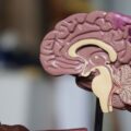
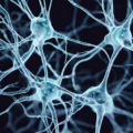
To use an analogy, the usefulness of a car obviously depends upon its engine. Mitochondria are the “engines” of each cell within our bodies, all 70 to 100 trillion cells that make up an adult body. They are known as organelles and are so small that their structure can only be seen with an electron microscope. But we can take this analogy further by comparing each cell to a different car model. A high powered car uses more gasoline than a low powered one and there are many models of each type of car. So some cells in the body require more energy than others, depending on the special function of the cell.  The most energy consuming cells are in the brain, the nervous system and the heart, followed by the gastrointestinal system and muscles. That is why those organs and tissues are most affected in the disease known as beriberi, the thiamine deficiency disease that we have discussed previously in other posts. The function of other organs is affected by the deficiency because of the changes in the control mechanisms originating in the brain through the autonomic (automatic) nervous system.

It has been pointed out that this disease in its early stages affects the autonomic nervous system by causing POTS. Beriberi and POTS, both being examples of dysautonomia (abnormal activity of the autonomic nervous system), can only be distinguished by finding evidence of thiamine or other nutrient deficiency as a cause. Thiamine is but one factor whose deficiency causes loss of cellular energy, resulting in defective brain metabolism and dysautonomia.  Although the relationship with vaccination is conjectural, some individuals with post Gardasil POTS were found to be thiamine deficient and had some relief of symptoms by taking supplementary fat soluble thiamine, an important derivative that occurs in garlic and has been synthesized. Not all of these thiamine deficient individuals have benefited to the same degree, suggesting that other deficiencies might also be involved. This post is to provide some information about more recent knowledge concerning the action of thiamine and the incredible, far-reaching effects of its deficiency, particularly in the brain. Experimental work in animals has shown that thiamine deficiency will damage mitochondria, a devastating effect for an acquired rather than a genetic cause. Far too much research has been devoted to genetic cause without sufficient attention to the way genes are influenced by diet and lifestyle.

The Importance of Enzymes to Mitochondrial Function

Before I provide this new information, let me remind the reader that enzymes, like cogwheels in a man-made machine, enable bodily function to occur. The importance of thiamine is that it is a cofactor to many of the enzymes that preside over energy metabolism. Without its cofactor an enzyme becomes inefficient. Perhaps it might be compared with missing teeth in a cog wheel. With missing teeth the cog wheel may still function but not nearly as well as it would with all of its component parts.

In previous posts we have discussed how thiamine deficiency can be caused by an excess of sugar in the diet. I have likened this to a “choked engine” in a car where an excess of gasoline, relative to insufficient oxygen concentration in the mixture, makes ignition of the gasoline extremely inefficient. Bad diet, one that is rich in sugary, carbohydrate laden foods may be one of the more common contributors to latent thiamine deficiencies. Excessive intake of processed fats and the concomitant changes to mitochondrial function and energy metabolism may be another important contributor.

Thiamine and Fat Metabolism

All the enzymes affected by thiamine deficiency have a vital part to play in obtaining cellular energy from food by the process of oxidation. Most of them have been known for many years but in the nineties a new enzyme was discovered. It has a very fancy name that has been simplified by calling it HACL1.  Only in recent years has it been found that HACL1 is dependent on thiamine as its cofactor. Although not reported, it may mean that it is also dependent on magnesium. This is exceedingly important because it introduces the fact that thiamine is involved in fat as well as carbohydrate metabolism, something brand new, even to biochemists.

Here I must digress again to describe another type of organelle called a peroxisome that occurs in our cells.  Like mitochondria, they are infinitesimally small. Their job is to break down fatty acids and they have a double purpose. One purpose is to synthesize very important substances that construct and maintain cells and their function: they are particularly important in the brain. The other purpose is what might be called fuel preparation. As the fatty acids, consisting of long carbon chains, are broken down, the resulting smaller fragments can be used by mitochondria as fuel to produce energy.  Failure to break down these fatty acids can result in the accumulation of natural components that may be toxic in the brain and nervous system or simply result in lack of one type of fuel. That is why feeding medium chain triglycerides by administration of coconut oil has been reported useful to treat early Alzheimer disease. They can be oxidized in mitochondria.

The Important Use of Fatty Acids in Mitochondrial Health

Here, I want to use another analogy. Imagine a lake that admits water to a river through a sluice gate that has to be opened and closed by a farmer who regulates the supply of water. If the gate is open the river will supply water to the surrounding fields. If however the gate is closed, the river will begin to dry up and the crops in the fields will suffer. Perhaps the farmer half closed the gate during a rainy period and has forgotten to open it when a dry period follows. High temperatures in the dry period results in insufficient water to meet the growth needs of the crops.

In this analogy, the lake represents food, the sluice gate is the HACL1 enzyme and the farmer who controls the gate represents thiamine. The water in the river represents the flow of fatty acids to the tissues for the double purpose of cellular construction and fuel for oxidation. The half open gate represents a minor thiamine deficiency, more or less sufficient for everyday life but not enough when there is greater demand. A high temperature that increases the water needs for crops represents Gardasil and many other medications as a stress factor, placing a greater demand on essential metabolic action.  The analogy also implicates the nature of the crops, some of which require more water than others. The crops, of course, represent body tissues and organs.

If we consider high performing individuals, whether academically or athletically, like high performance cars or crops that demand more nutrients, we can see how a previously unrecognized minor deficiency might trigger clinical disease by the stressful demands of a vaccine or medication. Some pharmaceuticals can attack thiamine directly, like Gardasil and the fluoroquinolones, while others attack different pathways within the mitochondria.

No matter the pathway, high performing individuals, with high energy needs not covered by diet, may be hit harder when a medication attacks mitochondrial energy.

The Outcome of Defective Fatty Acid Metabolism

Returning back to the HACL1 enzyme, we now know that HACL1 is the first thiamine dependent enzyme to be discovered in peroxisomes. It is research news of the highest importance, affecting us all. Its action is to oxidize a diet related fatty acid called phytanic acid and fatty acids with long carbon chains that cannot be used for fuel until they are broken down. Phytanic acid is obtained through consumption of dairy products, ruminant animal fats and some fish. People who consume meat have higher plasma phytanic acid concentrations than vegans. If the action of HACL1 is impaired because of thiamine deficiency the concentration of phytanic acid will be increased. The river in the analogy actually represents a series of enzymatic reactions that may be thought of as down-stream effects, whereas thiamine deficiency, being up-stream, affects all down-stream phenomena. One of the reasons thiamine deficiency is such an important contributor to illness is because its effects are broad.

These enzymatic reactions, known technically as alpha oxidation, involve four separate stages. It has been known for some time that if another enzyme at stage two is missing because of a gene defect, the result will be damage to the neurological system known as Refsum’s disease. Symptoms include cerebellar ataxia (also reported after Gardasil vaccination), scaly skin eruptions, difficulty in hearing, cataracts and night blindness. Other genetic mutations in alpha oxidation, resulting in various biochemical effects, result in a whole variety of different diseases. This places thiamine deficiency as a potential cause for all the down-stream effects resulting from defective alpha oxidation, for it has been shown in mice that this vitally important chemistry is totally dependent on presence of thiamine. Since its complete absence would be lethal, we have to assume that it is mild to moderate deficiency, equivalent to a partial closure of the sluice gate in the analogy.

Sources of Phytanic Acid: How Diet Affects Thiamine

In ruminant animals, our source of beef, the gut fermentation of consumed plant materials liberates phytol, a constituent of chlorophyll, which is then converted to phytanic acid and stored in fat. The major source of phytol in our diet is, however, milk and dairy products.  It raises several important questions. If thiamine deficiency is capable of causing an increase in phytanic acid in blood and urine, it might be a means of depicting such a deficiency in a patient with confusing symptoms. It might also explain why some individuals who have been shown to have thiamine deficiency by means of an abnormal transketolase test have symptoms that are not traditionally accepted as those of such a deficiency, perhaps because of loss of efficiency in HACL1.

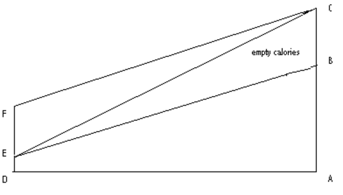
If an excess of sugar in the diet gives rise to a secondary (relative) thiamine deficiency, we are provided with an excellent view of the extraordinary danger of empty simple carbohydrate and fat calories, perhaps explaining much widespread illness in Western civilization. Interestingly, it would also suggest that something as benign as milk could give rise to abnormal brain action in the presence of thiamine deficiency, because of phytanic acid accumulation. Our problems with dairy products may go well beyond lactose intolerance and immune dysregulation.

In sum, the discovery of HCAL1 enzyme and its dependence upon thiamine suggests one more mechanism by which thiamine deficiency affects mitochondrial functioning. As emerging evidence indicates a myriad of environmental and pharmaceutical insults impair mitochondrial functioning, thiamine deficiency ought to be considered of prime importance. Deficits in thiamine evoke devastating health issues that can be treated easily if identified.  If, however, thiamine deficiency is not identified and the same old dietary habits continue, the latent thiamine deficiency can be guaranteed to produce a much more severe and difficult to treat chronic disease. Moreover, individuals with thiamine deficiency who do not respond sufficiently to thiamine replacement might also have aberrant fatty acid metabolism. This too should be investigated and dietary changes adopted.

Thresholds and Tipping Points in Thiamine Deficiency Syndromes

13997 views

I recently stumbled upon on a research paper published in 1968. It was not that long ago in the overall course of modern medicine, perhaps even its heyday, when all things were still possible and before the complete fealty to pharmaceuticals arrived. To the youngsters and to those coming of age in the last 20 years, however, anything published pre 1990 is ancient history.  What could such old paper tell me about medicine that is new and useful? It turns out an awful lot.

Back in the day, research was a little simpler and more focused, not on finding out which drug could be fit to which symptoms, but on how things worked. Good experimental design, answered mechanical questions, like if we apply X to Y or if we remove X from Y what happens?

In this paper, Encephalopathy of Thiamine Deficiency: Studies of Intracerebral Mechanisms, the researchers identified a very important component about Vitamin B1/thiamine deficiency – the time course of the disease process. That is, with a diet deficient in thiamine, how long does it take before symptoms emerge, what is the corresponding level of deficiency in the brain, and at what point, after supplementation, does recovery begin; important questions clinically.

Vitamin B1 – Thiamine Deficiency

Remember, vitamin B1 or thiamine deficiency at its worst is linked to severe decrements neurological functioning, like Wernicke’s Encephalopathy that include noticeable ataxic and gait disturbances (loss of voluntary control of muscle movements, balance and walking difficulties), aphasias (language comprehension and/or production difficulties), and if it persists, Korsakoff’s Syndrome (severe memory deficits, confabulations and psychosis). Thiamine deficiency was originally observed in only chronic and severe alcoholics or with severe nutritional deficits as seen in famine. Fortification of food stuffs was thought to relieve much of the nutritional risks for deficit, especially in impoverished regions. More recent research, however, indicates that thiamine deficiency has reared its ugly head once again and this time in modern, non-impoverished, regions where the food supply is ample. How can that be?

Non-Alcoholic Wernicke’s Encephalopathies

Thiamine deficits can be mediated by a number of factors, including by less obvious nutritional deficits where food supply is abundant but nutrition is lacking (a diet of highly processed, carbohydrate and fat laden foods), with thiamine blocking factors found in medications/vaccines, environmental toxicants and some foods, after bariatric surgery and in disease processes like AIDS. Over the course of our research, thiamine deficiency has been observed in previously healthy, young, non-alcoholic patients, post medication or vaccine, along with symptoms of dysautonomia.

What has always struck me about the thiamine deficits we observe is the differential expression and time course of the symptoms. In some people, the reaction leading to thiamine deficit appears linear, progressive and rapid. In others, the symptoms appear to wax and wane and to evolve more slowly. How is that possible? Certainly, individual predispositions come into play. Some individuals may be somewhat thiamine deficient prior to the trigger that initiates the full expression of symptoms, while others have higher baseline stores. Additionally, anti-thiamine environmental exposures and other medical conditions/medications may also come into play.  In the literature, however, the progression of symptoms from bad to worse is almost always direct and rapid, perhaps mistakenly so. Indeed, Wernicke’s Encephalopathy is a medical emergency necessitating immediate IV thiamine.  How is it then, that we see more chronic, remit and relapse patterns of thiamine deficiency, even in some cases where thiamine concentrations are being managed medically?

Cerebral Thiamine Deficiency: Crossing the Black Line

It turns out, there is black line with regard to thiamine deficiency, that when crossed overt symptoms emerge, and a similar black line, that demarks recovery. It is possible then that barring a continuous blockade of thiamine, one can move above and below those lines and the corresponding symptoms may wax and wane. The paper from 1968, cited above, found those black lines, in rodents, but we can extrapolate to humans.

The research. The investigators took three groups of female rodents, a paired group of thiamine deprived and thiamine supplemented, along with a group fed ad lib (as desired) and assessed the time course and concentrations of cerebral thiamine deficiency relative to the initiation and progression of the observable neurological symptoms associated with Wernicke’s encephalopathy in rodents (ataxia, loss of righting, opisthotonos –rigid body arching). The experiment lasted about 6 weeks.

Neither the control group (thiamine supplemented) nor the ad lib group demonstrated neurological deficits at any time during the study. The thiamine deprived group, on the other hand, demonstrated symptoms that began with weight loss, progressive anorexia, hair loss (recall our observations about hair loss) and drowsiness at about 2.5 weeks into the experiment. Interestingly, no neurological signs of thiamine deficiency were seen at that time.

The results. At 4.5 weeks in, the researchers noted a rapid progression of symptoms and decline of health over the course of the next 5 days (the black line). These symptoms included: incoordination with walking, impairment of the righting reflex, reluctance to walk, walking backwards in circles, imbalance, rigid posturing and eventually a total loss of righting activity and severe drowsiness.

One can imagine, if a similar deprivation of thiamine were observed in humans, the corresponding symptoms might also include the initial hair loose and weight loss, perhaps noticeable, perhaps not depending upon the time frame and severity of the thiamine deficiency. It would also include incoordination and difficulty with walking, balance and voluntary movement, perhaps tremors, excessive fatigue or sleepiness and the myriad of neuro-cognitive disturbances noted in Wernicke’s syndrome.

In the cited experiment, one injection of thiamine reversed these symptoms to a nearly normal, or apparently normal neurological state within 24 hours.

Brain Thiamine Thresholds

Animals from each of the groups were sacrificed and examined at each of the stages of the experiment. Brain thiamine and other markers of thiamine metabolism were assayed to determine the cutoff levels of thiamine that demark symptoms and recovery.  This is really interesting and the beauty of this entire study.  Neurological symptoms become apparent when cerebral thiamine concentrations reach 20% of normal.  Recovery begins when those concentrations climb to 26% of normal. At least in rodents, one has to deplete 80% of the brain thiamine stores before overt neurological symptoms become apparent; 80% – that is a huge deficit.  Similarly, it doesn’t appear to take much to right that deficit, only a 6% increase in thiamine concentration set the course for improvement.

If we extrapolate to humans, where life span, genetic and environmental factors likely moderate the degree of thiamine stores and consumption, we still contemplate a rather large thiamine deficit needed before overt symptoms of Wernicke’s emerge. Similarly though, it is also evident that a rather small change in thiamine can have enormous effects on neurological functioning. In the case of the rodents, a mere 6% point change reversed the symptoms. One might suspect equivalent deficit/recovery thiamine parameters in humans.

Waxing and Waning Symptoms:  A Case for Persistent Thiamine Deficiency

If we consider the possible course of non-alcoholic thiamine deficiency, where no extraneous variables like bariatric surgery or thiamine deficient parenteral feeding are present and where dietary thiamine varies daily and is not held constant as it is during experimental conditions or during famine, we can begin to see how thiamine related neurological symptoms may wax and wane. Different exposures and triggers may decrease thiamine periodically, even to the point where overt neurological symptoms present. When those exposures are removed and barring deficiencies in metabolism and diet, symptoms may abate, at least temporarily, and until the next trigger or until the black line is crossed anew and thiamine deficiency becomes the medical emergency observed in overt Wernicke’s.

In contrast, the more persistent or chronic thiamine deficits that do not cross the 80% depletion cutoff (or the human equivalent), may also wax and wane and show all the core neurological symptoms expected in overt Wernicke’s though to a much lesser degree. Additionally, as we have speculated, persistent thiamine deficiency might disable mitochondrial functioning in such a way that the patient presents with a myriad of seemingly unrelated symptoms, that are not typically attributed to thiamine deficiency, such as cardiac dysregulation, gastroparesis, autonomic instability, demyelinating syndromes and hormone irregularities, especially thyroid, but also reproductive hormones. These too may be related to thiamine deficiencies. Although, we cannot and should not rule out other causes as well, sub-optimal thiamine may be involved with a host of complex disease states and medication adverse reactions where neurological symptoms are present. Thiamine deficiency should be tested for and ruled out before more invasive therapeutic options are contemplated.

We Need Your Help

More people than ever are reading Hormones Matter, a testament to the need for independent voices in health and medicine. We are not funded and accept limited advertising. Unlike many health sites, we don’t force you to purchase a subscription. We believe health information should be open to all. If you read Hormones Matter, like it, please help support it. Contribute now.

Yes, I would like to support Hormones Matter. 

Of Oxygen, Spark Plugs, and Mitochondria

5149 views

Everyone knows that we need oxygen to live. Few know or care what the body does with it. Although everyone knows that it is extracted from the air by the lungs and carried by the blood to body tissues, it is left to scientists to understand what happens to it in the 50 to 100 trillion cells that make up an adult human body. The reaction that makes it possible for oxygen to maintain life is called oxidation. This reaction takes place in the mitochondria and produces energy that is used by each cell to carry out its program of function.  Perhaps we can understand this better by using an analogy.

Car Engines and Human Engines: Each Need Fuel

A car uses gasoline as a fuel. It is ignited by a spark plug that causes a controlled explosion in a cylinder. This drives a piston that passes the energy through a series of mechanical levers known collectively as the transmission. It is the conversion of chemical energy in gasoline to what Newton called kinetic energy that enables the car to move. The machine that does this is an engine. Perhaps it stretches the imagination to state that the body works on exactly the same principles. It is the details that make the difference. Oxidation is the equivalent of explosion in the cylinder. In other words, it is combustion. Now we have to compare it to the relatively simple mechanism of explosion.

First, combustion is merely the union of oxygen with a fuel. If we do not carry the reaction out in some controlled way, the energy is dissipated as heat into the surrounding air. In a car, the cylinder encloses the combustion and forces the energy into the transmission. In the body it is controlled in a much more complex way. Yes, heat is produced and is used to make us “warm blooded creatures” but there is no noise, fire or smoke as in the car engine. The energy is guided through an ingenious series of chemical reactions in what we might term “the engines of the cell”.

Mitochondria: The Engines in Our Cells

Each cell has a whole series of “engines” called mitochondria and it is in these organelles where oxidation occurs. A mitochondrion is so small that its structure can only be seen with the aid of an electron microscope and yet it is in each of the millions of cellular mitochondria where energy is produced for the use of each cell to perform its designed function. The usual fuel for this is glucose and it is not surprising that people have concluded that the consumption of sugar provides “quick energy”.

Good Sugar and Bad Sugar: Mitochondria Know the Difference

When sugar is ingested in its proper form, meaning as it is found in nature, it is stored in the liver and muscles as glycogen, a complex substance built up by sticking glucose molecules together, making something that looks like a miniature tree. As fuel is required, the glycogen is broken down and released as glucose into the blood. This requires an enzyme and there is an inborn error of metabolism where this enzyme is missing. The affected infant is found to have an enlarged liver stuffed with glycogen, together with low blood sugar, a situation that is not compatible with life and the patient dies in infancy.

Blood glucose is absorbed from the blood into cells under the influence of insulin and then goes into an ingenious “pipeline” that processes it. The beginning of this process requires a number of B group vitamins. There is a well known nutritional disease known as beriberi where the carbohydrate load is too great for this action to occur efficiently. It is now known that vitamin B1 is insufficient to meet the caloric demand and is the key to understanding the disease and how it is treated, a discovery that took many years to unravel.

Let us look again at the simpler method by which gasoline is ignited in a car. An electrically energized spark plug is used to ignite the fuel as it is passed into the cylinder by carefully controlled mechanisms. Some people will remember that cars once had a gadget called a choke, used for starting the cold engine. This allowed gasoline to flow into the cylinder with a relative deficiency of air, the so-called “rich” mixture. When the engine was warm the choke was automatically removed and the mixture weakened by allowing more air and less gas into the cylinder. If the choke mechanism stuck, there would be an excess of black smoke issuing from the exhaust pipe and the engine would not run properly. The smoke represents the hydrocarbons in gas that have not been ignited and a simple equation shows us why:

Fuel + Oxygen + Catalyst = Energy

empty calories

The Figure shows the ratio of calories to B vitamins in a healthy diet. The line AB represents the calorie intake (protein, fat and carbohydrate) and the line ED the vitamin intake that enables its efficient processing. If the line AB is extended to C (line AC) without the increase in vitamin intake, the triangle BCE represents “empty calories” equivalent to a “choked engine”. The remedy is obvious: we can extend line DE to F, thus restoring the ratio as in line FC, reduce the calories back to line AB, or meet each other half way (not shown).

Beriberi: Bad Sugar and Empty Calories

Beriberi is caused by consuming empty calories (triangle BCE), where the line AC represents carbohydrate calories and ED the corresponding ingestion of vitamin B 1. (thiamin).  The disease, throughout history, has been primarily in Eastern countries where the diet has been white rice based, particularly in times of greater affluence. This is because the grain in rice is starch and the cusp contains the necessary vitamins. When the Chinese peasants became more affluent they would take their rice to a rice mill where white rice was produced by removing the cusps. This was because it looked better when served to their friends, thus demonstrating their new found affluence. Outbreaks of beriberi were always associated with an increase in consumption of white rice.

What is the lesson to be learned from this in our modern age where diseases like beriberi have been thought to be of only historical interest?  Think of the enormous load of simple carbohydrate consumed by millions in the U.S. Everything supplied by the food industry is sweetened or it would not sell. White bread (the equivalent of white rice), cookies, pastry in general, ice cream, soft drinks, desserts, tomato ketchup——— the list goes on and on! Even the vitamin enrichment indicated on the label is insufficient. Obesity, often associated with inflammatory disease, is affecting millions. Our health bills are threatening us with national bankruptcy and we wonder why we are being “hit” with so many diseases and health catastrophies. Pockets are being lined with money made from a variety of reducing diets and pills.

Diet is Everything: Feed your Mitochondria

That is why I have a standard answer to every query that I get about diet. Eat only nature made food and the less that it is handled by mankind the better. The balance of calories and vitamins is automatically produced. If the food had not been available when life started on Earth animal evolution could not have occurred and we would never have survived. Granted, unfortunately with population explosion, fresh food of this nature is expensive and we have all given up back yard gardening The First Lady has shown the example. Will we take a “leaf from her book” and acknowledge that a lot of our health is in our own hands.

We Need Your Help

More people than ever are reading Hormones Matter, a testament to the need for independent voices in health and medicine. We are not funded and accept limited advertising. Unlike many health sites, we don’t force you to purchase a subscription. We believe health information should be open to all. If you read Hormones Matter, like it, please help support it. Contribute now.

Yes, I would like to support Hormones Matter. 

Image by Sumanley xulx from Pixabay.

The Paradox of Modern Vitamin Deficiency, Disease, and Therapy

13297 views

In order to understand why this article is about “paradox”, the concept of vitamin therapy must be appreciated. Hence, the explanation of the title is deferred to the end. Although vitamin deficiency disease is believed by most physicians to be only of historical interest, this is simply not true. When we think of a vitamin deficiency disease, we envision an individual living in a third world country where starvation is common. Such an individual is imagined as being skeletal, whereas an obese person is considered to be well fed with vitamin enriched foods. For this reason, common diseases, some of which are associated with obesity, are rarely, if ever, seen as potentially vitamin deficient.

The Calorie Rich and Nutrient Sparse Modern Diet

Our food is made up of two different components, the caloric and the non-caloric nutrients.  When we ingest high calorie foods (e.g. a doughnut) without even a vestige of non-caloric nutrients, we refer to this as “empty” or “naked” calories.  For our food to be processed into energy that enables the body and brain cells to function, there must be a ratio of the calorie bearing component to that of the non-caloric nutrients.  When we load the calories together with an insufficiency of non-caloric nutrients, we alter this ratio and produce a relative vitamin deficiency.  The trouble with this is that it does not result in the formation of the classic vitamin deficiency diseases as recorded in the medical literature. There is a gradual impairment of function, resulting in many different symptoms. Because modern medicine seeks to make a diagnosis by the use of imaging techniques and laboratory data and because of the physician’s mindset, if the tests used are normal, the possibility of a relative vitamin deficiency is ignored.

The Brain as a Chemical Machine

We have two different nervous systems. One is called “voluntary” that enables us to do things by will-power.  This is initiated and controlled by the upper brain, the part of the brain that thinks. The other system is known as the autonomic nervous system (ANS).  This is initiated and controlled by the lower part of the brain, the limbic system and brainstem.  This system is controlled automatically.  Although it collaborates with the other system, it is not normally under voluntary control. The limbic system and brainstem are highly sensitive to oxygen deficiency, but since the oxygen is useless without the non-caloric nutrients, their absence would produce the same kind of phenomena as oxygen deficiency. Thiamine (vitamin B1) has been found to be of extreme importance as a member of the non-caloric nutrients. The brain, and particularly the limbic system and brainstem, is highly sensitive to its deficiency.

Since the ANS is automatic, we are forced to think of the limbic system and brainstem as a computer.  For example, when it is hot, you start to sweat.  Evaporation of the sweat from the skin produces cooling of the body, representing an adaptive response to environmental hot temperature. When it is cold, you may start to shiver. This produces heat in the muscles and represents an adaptive response to environmental low temperature. If you are confronted by danger, the computer will initiate a fight- or- flight reflex.  This is a potential lifesaving reflex.  It is designed for short term use, consumes a vast amount of energy and prepares you to kill the enemy or flee from the danger.  Any one of these reflexes may be modified by the thinking brain. For example the lower brain, also known as the reptilian system, initiates the urge to copulate.  It is modified by the upper brain to “make love”.  The reptilian system, working by itself, can convert us into savages. There is an obvious problem here because our ancestors were faced with the dangers of short term physical stress associated with survival.  In the modern world the kind of stress that we face is very different for the most part.  We have to contend with traffic, paying bills, business deadlines and pink slips. The energy consumption, however is enormous, continues for a long time and it is hardly surprising that it is associated with fatigue, an early sign of energy depletion. It has been shown in experimental work that thiamine deficiency causes extensive damage to mitochondria, the organelles that are responsible for producing cellular energy.

Autonomic Function

The autonomic nervous system, controlled by the lower brain, uses two different channels of neurological communication with the body. One is known as the sympathetic system and the other is the parasympathetic. There are also a bunch of glands called the endocrine system that deals with the brain-controlled release of hormones.

We can think of the sympathetic branch of the ANS as the action system. It governs the fight-or-flight reflex for personal survival and the relatively primitive copulation mechanisms for the survival of the species. It accelerates the heart to pump more blood through the body.  It opens the bronchial tubes so that the lungs may get more oxygen. It sends more blood to the muscles so that you can run faster and the sensation of fear is a normal part of the reflex. When the danger is over and survival has been accomplished, the sympathetic channel is withdrawn and the parasympathetic goes into action. Now in safety and under its influence, body functions such as sleep and bowel action can take place.  That is why I refer to the parasympathetic as the “rest and be thankful system”.

Dysautonomia, Dysfunctional Oxidation and Disparate Symptoms 

When there is mild to moderate loss of efficiency in oxidation in the limbic system and/or brainstem they become excitable. This is most easily accomplished by ingesting a high calorie diet that is reflected in relative vitamin deficiency.  The sympathetic action system is turned on and this can be thought of as a logical reaction from a design point of view.  For example, if you were sleeping in a room that was gradually filling with carbon dioxide, the gradual loss of efficiency in oxidation would be lifesaving by waking you up and enabling you to exit the room. In the waking state, this normal survival reflex would be abnormal.

High calorie malnutrition, by upsetting the calorie/vitamin ratio, causes the ANS to become dysfunctional. Its normal functions are grossly exaggerated and reflexes go into action without there being any necessity for them. Panic attacks are merely fragmented fight-or-flight reflexes.  A racing heart (tachycardia) may start without obvious cause.  Aches and pains may be initiated for no observable reason. Affected children often complain of aching pain in the legs at night. Unexplained chest and abdominal pain are both common. This is because the sensory system is exaggerated. One can think of it as the body trying to send messages to the brain as a warning system.

Nausea and vomiting are both extremely common and are usually considered to be a gastrointestinal problem rather than something going on in the brain. Irritable bowel syndrome (IBS) is caused by messages being conveyed through the nervous system of the bowel, increasing peristalsis (the wave-like motion of the intestine) and often leading to breakdown of the bowel itself, resulting in colitis.  Of course, the trouble may be in the organ itself but when all the tests show that “nothing is wrong”, the symptoms are referred to as psychosomatic. The patient is often told that it is “all in your head”.

Emotional instability seems to be more in keeping with psychosomatic disease because emotional reactions are initiated automatically in the limbic system and thiamine deficient people are almost always emotionally unstable. A woman patient had been crying night and day for three weeks for no observable reason. A course of intravenously administered vitamins revealed a normal and highly intelligent person.  Intravenously administered vitamins are often necessary for serious disease because the required concentrations cannot be reached, taking them by mouth only.

The Vitamin Therapy Paradox

The body is basically a chemical machine.  But instead of cogwheels and levers, all the functions are manipulated through enzymes that, in order to function efficiently, require chemicals called “cofactors”. Vitamins are those essential cofactors to the enzymes.  If a person has been mildly to moderately deficient in a given vitamin or vitamins for a long time without the deficiency being recognized, the enzyme that depends on the vitamin for its action appears to become less efficient in that action.  A high concentration of the vitamin is required for a long time in order to induce its functional recovery.

Although the reason is unknown, doctors who use nutritional therapy with vitamins have observed that the symptoms become worse initially.  Because patients expect to improve when a doctor does something to them and because drugs have well-known side effects, it is automatically assumed by the patient that this worsening is a side effect of the vitamins. If the therapy is continued, there is a gradual disappearance of those symptoms and overall improvement in the patient’s well being. Unless the patient is warned of this possibility he or she would be inclined to stop using the treatment, claiming that vitamins have dangerous side effects and never getting the benefit that would accrue from later treatment.  This is the opposite effect that the patient expects. This is the paradox of vitamin therapy. 

If we view dysautonomia as an imbalance in the functions of the ANS and the vitamin therapy as assisting the functional recovery by stimulating energy synthesis, we can view this initial paradoxical as the early return of the stronger arm of the ANS before the weaker arm catches up, thus worsening an existing imbalance. However, this is mere speculation. I did not learn of the “paradox” until I actually started using mega dose vitamins to treat patients.

The Paradox and Thiamine

In this series of posts, we are particularly concerned with energy metabolism and the place that thiamine holds in that vital mechanism.  It is, of course, true that worsening of serious symptoms is a fact that has to be contended with and vitamin therapy should be under the care of a knowledgeable physician. The earlier the symptoms of thiamine deficiency are recognized, the easier it is to abolish them. The longer they are present the more serious will be the problem of paradox and a clinical response will also be much delayed and may be incomplete.

Beriberi and Thiamine Deficiency

I will illustrate from the early history of beriberi when thiamine deficiency was found to be its cause.  Many of the patients had the disease for some time before thiamine was administered, so the danger of paradox was increased. It was found that if the blood sugar was initially normal, the patient recovered quickly. If the blood sugar was high, the recovery was slow.  If the blood sugar was low, the patient seldom recovered.  In the world of today, an abnormal concentration of glucose in the blood would make few doctors, if any, think of thiamine deficiency as a potential cause. It is no accident that diabetes and thiamine metabolism are connected. Education of the doctor and patient are both absolutely essential. I believe that the ghastly effects of Gardasil, and perhaps some other medication reactions covered on Hormones Matter, can only be understood by thinking of the body as a biochemical machine and that the only avenue of escape is through the skilled use of non caloric nutrients.

We Need Your Help

More people than ever are reading Hormones Matter, a testament to the need for independent voices in health and medicine. We are not funded and accept limited advertising. Unlike many health sites, we don’t force you to purchase a subscription. We believe health information should be open to all. If you read Hormones Matter, like it, please help support it. Contribute now.

Yes, I would like to support Hormones Matter. 

Image by Rochak Shukla on Freepik

How Dietary Mayhem Causes Disease: The Choked Engine Syndrome

11451 views

Over the past year, I have written extensively about thiamine deficiency post Gardasil vaccination (here, here, here, here). We now have five cases where thiamine deficiency was identified and clinical symptoms remediated with supplementation. Many more are suspected but recognition and testing have been slow. Thiamine deficiency may not be limited to the post Gardasil population, although that is where we first recognized it. Symptoms of thiamine deficiency and dysfunctional oxidative metabolism have been observed amongst the post fluoroquinolone and post Lupron populations and likely other populations adversely affected by a vaccine or medication, though data are limited. For the current paper, I should like to offer an explanation of the effect of thiamine deficiency in relationship to the stress of the vaccination or medications.

Thiamine Deficiency and Diet

With the widespread ingestion of simple carbohydrates that is almost a hallmark of Western civilization I suggest that the Gardasil vaccination and certain other medications represent “the last straw to break the camel’s back”.  I have included a case report, from my clinical practice, as an example of the effect of a simple nutritional stressor – sugar – imposed on an individual who’s oxidative metabolism was marginal at the time. I have included the references for anybody that wishes to check on how much of this is published.

Cellular Energy and Diet

Present knowledge indicates that cellular energy arises only from oxidation of food sources. The prevalently common form of nutritional mayhem in the U.S. is a high calorie content from simple carbohydrates with insufficient vitamin/mineral content to catalyze efficient oxidation. This form of malnutrition might be compared with functional decline in a choked internal combustion engine. Evidence presented in this case report presented below indicates that simple carbohydrate ingestion can have far-reaching consequences.  A review indicates that a common manifestation of its effect is oxidative stress in the brain, particularly in the limbic system where emotional reflexes originate and where the controls of the autonomic and endocrine systems react automatically to sensory input. Beriberi is the classic example of high calorie carbohydrate malnutrition and is the prototype for dysautonomia (abnormal function of the autonomic nervous system [ANS] ) in its early stages. A later stage results in degeneration of autonomic ganglia and irreversible disease. Symptoms arising from thiamine deficiency or abnormal homeostasis are protean and diverse in nature.

Dysautonomia, Oxidative Stress and Thiamine

Dysautonomia, a common presentation of functional disease and often associated with variable organic diseases caused by loss of oxidative efficiency in the brain, has been reviewed. A hypothesis was presented that there is a combination of genetic risk, different forms of sensory input defined as stress, particularly those imposed by present civilization, and high calorie malnutrition that are collectively responsible. This was presented diagrammatically by the degree of overlap in the “three circles of health, named genetics, stress and nutrition” (1).  It is also known that mitral valve (a heart valve) prolapse (MVP) is widespread in the population and is associated with dysautonomia, although the cause and effect relationship is said to be unknown (2-4). MVP is associated with adrenergic overdrive (the well-known adrenalin rush) in the normally balanced adaptive reactions of the autonomic/endocrine axis (5-8). (The autonomic nervous system and the glands of the endocrine system are under the control of the brain).  Panic disorder, also sometimes associated with MVP, is seen as an example of falsely triggered fight-or-flight reflexes engendered in the limbic brain.  Pasternac and associates (6) showed that symptomatic patients with MVP demonstrated increased resting sympathetic tone and that supine bradycardia (slow heart rate) suggested increased vagal (the vagus is a nerve that runs from the brain to many parts of the body) tone at rest. Davies and associates (7) demonstrated physiologic and pharmacologic hypersensitivity of the sympathetic system in a group of patients with MVP. Sympathoadrenal responses were noted in rats exposed to low oxygen concentration (9) and impaired cerebral autoregulation has been reported in obstructive sleep apnea in human subjects (10). It has also been shown that thiamine deficiency produces traditionally accepted psychosomatic or functional disease (11,12).  A low oxygen concentration results in changes in brain structures similar to those induced in thiamine deficiency (13).

A Case Study of Thiamine Deficiency and Dietary Influence: The Sugar Problem

The Table below shows laboratory results from an 84-year old man who had begun to experience severe insomnia for the first time in his life. He also had painful tenosynovitis (also known as “trigger finger”) in the index finger of the left hand.  He had edited a journal for some 14 years and for several years, had been a member of a bell choir in which he played a heavy base bell in each hand, involving repetitive trauma to the index fingers.  He did not crave sugar, his ingestion of simple carbohydrates being minimal to moderate. The only treatment offered was complete withdrawal from all forms of simple carbohydrates.

Serial laboratory studies revealed a gradual improvement over six months and his weight decreased from 182 to 170 pounds without any other change in diet. Insomnia and tenosynovitis gradually improved. The Table shows that serial laboratory tests over a period of six months, from February to August, showed continued gradual improvement. In September, the day after a minimal ingestion of simple carbohydrate, there was an increase in triglycerides and TPPE.

Understanding the Labs

Notice that the triglycerides dropped from 206 in February to 124 in August, then rose again in September only one day after a minimal amount of sugar.  Triglycerides are part of the routine lipid profile test done by doctors and are well known to be related to the ingestion of simple carbohydrates.  Fibrinogen and HsCRP are both recognized as markers of inflammation.  Notice that both of them decreased between February and August but HsCRP rose again in September like the triglycerides.  The TPPE is the important part of the transketolase test.  The higher the percentage, the greater is the degree of thiamine deficiency.  Notice that it dropped from 35% to zero between February and August, but that it jumped to 8% in September, the day after the ingestion of sweets.  I have provided the normal laboratory values for the discerning reader.

  TABLE 1
Month

Cholesterol

Triglycerides

Fibrinogen

HsCRP

TKA

TPPE

February

169

206

412

7

65

35%

March

155

165

55

25%

May

160

152

312

0.9

85

2%

August

166

124

0.3

59

0%

September*

169

165

220

1

62

8%

Consecutive laboratory blood tests

Cholesterol N <200 mg/dL. Triglycerides N< 150 mg/dL. Fibrinogen N 180-350,g/dL
HsCRP N 0.1-1.0 mg/L. TKA 42-86mU. TPPE 0-18%. *Next day after ingestion of simple carbohydrate.


The abnormal TPPE indicated thiamine deficiency in this patient (14). The increased triglycerides and their steady decrease over time indicated that sugar ingestion was a potent cause of his symptoms. An increase in fibrinogen and hypersensitive CRP are both laboratory markers of inflammation, although the site is not indicated.  Recent studies in mice (15) have shown that high calorie malnutrition activates a normally silent genetically determined mechanism in the hypothalamus, causing either obesity, inflammation or both. The potential association of thiamine with electrogenesis (formation of electrical energy) (16) may have some relationship with brain metabolism and the complex functions of sleep.

Compromised Oxidative Function: Thiamine Deficiency, Beriberi and Diet

It has long been known that beriberi is a classic disease caused by high consumption of simple carbohydrate with insufficient thiamine to process glucose into the citric acid cycle. (This complex chemistry represents the engine of the cell, meaning that it produces the energy for function).  Widespread thiamine deficiency has been reported in many publications(17-20), producing the same brain effects as low oxygen concentration (13,21). In rat studies, this produces an imbalance in the autonomic nervous system (9). Thiamine  deficiency is easily recognized in a clinical laboratory by measuring TKA and TPPE (14).

Thiamine and the Brain

Thiamine triphosphate (TTP) (this is synthesized from thiamine in the brain) is known to be important in energy metabolism. Although its action is still unknown, the work with electric eels has revealed that the electric organ has a high concentration of TTP and may have a part to play in electrogenesis, the transduction of chemical to electrical energy (16,22). The energy for its synthesis from thiamine comes from the respiratory chain. This is also complex chemistry in the formation of energy synthesized within mitochondria, the “engines” of the cell (23), so that any form of disruption of mitochondria would be expected to reduce adequate synthesis of this thiamine ester. Although slowing of the citric acid cycle appears to be the main cause of the biochemical lesion in brain thiamine deficiency (24), the part played by TTP is not yet known. Alzheimer’s disease has been helped by the use of therapeutic doses of thiamin tetrahydrofurfuryl disulfide (TTFD) (25), a more efficient method of administering pharmacologic doses of thiamine (26).

Acetylcholine, the neurotransmitter used by both branches of the autonomic nervous system, is generated from glucose metabolism, requiring  B vitamins, particularly thiamine. Choline is a “conditional nutrient”, meaning that it is derived mainly from diet but is also made in the body. The presence of all these nutrients leads to the synthesis of this neurotransmitter.  It’s depletion would affect both branches of the autonomic nervous system, resulting in dysautonomia.

There is evidence that high-dose thiamin increases the effect of acetylcholine (27). Animal studies have shown that TTFD improves long term memory in mice (28) and it has been shown that it extends the duration of  neonatal seizures in DBA/J2 mice, seizures that normally cease in a few days with normal maturation (29).  These seizures are naturally related to a prolonged effect of this neurotransmitter in this strain of mouse.  The experimental prolongation of the seizures by administration of TTFD indicated that it enhanced the effect of the neurotransmitter. A pilot study in autistic spectrum disorder showed clinical improvement in 8 of the 10 children treated with TTFD (30), a disease that has been shown to have reduced  parasympathetic activity in the heart (31,32). Neural reflexes regulate immunity (33).  Dysautonomia was found in a large number of patients with cancer at Mayo Clinic (34).

Dysautonomia and Thiamine Deficiency         

Evidence has been presented that a common connection exists between dysautonomia, inefficient oxidative metabolism produced mainly by high calorie malnutrition, and organic disease (1). Thiamine enters the equation in terms of its relationship with carbohydrate ingestion and its use by the brain as fuel (35). Decreased transketolase activity in brain cells induced by thiamine deficiency contributes to impaired function of the hippocampus (36) each, part of the limbic system control mechanisms that affect autonomic sympathetic/parasympathetic balance. Erythrocyte (red cells) transketolase indicates abnormal thiamine homeostasis that is commonly achieved by carbohydrate ingestion and deficiency of vitamin B (14).  Beriberi gives rise to functional changes in the autonomic nervous system in its early stages and produces irreversible degeneration in its later stages (37). This, because it represents a largely forgotten aspect of disease, might equate with the wide use of simple carbohydrates in Western civilization. Deficiency of other essential non-caloric nutrients has been associated with dysautonomia (1).

The Role of Nutritional Stress in Post Vaccination and Medication Reactions

Two results of post- Gardasil vaccination have been reported, Postural Orthostatic Tachycardia Syndrome (POTS) and cerebellar ataxia.  POTS, a disease easily confused with beriberi, is one of the many syndromes reported under the general heading of dysautonomia and stress related intermittent episodes of cerebellar ataxia were reported in thiamin dependency (38).  Since the inflammatory reflex has recently been found to be involved with the sympathetic branch of the ANS (39), enhancement of its dysfunction by TD might explain some of the Gardasil affected illnesses.

Conclusion

Thiamine deficiency is now accepted as the major cause of the ancient scourge of beriberi. The underlying mechanisms are still not fully understood for we do not yet know the complete roles of thiamine. The clinical effects are protean and unpredictable. It is, however, clear that thiamine has a vital effect on many aspects of oxidative metabolism and its deficiency can be used as a model for the clinical effects produced by disruption in energy synthesis. It can be summed up under the general heading of dysoxegenosis and thiamine is certainly not the only component that governs this vital life process. The example of beriberi indicates that the brain, peripheral nervous system and the heart are the tissues most affected by the disease, the tissues that rapidly consume oxygen.

The limbic system is a complex computer that organizes all our adaptive survival reflexes and its sensitivity to hypoxia is well known. It is evident that non-caloric nutrient deficiency, especially thiamine, gives rise to the same symptoms and histopathology as mild to moderate hypoxia (oxygen deficiency) and that the leading symptomology is that of dysautonomia. Since the limbic system gives rise to emotional reflexes and mild to moderate hypoxia enhances sympathoadrenal response, it can be expected that an affected individual would be more aggressive and more likely to experience exaggerated fight-or-flight reflexes. A “nursed” emotional grievance might be expected to explode in violence that would otherwise be curtailed or suppressed by normal brain metabolism. It suggests that high calorie malnutrition, particularly that provided by excessive consumption of simple carbohydrates, gives rise to uncontrolled pathophysiological actions that might explain some of the widespread incidence of emotional and psychosomatic disease in contemporary society. It may also explain some of the “hot” juvenile crime and vandalism, much of which is poorly understood in our present civilization. It is also hypothesized that a marginal state of oxidative metabolism, perhaps asymptomatic or with only mild symptoms that are ignored, might be precipitated into clinical expression with a mild degree of stress imposed by a vaccination. The individual in the case reported above appeared to be unusually sensitive to sugar ingestion and this may be an additional genetically determined risk.

We Need Your Help

More people than ever are reading Hormones Matter, a testament to the need for independent voices in health and medicine. We are not funded and accept limited advertising. Unlike many health sites, we don’t force you to purchase a subscription. We believe health information should be open to all. If you read Hormones Matter, like it, please help support it. Contribute now.

Yes, I would like to support Hormones Matter. 

Image created using Canva AI.

References

  1. Lonsdale D. Dysautonomia, a heuristic approach to a revised etiology for disease. eCAM 2009;6(1):3-10.
  2. Orhan A L, Sayar N, Nurkalem Z, Uslu N, Erdem I, Erdem E C, Assessment of autonomic dysfunction and anxiety levels in patients with mitral valve prolapase. Turk Kardiyol Dern Ars 2009;37(4):226-233.
  3. Alpert M A, Murkerji V, Sabeti M, Russell J L, Beitman B D. Mitral valve prolapse, panic disorder, and chest pain. Med Clin North Am 1991;75(5):1119-1133.
  4. Raj A, Sheehan D V. Mitral valve prolapse and panic disorder. Bull Menninger Clin 1990;54(2):199-208.
  5. Di Salvo G, Pergola V, Ratti G, Tedesco M A, Giordano C, Scialdone A, et al. Atrial natriuretic factor and mitral valve prolapse syndrome. Minerva Cardioangiol 2001;49(5):317-325.
  6. Pasternac A, Tubau J F, Puddu P E, Krol R B, de Champlain J. Increased plasma catecholamine levels in patients with symptomatic mitral valve prolapse. Am J Med. 1982;73(6):783-790.
  7. Davies A O. Mares A, Pool J L, Taylor A A. Mitral valve prolapse with symptoms of beta-adrenergic hypersensitivity. Beta 2-adrenergic receptor supercoupling with desensitization on isoproterenol exposure. Am J Med 1987;82(2):193-201.
  8. Boudoulas H, Wooley C F. Mitral valve prolapse syndrome: neuro-endocrinological aspects. Herz 1988;13(4):249-258.
  9. Johnson T S, Young J B, Landsberg L, Dana C A. Sympathoadrenal responses to acute and chronic hypoxia in the rat. J Clin Invest 1983;71:1263-1272.
  10. Urbano F, Roux F, Schindler J, Mohsenin V. Impaired cerebral autoregulation in obstructive sleep apnea. J Appl Physiol 2008;105(6):1852-1857.
  11. Lonsdale D, Shamberger R J. Red cell transketolase as an indicator of nutritional deficiency. Am J Clin Nutr 1980;33:205-211.
  12. Lonsdale D. Three case reports to illustrate clinical applications in the use of erythrocyte transketolase. eCAM 2006;4(2):247-250.
  13. Macey P M, Woo M A, Macey K E, Keens T G, Saeed M M, Alger J R et al. Hypoxia reveals posterior thalamic, cerebellar, midbrain, and limbic deficits in congenital hypoventilation syndrome. J Appl Physiol 2005;98(3):958-969.
  14. Massod M F, McGuire S L, Werner W R. Analysis of blood transketolase activity.Am J ClinPathol 1971;55:465-470.
  15. Zhang X, Zhang G, Zhang H, Karin M, Bai H, Cai D. Hypothalamc 1KKbeta/N-kB and ER stress link overnutrition to energy imbalance and obesity. Cell   2008;135(1):61-73.
  16. Bettendorff L, Michel-Cahay C, Grandfils C, De Rycker C, Schoffeniels E. Thiamine triphosphate and membrane-associated thiamine phosphatases in the electric organ of Electrophorus electricus. J Neurochem 1987;49(2):495-502.
  17. O’Keefe S T, Tormey W P, Glasgow R, Lavan J N. Thiamine deficiency in hospitalized elderly patients. Gerontology 1994;40(1):18-24.
  18. Macias-Matos C, Rodriguez-Ojea A, Chi N, .Zulueta D, Bates C J. Biochemical evidence of thiamine depletion during the Cuban neuropathy epidemic, 1992-1993. Am J Clin Nutr 1996;64(3):347-353.
  19. Mazavet D. Vassilev K, Perrigot M. Neuropathy with non-alcoholic thiamine deficiency: two cases of bladder disorders [article in French]. Ann Readapt Med Phys 2005;48(1):43-47.
  20. Hazell A S, Butterworth R F. Update of cell damage mechanisms in thiamine deficiency: focus on oxidative stress, excitotoxicity and inflammation. Alcohol Alcohol 2009;44(2):141-147.
  21. Vortmeyer A O, Hagel C, Laas R. Hypoxia-ischemia and thiamine deficiency. Clin Neuropathol 1993;12(4):184-190.
  22. Nghiem H O, Bettendorff  L,Changeux J P. Specific phosphorylation of Torpedo 43K raspsyn by endogenous kinase(s) with thiamine triphosphate as the phosphate donor. FASEB J 2000;14(3):543-554.
  23. Gangolf M, Wins P, Thiry M. Thiamine triphosphate synthesis in the rat brain is mitochondrial and coupled to the respiratory chain. J Biol Chem 2010;285(1):583-594.
  24. Bettendorff  L, Sluse F, Goessens G,  Wins P, Grisar T. Thiamine deficiency-induced partial necrosis and mitochondrial uncoupling in neuroblastoma cells are rapidly reversed by addition of thiamine. J Neurochem 1995;65(5):2178-2184.
  25. Mimori Y, Katsuoka H, Nakamura S. Thiamine therapy in Alzheimer’s disease. Matab Brain Dis 1996;11(1):89-94.
  26. Lonsdale D. Thiamine tetrahydrofurfuryl disulfide: a little known therapeutic agent. Med Sci Monit 2004;10(9):RA199-203.
  27. Meador K J. Nichols M E, Franke P, Durbin M W. Evidence for a central cholinergic effect of high-dose thiamine. Ann Neurol 1993;34:724-726.
  28. Micheau J, Durkin D P, Destrade D C, Rolland Y, Jaffard R. Chronic administration of sulbutiamine improves long term memory formation in mice: possible cholinergic mediation.  Pharacol Biochem Behav 1985;23(2):1
  29. Lonsdale D. Effect of thiamine tetrahydrofurfuryl disulfide on audiogenic seizures in DBA/2J mice. Dev Pharmacol Ther 1982;4(1):28-36.
  30. Lonsdale D, Shamberger R J, Audhya T. Treatment of autism spectrum children with thiamine tetrahyhdrofurfuryl disulfide: a pilot study. Neuro Endocrinol Lett 2002;23(4):303-308.
  31. Ming X,  Julu P O O, Brimacombe M, Connor S, Daniels M L. Reduced cardiac parasympathetic activity in children with autism. Brain Dev 2005;27:509-516.
  32. Palmieri L Persico A M. Mitochondrial dysfunction in autism spectrum disorders: cause or effect? Biochem Biopys Acta 2010; May 1 [Epub ahead of print].
  33. Rosas-Ballina M, Tracey K J. The neurology of the immune system: neural reflexes regulate immunity. Neuron 2009;64(1):28-32.
  34. McKeon A, Lennon V A, Lachance D H, Fealey R D, Pittock S J. Ganglionic acetylcholine receptor autoantibody: oncological, neurological and serological accompaniments. Arch Neurol 2009;66(6)(:735-741.
  35. Elmadfa I Majchrzak D, Rust P Genser D. The thiamine status of adult humans depends on carbohydrate intake. Int J Vitam Nutr Res 2001;71(4):217-221.
  36. Zhao Y, Pan X, Zhao J, Wang Y, Peng Y, Zhong C. Decreased transketolase activity contributes to impaired hippocampal neurogenesis induced by thiamine deficiency. J Neurochem 2009;111(2):537-546.
  37. Inouye K, Katsura E. Etiology and pathology of beriberi. In: Thiamine and
  38. Beriberi. Igaku Shoin Ltd. Tokyo;1965:1-28.
  39. Lonsdale D. Faulkner W R, Price J W, Smeby R R. Intermittent cerebellar ataxia associated with hyperpyruvic academia,hyperalaninemia and hyperalaninuria. Pediatrics 1969;43:1025-34.
  40. Martelli D, Yao S T,McKinley M J,McAllen R M. Reflex control of inflammation by sympathetic  nerves, not the vagus. J Physiol 2014; Jan 13 [Epub ahead of print].


Cerebellar Ataxia and the HPV Vaccine – Connection and Treatment

6257 views

Anecdotal evidence points to a connection between Gardasil and Cervarix, the HPV vaccines, and cerebellar injury. Here, from the journal Neuropediatrics comes the first published report linking the HPV vaccine to cerebellar ataxia: Association of Acute Cerebellar Ataxia and Human Papilloma Virus Vaccine: A Case Study.

I should note, from our research we’re also seeing cases of cerebellar ataxia post fluoroquinolone reaction and related to Hashimoto’s thyroiditis. The cerebellum appears to be particularly sensitive to insult from environmental toxins – to functional mitochondrial injuries, perhaps because it collects the millions of peripheral nerves coming from the body that control sensation and movement, as they pass to higher brain centers. As such, the cerebellum demands high levels of oxygen and nutrients.

For those of our readers new to neuroanatomy, the cerebellum is the cauliflower looking section at the base of the brain that controls motor coordination – the ability to perform coordinated tasks such as walking, focusing on a visual stimuli and reaching for objects in space. The walking and balance disturbances associated with cerebellar damage or degeneration have a very distinct look, a wide gait, with an inability to walk heal to toe – very much like a drunken sailor. Videos of cerebellar ataxia can be seen here.

The Case Details: Acute Cerebellar Ataxia Post HPV Vaccine

Approximately, two weeks after receiving the HPV vacccine, Cervarix, a previously healthy 12.5 year old girl developed nausea and dizziness with severe cerebellar ataxia, tremors and nystagmus. Initial tests came back normal and she was hospitalized on day 20 post HPV vaccine. Though she could sit on her own, she could not stand or walk unaided and the nystagmus prevented her from focusing on TV, reading or other activities. She had no fever. Heel-knee-shin and finger-nose tests indicated ataxia with terminal intention tremor and dysmetria (see videos: horizontal nystagmus or here for multiple types of nystagmus, heel-knee-shin test, finger-nose test).

All blood tests, cerebral spinal fluid tests and imaging tests were normal, with the exception of testing positive for IgG and varicella zoster virus – chicken pox and shingles – indicating earlier exposure. Tumors, paraneoplastic disease, cardiovascular disease, metabolic conditions and labyrinthitis (inner ear disturbance) were all ruled out. Her symptoms did not remit as was expected with acute cerebellar ataxia.

Treatment Options for Acute Cerebellar Ataxia

Beginning on day 25 post HPV vaccine, pulsed IV methylprednisone (1000mg/d) was administered for three days. Her symptoms persisted. On day 44 post HPV vaccine, IV immunoglobulin (IVIG) at 400mg/kg was initiated and run for 5 days. Her symptoms persisted.

At day 65 post vaccine, with no indication of improvement, immunoadsorption plasmapharesis was begun at a rate of seven times per month. The physicians report a gradual improvement of the nystagmus after two treatments with a full resolution of symptoms after 19 courses of treatment (day 134 post HPV vaccine). The improvement was short-lived, however, and beginning at day 220 post HPV vaccine, the symptoms began to return, gradually at first with nystagmus, and then completely. Immunoadsorption plasmapharesis was begun anew on day 332 post HPV vaccine. After five courses of treatment, the patient’s symptoms again remitted.

Immunoglobulin G (IgG) and Cerebellar Ataxia Symptoms

Of interest, symptom severity corresponded to IgG levels. Her initial IgG levels were not reported, but after 19 treatments, when symptoms disappeared completely for the first time, her IgG levels were 354 mg/dL (day 134). When the symptoms appeared again (day 332) her IgG levels were elevated at 899 mg/dL. Upon treatment, her IgG levels dropped to 503 mg/dL as the nystagmus abated and then to 354 mg/dL upon complete remission, for the second time, at day 332 post HPV vaccine.

HPV16L and Post HPV Vaccine Reactions and Death

The researchers from this study, speculate a connection between the IgG response, and an as of yet, undetermined antibody. Testing for a variety of known antibodies were negative. Since the HPV16L is molecularly  similar to certain cell adhesion molecules, enzymes, transcription factors and neural antigens, it is possible that the HPV16L particles triggered the response.

In separate studies, autopsies of girls who died suddenly post HPV vaccine have found non-degrading HPV16L particles linked to the deaths. In the first case, researchers performed secondary postmortem immunochemistry of two girls who died suddenly after receiving Gardasil. They found evidence of cerebral vasculitis linked to the HPV16L particles throughout the cerebral vasculature.

Similarly, a postmortem exam of another girl who died from the HPV vaccine, found HPV16L DNA particles in the blood and spleen.  The researcher reported that the DNA fragments were found in the macrophages, and protected from degradation because of the tight binding of the HPV16L gene fragments to the aluminum adjuvant. The fragments underwent a conformational change rendering them more ‘stable’ and resistant to degredation, perhaps explaining their presence in the blood and spleen six months post vaccine. This has been contended.

Methods in both of the above studies have been controversial and questioned and should be interpreted with caution. However, researchers from Italy compared HPV16 proteome in the vaccine to the human to proteome and found 84 identical proteins involved in cell differentiation and neurosensory regulation. According to these researchers, the homology between the vaccine and the human proteome, bound to aluminum adjuvant

“make the occurrence of side autoimmune cross-reactions in the human host following HPV16-based vaccination almost unavoidable”.

Whatever the exact culprit, in this case the cerebellar ataxia was acute and temporally related to the HPV vaccine. The favorable response to immunoadsorption and consequent reduction in IgG levels, indicates an auto-immune response.

Mitochondrial Injury, Thyroid, Thiamine and Cerebellar Ataxia

With a more slowly developing cerebellar ataxia and related symptoms, it is possible a medication induced mitochondrial injury, related to a depletion of thiamine is present. Thiamine is critical for mitochondrial function. Similarly, patients have reported cerebellar ataxias related to Hashimoto’s. Generally, when testing for both thiamine deficiency and Hashimoto’s is undertaken, both are confirmed.

Final Thoughts

This report represents one of the first clear linkages between the HPV vaccine and acute cerebellar ataxia. More importantly, it suggests a treatment opportunity when caught early. With so little data available, it is not clear whether immunoadsorption would work for more chronic cases. However, there is evidence of its success in Guillian Barre, Myasthenia Gravis and other autoimmune conditions. When combined with the early data pointing to Hashimoto’s and thiamine deficiency, paths forward post injury are emerging.

Participate in Research

Hormones MatterTM is conducting research on the side effects and adverse events associated with Gardasil and its counterpart Cervarix. If you or your daughter has had either HPV vaccine, please take this important survey. The Gardasil Cervarix HPV Vaccine Survey.

We are also conducting research adverse reactions associated with the fluoroquinolone antibiotics, Cipro, Levaquin and Avelox: The Fluoroquinolone Antibiotics Side Effects Study.

To take one of our other surveys, click here.

To sign up for our newsletter and receive weekly updates on the latest research news, click here.

What Else Can I Do To Help?

Hormones MatterTM is completely unfunded at this juncture and we rely entirely on crowdsourcing and volunteers to conduct the research and produce quality health education materials for the public. If you’d like help us improve healthcare with better data, get involved. Become an advocate, spread the word about our site, our research and our mission. Suggest a study. Share a study. Join our team. Write for us. Partner with us. Help us grow. For more information contact us at: info@hormonesmatter.com.

To support Hormones Matter and our research projects – Crowdfund Us.


My Son’s Gardasil Story and Thiamine Deficiency

4827 views

On June 16th 2012 my son complained of ear pain, so I took him to his doctor thinking he had an ear infection. He had no infection but his doctor suggested doing a physical exam since he had not been in for a couple of years. My son had just turned 18 years old three weeks prior and just graduated from high school. He was happy, healthy, and active. After the exam I was called into the room. His doctor said he was in good health and observed no problems, but since he would be going off to college in the fall, he recommended that he should receive the meningococcal vaccine along with the Gardasil vaccine. In his words, “HPV is rampant in colleges and he should have this vaccine.” This had been my son’s physician since birth, and having no prior knowledge of the Gardasil vaccine controversy, I trusted him and agreed to these two vaccines that day.

There was absolutely no discussion of possible harmful side effects.

My son did not have any immediate reactions that I can remember, but on July 30th 2012 that all changed. We were out to lunch and when his food arrived he looked at me with a very strange look on his face and said that he just didn’t feel right, something was wrong. He could not eat that day even though he was hungry just prior. He would complain of severe stomach pain that came and went over the next few weeks.

On August 7th 2012 he received the second dose of Gardasil. His stomach pain increased in severity, but we still did not make the Gardasil connection. Who would think that a vaccine for HPV would cause stomach aches?

Just nine days after that second injection, he felt he needed to go in and see his doctor. The pain was becoming unbearable. The doctor prescribed antacids but this only made his problem worse, so he then suggested an endoscopy. The endoscopy came back completely normal. At this point his doctor felt that his stomach pain was due to stress and anxiety because he was going off to college. The doctor suggested that he should “go talk to someone.” I knew for a fact that the pain was not in his head or simply due to stress. It was real. Now, almost a year later, and with the knowledge of the possible side effects of the Gardasil vaccine, I am very angry that his doctor did not recognize “severe stomach aches” as being one of the Gardasil side effects. How did he not connect those dots, especially given the fact that my son was in his office just nine days after receiving the second dose complaining of that very thing? This recognition would have prevented him from getting that dreadful final dose.

My son left for college and soon after began developing other symptoms, mainly extreme fatigue and brain fog. He made it through the quarter and came home for Winter break. On December 27th he received the 3rd and final dose of Gardasil. The very next evening he became extremely sick. All the symptoms he had been experiencing along with many others became instantly worse. I was finally able to make the Gardasil connection. Since then he has had more symptoms than I can list, sinus headaches, pain at the base of his skull, fever, chills, hair loss, vision changes, gallbladder pain/gallstones, sleep disturbances, tingling, numbness, no appetite, weight loss, anxiety, excessive thirst, salt cravings, kidney issues, liver issues, heart palpitations, slow heartbeat, fast heartbeat, dizzy, rashes, mouth sores, yeast issues, low stomach acid… the list goes on. To this day he still suffers from many of these symptoms.

What has followed are many doctors and  many, many tests; most of which have come back normal with the exception of his most recent test. After reading Dr. Lonsdale’s article on thiamine deficiency and his recommendation for Gardasil injured to have a red cell transketolase blood test,  I immediately requested one for my son. I researched the symptoms of thiamine deficiency and he pretty much had every single one. The test came back strongly positive. He was severely thiamine deficient.

This is where we are today. We started immediate supplementation with oral alliathiamine and we are looking into possible IV supplementation, for perhaps, a quicker, more thorough improvement. I sincerely hope that this discovery might be the key to my son finally being well again and that this devastating nightmare may finally come to an end.

Participate in Research

Hormones MatterTM is conducting research on the side effects and adverse events associated with Gardasil and its counterpart Cervarix. If you or your daughter has had either HPV vaccine (we hope to launch the male version soon), please take this important survey. The Gardasil Cervarix HPV Vaccine Survey.

To take one of our other Real Women. Real Data.TM surveys, click here.

To sign up for our newsletter and receive weekly updates on the latest research news and research participation opportunities click here.

To share your health story with our community, click here.

If you think what we do at Hormones Matter is worthwhile, contribute to our research programs and our continued health research reporting. Hormones Matter is totally unfunded and can use your help to continue operations. Crowdfund Hormones Matter – Buy an Unsubscription.


Thiamine Deficiency Testing: Understanding the Labs

57918 views

It has recently been found that a number of individuals who have experienced adverse reactions to the Gardasil or Cervarix vaccines and some medications have had a blood test that indicated thiamine deficiency (TD), or its abnormal chemistry (TAC) in the body. This article reviews the methods by which TD or TAC can be detected.

Blood Thiamine – Vitamin B1 Concentrations

Measuring blood thiamine or B1 concentrations is the laboratory test that is commonly offered by doctors. It is only helpful in extreme cases and is usually in the normal range even when there is clinically demonstrable abnormal body chemistry. The reason for this is that thiamine does its work inside cells and has no effect outside them. When we get this vitamin from our natural food, it goes through a very important genetically determined process to enter our cells. There can be something wrong with this system so that even a dietary sufficiency will not be effective and the concentration in the blood will be in the “normal” range. When the B1 is inside a cell, it has to be treated by a biochemical process known as phosphorylation to become an active vitamin. Failure of this mechanism will result in a “normal” blood level but no vitamin activity. We therefore have to use a method that actually detects this “vital” activity.

Erythrocyte Transketolase: The Test of Choice for Assessing Thiamine Deficiency

Erythrocyte is the technical name for red cells. These are the cells that carry oxygen to our tissues and they contain a complex mechanism that depends on a series of biochemical processes, each of which requires an enzyme. Transketolase is one of these enzymes. Its activity can be detected by a laboratory test and measuring transketolase is the only way of showing that the activity of thiamine is normal. The reason for this is that all the enzymes in body and brain cells require one or more “cofactors” that enable the enzyme to function properly. Vitamins and some minerals are cofactors and that is why they are so important. We have long known that they have to be obtained from our diet, but the reasons given above make it clear that dietary intake may be normal and still result in poor function of the enzyme in question.

Transketolase requires two cofactors, thiamine and magnesium and the laboratory test is designed to show their deficiency or abnormal chemistry by detecting the activity of the enzyme. Because thiamine is vital to cellular energy production, its deficiency affects first the tissues that are the most active oxygen using tissues, the brain, nervous system and heart.

Method of Performing Erythrocyte Transketolase Test

First, the baseline (as it exists in the patient’s red cells) activity of the enzyme is detected by measuring the rate at which it synthesizes its product, the chemical substance next in line in the series of biochemical reactions that are referred to as a “pathway” to the final end product. This is reported as TKA and it has a normal range. In moderate thiamine deficiency the TKA can still be in the normal range but if it is low it indicates that the enzyme is not doing its job efficiently.

The next step is to repeat the test after the addition of thiamine pyrophosphate (the biologically active form of the vitamin) to the test tube reaction. If there is an acceleration of the product synthesis, it indicates that the enzyme needed its cofactor to become efficient in its job. This is reported as a “percentage increase in activity over baseline”. This is called TPPE (thiamine pyrophosphate effect); the higher the TPPE, the greater the deficiency.  A “normal” range for TPPE is allowed up to 18% and this was drawn from people that were supposedly “healthy”, meaning free of symptoms.

In essence the TPPE should be zero, indicating that the enzyme is fully saturated with its cofactor. If a person is (unknowingly) sensitive to sugar, this test may be abnormal and show the effect of sugar in that individual. This is because thiamine is vitally necessary to metabolism of ALL simple sugars. That is the reason why sugar caused disease is so common in our world today.

In order to test thiamine deficiency, one must request transketolase testing. Not all labs can perform this test and so many will substitute the simple blood vitamin B1 testing. This test is insufficient for detecting thiamine deficiency for the reasons stated above. In this case, you may have to advocate on your own behalf and find the appropriate lab testing service.

Additional Labs

Since the publication of this article, the US lab performing these tests has closed. We have just learned a lab in London offers transketolase testing: Biolab Medical Unit. As we learn of additional labs offering the appropriates tests we will post them here.

Health Diagnostics and Research Institute in New Jersey also apparently will test for thiamine pyrophosphate (TPP) and erythrocyte transketolase (ETKA), but these tests are not listed on their menu and have to be requested.

In Germany – SYNLAB MVZ Leinfelden-Echterdingen GmbH
Labor Dr. Bayer
Nikolaus-Otto-Str. 6
70771 Leinfelden-Echterdingen / Germany

In Spain – Estudios Analiticos – Avenida nuevo mundo 11 (Madrid)
http://www.eaac.es
Email: info@eaac.es
Telephone: 916334223
Fax: 91 533 10 44 / 91 632 44 17
Information: Monday to Friday: 8:00 a.m. to 8:00 p.m. Saturdays: 9:00 a.m. to 10:30 a.m.

We Need Your Help

More people than ever are reading Hormones Matter, a testament to the need for independent voices in health and medicine. We are not funded and accept limited advertising. Unlike many health sites, we don’t force you to purchase a subscription. We believe health information should be open to all. If you read Hormones Matter, like it, please help support it. Contribute now.

Yes, I would like to support Hormones Matter. 

Image by Tatiana from Pixabay.