maternal thiamine deficiency

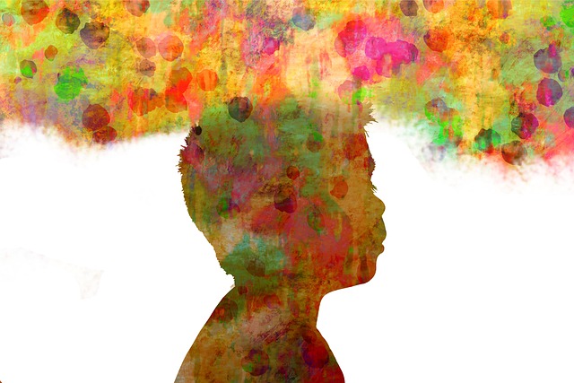
Childhood Trauma, Diet, and Behavior

5742 views

Adverse Childhood Experiences and the Diet Variable

Nearly one in eight children (12%) are reported to have had three or more negative life experiences associated with levels of stress that can harm their health and development. In 2011, nearly 60% of children age 17 and younger were exposed to violence within the year, either directly as victims or indirectly as witnesses. Twenty-one per cent of children in the United States suffer from mild behavioral health problems and an additional 11% struggle to overcome significant behavioral health, according to the Adverse Childhood Experiences study. This estimate translates into a total of 4 million youth who suffer from major mental illness (US Department of Health and Human Services 1999, 123-124). An estimated 26% of Americans aged 18 and older (about one in four or over 57 million adults) suffer from a diagnosable mental disorder in a given year.

As if this were not enough, the Children’s Crisis Treatment Center in Philadelphia indicated that “in 2011, 29% of students in grades 9 through 12 reported feeling sad or hopeless almost every day for two or more weeks in a row in a year. Up to 70% of children and teenagers in the juvenile justice system have a diagnosable mental health disorder and up to 44% of high school students suffering from behavioral health issues drop out of school”.  If these statistics are accurate, they are deplorable.

Malnutrition as a Major Cause of Brain Disease

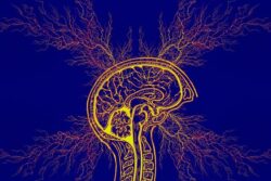
With these huge numbers involved, of many potentially causative issues, there is considerable evidence that bad nutrition may dominate them. It is interesting that, many years ago, a Probation Officer in Cuyahoga Falls in Ohio persuaded a judge to hand over to her care all the juvenile criminals that stood trial in his court. She regulated their diet and supervised it. The recidivism (habitual lapsing back into crime) dropped to almost zero. Unfortunately, good nutrition is commonly overcome by hedonism (love of pleasure) and is usually governed by the sweet taste. There are two aspects to this. Sugar in all its different forms precipitates thiamine deficiency and the signal from the tongue to the brain is responsible for its addictive qualities. There is little doubt that thiamine deficiency is heavily responsible for much of the mental disease that is so commonly represented in our culture. It is especially damaging to the lower part of the brain that governs our emotional responses and our ability to adapt to a hostile environment. Because thiamine deficiency produces an effect similar to that of oxygen deficiency (hypoxia) it has been seen as a cause of pseudo-hypoxia (false hypoxia). Either true or false hypoxia is interpreted by the brain as a potentially dangerous threat to the organism.

Some years ago I had the opportunity to visit the Philadelphia Crisis Treatment Center that then existed under another name. I learned of a neurosurgeon who had been deeply involved with its original inception. He had suggested that seizures (epilepsy) were caused by a deficiency of oxygen (hypoxia) in the brain, perhaps explaining the usual resistance of juvenile seizures to drug treatment. Although the statistics above did not specify the nature of “major mental illness” in 4 million youths, I pondered over the years whether hypoxia or any part of its equivalent mechanisms (pseudo-hypoxia) could be the underlying cause common to brain disease in a variety of different expressions, including even epilepsy in some cases.

After my visit to Philadelphia, I had an opportunity to test the neurosurgeon’s suggestion by treating a 12-year-old boy in “status epilepticus” after his current medication had been suddenly withdrawn. I gave him an intravenous injection of thiamine tetrahydrofurfuryl disulfide (TTFD), a synthetic derivative of thiamine) and this quickly stopped the continuous seizuring. I then started TTFD by oral administration but it was discontinued by a neurologist who saw the incident as “spontaneous remission and nothing to do with vitamin therapy”. Unfortunately, I did not have any data to be able to publish the case. Status epilepticus is the name given to a situation where the seizuring is continuous and often very difficult to stop. It usually occurs when a medication is suddenly withdrawn. Many years later, I discovered that thiamine deficiency could produce the same symptoms as brain hypoxia, thus giving rise to describing this deficiency as pseudo-hypoxia (false hypoxia). I had evidently treated the SE by relieving the pseudo-hypoxia in the brain cells responsible for this patient’s potentially fatal illness.

Maternal Diet and Neurological Development

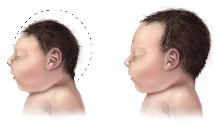
There is growing concern about the long-term neurologic effects of prenatal exposure to maternal overweight and obesity, a result of malnutrition. The causes of epilepsy are poorly understood and in more than 60% of the patients no definite cause can be determined. Authors from the well-known Karolinska Institute showed that there was indeed a relationship between obesity in pregnancy and the risk of epilepsy in the offspring. Although the mechanism is not articulated, micronutrient deficiency may be culpable. Increasingly, it has become clear that a person’s weight does not correspond to their nutritional status. Indeed, in many cases, obesity is associated with a state malnutrition; a malnutrition we call high calorie malnutrition.

Clinical thiamine deficiency is defined by both consistent clinical symptoms and either a low whole-blood thiamine concentration, significant improvement, or resolution of consistent clinical symptoms after receiving thiamine supplementation. Of 400 obese patients, 66 (16.5%) were shown to have clinical thiamine deficiency. Their symptoms included gastrointestinal, cardiac, peripheral neurologic and neuropsychiatric manifestations, the characteristic symptoms of beriberi. Hypoxia threatens brain function during the entire life span, starting from early fetal age up to senescence. A relatively common condition in newborns is lack of adequate oxygen supply to the brain and is known as hypoxic-ischemic encephalopathy. This has been shown to correlate with multiple organ dysfunction and must surely be a severe legacy in the affected child.

The outstanding question then is whether poor diet, perhaps coupled with genetic risk in some cases, could be a substantial causative factor in widespread brain illness. Dr. Marrs and I have published considerable evidence that high calorie malnutrition, by inducing thiamine deficiency, is widespread throughout America and is responsible for a variety of brain related symptoms. With an excess of simple carbohydrate calories, the action of thiamine in burning those calories is overwhelmed. Thiamine might well be in a sufficient concentration for a healthy diet but insufficient for an excess of empty calories. It is the proper calorie/thiamine ratio that results in oxidative efficiency. Unfortunately, the many symptoms produced by thiamine deficiency in the brain are not recognized by the vast majority of physicians for what they represent. If thiamine deficiency is even suspected, they find a “normal” blood level of thiamine that is the usual result in moderate deficiency. The symptoms are falsely attributed to “a more acceptable diagnosis”. No appropriate laboratory tests are usually performed and in many cases the patient is diagnosed with psychosomatic disease, without even considering a necessary underlying mechanism.

High Calorie Malnutrition and Emotional Lability

This kind of malnutrition can severely affect emotional reactions, resulting in a variety of manifestations that include persistent anxiety, depression and bizarre behavior. We have even suggested that poor emotional control can lead to expressions of violence that hitherto have had no explanation for their almost daily occurrence in America. Poverty, poor education, environmental pollution and hedonism are all components that are predictable causative agents. When energy production in the brain is compromised by inefficient use of oxygen (oxidation), the affected person is unable to muster an adequate biological response in the process of adapting to virtually any form of stress. The affected patient is also wide open to succumbing from infection by any microorganism. Brain function becomes abnormal from lack of energy drive.

This may explain the breakdown in health in the children exposed to the stress of active (physical) or passive (mental) violence referred to at the beginning of this post. The well-known saying that “we are what we eat” should be broadened to “we behave according to what we eat”. So many books have advocated the principles of healthy nutrition, without producing much overall health improvement. There is a fairly consistent refusal to “give up” sugar, mainly because its ubiquitous consumption makes it hard to understand its inherent danger to complete health and its addictive properties. Perhaps a more logical attitude might be required to the use of nutritional supplements. The pharmaceutical industry has most people attuned to consumption of pills, so that a transfer of principle would probably be easy. However, it also demands a realization that disease can be reduced to an understanding of energy deficiency as the root cause.

We Need Your Help

More people than ever are reading Hormones Matter, a testament to the need for independent voices in health and medicine. We are not funded and accept limited advertising. Unlike many health sites, we don’t force you to purchase a subscription. We believe health information should be open to all. If you read Hormones Matter, like it, please help support it. Contribute now.

Yes, I would like to support Hormones Matter.

Image by Gerd Altmann from Pixabay.

This article was published originally on January 13, 2020. 


Maternal Thiamine Deficiency and Fetal Brain Damage

8038 views

Over the last several months Dr. Lonsdale and I have been working on a book about thiamine deficiency and dysautonomia. Last week I wrote about the presumed connection between the Zika virus and microcephaly where I hinted at a thiamine connection. One might say, that I have thiamine on the brain and that would be a fair assumption. The old adage, ‘if one has a hammer, everything becomes a nail‘, may apply. I may be focusing too much on thiamine and its role on mitochondrial health. Alternatively, it could be that thiamine is just that important. After all, it sits atop at least two of the four energy producing pathways that give us ATP and is deeply embedded within the remainder of the oxidation process. The consequences of impaired oxidative metabolism in the brain are vast and include a range of disease processes like Alzheimer’s disease, amyotrophic lateral sclerosis (Lou Gerhig’s disease), Parkinson’s disease, multiple sclerosis, alcoholic brain disease, and stroke.  Without thiamine, the mitochondrial factories stop producing energy or ATP and without ATP, stuff slows and then dies. So yes, thiamine is critical to health.

It is not difficult to imagine what happens to energy levels when thiamine concentrations diminish even slightly in an adult. An unrelenting fatigue is one of the early symptoms of struggling mitochondria and thiamine deficiency. More fundamentally, however, all the organs tasked with maintaining life, demand energy. When energy stores diminish, those organ systems struggle. The organ systems requiring the most energy, like the brain and the heart, are hit hardest. Maintain a slight deficiency chronically and damage ensues. In Cuba, for example, trade embargo policies resulted widespread thiamine deficiency in the population, which in turn initiated an epidemic of neuropathy – nerve damage. Over 50,000 Cubans were reported to have developed optic neuropathy, deafness, myelopathy, and sensory neuropathy related to embargo imposed dietary changes. In contrast to the more insidious damage initiated by chronically low thiamine concentrations, severe and acute thiamine deficiency is life-threatening, especially in children, but also in pregnant women.

With low maternal thiamine concentrations, the effects on fetal development, especially fetal brain development that requires enormous amounts of energy, are likely to be devastating. And indeed, they are. But we don’t study that very often, even in rats. Do a search on the subject and there is not much research out there. Sure, some researchers have investigated maternal thiamine deficiency in fetal alcohol syndrome (FAS), postulating thiamine might be the mechanism by which FAS develops, but that is about it. Given how critical it is to fetal development, I expected more research.

It is not just alcoholics who are at risk of thiamine deficiency. An increasing percentage of Western populations are likely thiamine deficient. Thiamine depletion occurs with numerous medications and vaccines via multiple mechanisms, many of which are just beginning to be understood. Conventional farming practices use herbicides and pesticides that block vitamin B absorption and so even diets presumed healthy may not be as nutrient dense as in the past. Poor absorption from altered gut microbiomes may be another common mechanism for thiamine deficiency and emerging evidence finds that Type 1 and Type 2 diabetics excrete significantly more thiamine than non diabetics, making them thiamine deficient as well. Not studying this more broadly is leaving millions of folks to suffer with entirely preventable disease processes. During pregnancy, however, this lack of recognition and research is just downright negligent, especially when we consider fetal brain development.

Thiamine During Pregnancy

Thiamine is absolutely critical for both maternal health and fetal development. Women with hyperemesis gravidarum, excessive vomiting during pregnancy, are at a particularly high risk for thiamine deficiency and though there is increasing awareness of maternal Wernicke’s encephalopathy during pregnancy, a condition typically associated with thiamine deficient alcoholics, the full scope of damage associated with maternal thiamine deficiency is insufficiently understood. There is little to no appreciation of the long term effects on maternal health and even less recognition of how the deficiency impacts fetal development in either the short or long term.

Provided mom survives a thiamine deficient pregnancy, what happens to the growing fetus? In 37% of the cases of severe maternal thiamine deficiency, spontaneous fetal loss occurs. If thiamine is critical for mitochondrial energy production, and fetal development requires exorbitant amounts of mitochondrial energy, what happens if one of the key components to that energy production process is lacking? All sorts of things, it turns out, including microcephaly. Beyond a rare congenital defect in thiamine transport believed to affect only consanguineous Amish, there are very few studies that have considered the effects of epigenetic and more functional maternal or fetal thiamine deficits. We know from the Amish cases, that when the fetal thiamine transporters are impaired, microcephaly ensues. Is it so hard to imagine that we might impair those transporters epigenetically or reduce maternal thiamine concentrations functionally by dietary choices, medications or environmental toxicants that leach nutrients and/or by malabsorption?  And yet, as I dig into this, I find only a few studies that have addressed maternal thiamine and fetal brain development. Here they are.

Maternal Thiamine Deficiency and Fetal Brain Damage

A 2005 study from researchers in West Africa showed that the pups from thiamine deficient dams, had significantly smaller brains by weight. Digging deeper, they found far fewer neurons in the hippocampus, the region of the brain responsible for memory consolidation and retrieval, than the pups from thiamine sufficient diets. Brain damage in the offspring could be induced by maternal thiamine deficiency either leading up to, during, or after pregnancy (while lactating) but varied in scope, severity, and pattern. The most significant damage occurred when the dams were deficient during pregnancy.

In the offspring from perinatal thiamine deficiency, hippocampal volume was reduced by almost a third due to neural cell death.  The neurons that survived were smaller than normal and misshapen. The hippocampus is critical to memory. Hippocampal damage in human adults causes all manner of amnesias and aphasias (speaking and language comprehension deficits) and is found in neurodegenerative disorders like Alzheimer’s disease.

The neurons affected most by the thiamine deficiency, the CA1 neurons, are especially susceptible to oxidative damage and insult. Thiamine is integral to brain oxidation and so this makes sense. What we have to remember though, is that in a fully developed human brain, oxidative damage to the CA1 region is associated with hippocampal ischemia, limbic encephalitis, status epilepticus, and transient global amnesia – very serious conditions. To a developing brain, requiring vast amounts of energy to grow, the consequences of hippocampal deficits are largely under-recognized except again in fetal alcohol syndrome.

Another animal study looked at the effects of maternal thiamine deficiency to the cerebellum of the offspring. The cerebellum is the region of the brain responsible for balance and coordinated motor movements. Here again, the damage was severe with a significant reduction of size, loss of neuron viability, and conduction. There have been a smattering of studies across the decades (here, and here, for example) looking at thiamine deficiency and brain damage in non-pregnant rats, but that’s about it.

Not much else is out there.

From these few animal studies, the work on Amish microcephaly and the work connecting neurodegenerative disorders to thiamine deficiency, we can surmise that thiamine is essential to brain development. More specifically, in pregnancies where thiamine concentrations are low, cerebral development of the offspring will be impaired in some pretty significant ways. Namely, the number and size of neurons is reduced, and as a consequent, total brain volume is reduced. If the deficiency is severe enough, microcephaly is possible and has been identified in the two of studies mentioned above. I think this is what is happening in Brazil. That is, a combination of seemingly unrelated factors, coalesce to produce fetal thiamine deficiency which results in microcephaly and other sorts of brain damage. The questions that remain include:

  1. By what mechanisms specifically is thiamine deficiency produced?
  2. What are the risks for maternal thiamine deficiency in other regions?

One of the most direct routes to thiamine deficiency during pregnancy is hyperemesis gravidarum, excessive vomiting. Case studies abound where it is often not recognized until the mother is in critical condition. It is considered a rare complication, but is it? Unless and until those questions are answered more fully and physicians recognize maternal thiamine deficiency as a potential problem, women and children will continue to be at risk for what are entirely preventable complications of pregnancy.

We Need Your Help

More people than ever are reading Hormones Matter, a testament to the need for independent voices in health and medicine. We are not funded and accept limited advertising. Unlike many health sites, we don’t force you to purchase a subscription. We believe health information should be open to all. If you read Hormones Matter, like it, please help support it. Contribute now.

Yes, I would like to support Hormones Matter. 

Image credit: Centers for Disease Control and Prevention, Public domain, via Wikimedia Commons

This article was first published on June 16, 2016. 

Sudden Infant Death Syndrome, Autism, and Maternal Thiamine Deficiency

5830 views

I have addressed this problem before on this website. However, in the Wall Street Journal on Tuesday, March 27: an article appeared entitled “A Study of Sudden Infant Deaths Divides Doctors“. It goes on to say “The finding on sudden and unexpected infant deaths is surprising, says Joel Bass, of the pediatrics department at Newton Wellesley Hospital in Newton Mass”, the first author of the study, who   said “that’s more than one newborn baby dying of SUID [sudden and unexpected infant death] a day”. Oddly enough, the article indicates that “among the possible causes, some researchers point to the promotion of certain hospital practices to encourage breast-feeding. But some doctors vehemently disagree with that theory”. Apparently, the theory alluded to the practice of encouraging skin-to-skin care between mother and infant during breast feeding. I had not read the article so I do not understand how this connection was made. What arrested my attention was the reference to breast-feeding, long encouraged by pediatricians as the best way to feed newborn infants. How could skin-to-skin contact be involved? However, an article was published in 2011 in which the author had carefully examined the nutrient content of breast milk in the United States (Shamberger, 2011). It reminded me of an extremely important article that was written in the 1940s.

Breast Milk Toxicity Syndrome

It may be remembered that Hong Kong at that time was a British protectorate. A medical officer of health was sent out from Britain to Hong Kong to investigate a relatively common occurrence of sudden death in breast-fed infants of Chinese mothers. This death commonly occurred between three and four months of age and happened in infants that, from their appearance, were considered to be the healthiest in the family. Fehily, the author, was able to show that the breast milk was deficient in vitamin B1 and it was well known by early researchers of beriberi that this form of infant death was virtually pathognomonic (indication of cause) of the infantile form of this disease. In fact it was well known by these early researchers that there was no other disease of infancy that behaved like this. Fehily herself was struck by the exact likeness to “cot death”, the term used in England for what we call Sudden Infant Death Syndrome in the United States today.

Autism Rates for Each State Connected to Maternal Nutrient Status

The paper published in 2011, referred to above, was in consideration of the cause of autism, not SIDS. The author had studied the rate of autism associated with nutrition. The paper stated that autism rates in the United States are increasing at a rate of 15% per year. The study abstract reported that the design of the study used nutritional epidemiology and an ecologic study design. In other words, he studied public health data and the nutrient content of breast milk. The objective was to try to link the possible cause of autism to nutrition by creating autism rates for the 50 states of America and comparing them with published measures of infant nutrition. These included the duration of exclusive breast-feeding and participation in the Women, Infants and Children (WIC) program. The results were impressive. The states with the highest WIC participation have significantly lower autism rates (P <0.02).

In contrast, there was a direct correlation with the increasing percentage of women exclusively breast-feeding from the years 2000 to 2004. Infants who were solely breast-fed had diets that contained less thiamine, riboflavin, and vitamin D than the minimal daily requirements. Although the author was studying the rates of autism and was not in the least interested in SIDS, his study supports the finding of vitamin deficiency in breast milk, in turn supporting the possible relationship between breast-feeding and SIDS. Of course, the modern medical model would find it objectionable to hypothesize a cause common to two diseases, although both autism and SIDS have a slight male dominance and are diseases that occur during rapid growth, particularly of the brain.

My Particular Interest in SIDS

Many years ago, because of clinical experience, I became interested in SIDS. At about that time, the idea of threatened SIDS had become an acceptable diagnosis, whereas previously it had been considered that the death was truly sudden and completely unpredictable. I can remember two parents who had brought their infant to see me. They had observed him in his crib when he had stopped breathing. When one of them picked him up he started breathing again. They took him to the nearest emergency room where he was pronounced completely fit and the situation was dismissed. The parents were so scared that one of them took turns to sit up all night to watch in case this thing should happen again.

This is exactly the clinical situation that I began to experience with other infants. A monitor had been invented that could be attached to an infant that would sound an alarm if either his heart slowed or his breathing stopped. It had become well-known that picking the infant up or giving him a little slap on the buttock would instantly return him to a normal state (SIDS is more common in male infants). To cut a long story short, my colleagues and I performed a lot of clinical research on these threatened SIDS babies. We had found that a machine known as brainstem auditory evoked potential (BAEP) gave abnormal readings . This strongly suggested that the mechanism was an electrochemical dysfunction in the brainstem. This is the part of the brain that connects with the spinal cord and it contains the controls for automatic breathing. Let me explain this a little.

Most of us are not aware that our essential breathing is controlled by centers in the brainstem when we are unconscious, as in sleep. The condition known as sleep apnea is a failure of this mechanism (apnea is a temporary cessation of breathing). It was during my library research that I discovered the 1944 paper by Fehily. It seemed only to be common sense to look at the possible association of thiamine deficiency as the underlying biochemical cause. We treated several infants with thiamine injections that seemed in every case to stop their episodes of apnea. I received a visit from a university researcher in Australia. His group had been studying SIDS and they had come to the same conclusion. I was so impressed by their work that I took sabbatical leave in Australia with Dr. Read who was the leader of the group. Abnormal brainstem responses in infants at risk of SIDS was reported at about the same time. The authors said that this suggested immature development of brainstem. A recent publication found an association between maternal alcohol consumption and SIDS. The relationship of thiamine deficiency in brain and alcohol consumption is well-known. It is not too difficult to imagine that the concentration of alcohol in the blood of the mother would have an exaggerated effect in the brain of the fetus, perhaps injecting a risk factor for the infant after birth.

Conclusion

Can a parent who has not experienced the sudden death of a precious infant at the age of 3 to 4 months even imagine the horror and the enormous stress imposed on such a parent? We now know that there are quite nonspecific symptoms that may appear in an infant who threatens SIDS. They are sometimes unusually irritable and a “runny nose” may be mistaken for a cold, but such symptoms may be so slight as to be ignored. I must emphasize that the lower part of the brain that organizes automatic body functions, including breathing, is peculiarly sensitive to thiamine deficiency. If the infant is breast-fed with thiamine deficient breast milk as the 2011 study suggests, studies that I have reported here would make sense. Even if only a few of the cases were due to this biochemical phenomenon, giving some thiamine on suspicion can do no harm.

A diet history from the mother might offer clues, particularly emphasizing whether there is ingestion of sugar in all its different forms. If I were a physician in charge of such a pregnancy, I would not hesitate to add thiamine supplementation, starting as early as three months of gestation, as advised in the book written by an American obstetrician/gynecologist by the name of John B Irwin, M.D. He had found that supplementary thiamine removed the complications of pregnancy such as toxemia and even prevented premature delivery. It might well provide a nutritional legacy for the infant. Of course, I am not suggesting that alcohol ingestion by the mother is THE cause of SIDS. However, I am suggesting that the present widespread use of unhealthy nutrition may well be at fault for both SIDS and autism. Genetic risk, coupled with some form of stress (e.g. mattress flame retardants, a cold virus) and vitamin deficiency breast-feeding, might be more or less important individually, or more than one of the three items collectively.

We Need Your Help

More people than ever are reading Hormones Matter, a testament to the need for independent voices in health and medicine. We are not funded and accept limited advertising. Unlike many health sites, we don’t force you to purchase a subscription. We believe health information should be open to all. If you read Hormones Matter, like it, please help support it. Contribute now.

Yes, I would like to support Hormones Matter. 

Autism and Maternal Nutrients: An Unrecognized Connection

3459 views

Autism is a neurodevelopmental disorder characterized by impaired social interaction, verbal and non-verbal communication and restricted and repetitive behavior. Parents usually notice signs in the first two years of their child’s life. Autistic Spectrum Disorder (ASD), although classified as a distinct entity, probably represents “variations on a symphonic theme”. On February 22, the School of Public Health, Columbia University reported a study that women actively infected with genital herpes during early pregnancy had twice the odds of giving birth to a child later diagnosed with ASD. Researchers for the Kaiser Permanente health-care group examined the medical records of 594,638 children. The lead author reported that, of 6,255 who had been diagnosed with autism, 37% had experienced complications during pregnancy or delivery. Issues during birth, such as oxygen deprivation, increased the risk by 10%. Infants born to mothers who had pre-eclampsia or other pregnancy-related complications had a 22% greater risk. For those who experienced complications both before and during delivery, the risk increased by as much as 44%.

Preeclampsia, Vitamin B1 Deficiency, and Autism

The reference above to pre-eclampsia or other pregnancy -related complications caught my attention. About two years ago, I received a three-page letter from an extremely frustrated specialist in OB/GYN by the name of John B. Irwin. He also enclosed what I consider to be an extremely important book that has almost certainly been ignored by the medical profession. He opened his letter by saying “I am writing to you because I have found another mortal being who is interested in the biological activities of thiamine” (vitamin B1). In his letter he mentioned that for 25 years, during his retirement he had been concentrating on the use of megadose thiamine (100 mg/day) in pregnancy. He had hired himself out to the government of the Commonwealth of the Northern Mariana Islands to improve on their system of obstetrical care. His first job was to attend a meeting with a group of island doctors who were all American Board-Certified in OB. Having heard of Dr. Irwin’s interest in thiamine they introduced him to a young woman in the 36th week of pregnancy. She had severe preeclampsia, heart disease and early signs of premature separation of the placenta. Spontaneous labor and delivery would most likely cause loss of mother and infant and it was impossible to lay her flat for cesarean section because of her difficulty in breathing. In short, the island doctors were testing him. In a private huddle “the doctors apparently decided that if the patient died while they were holding me up, they would be found solely guilty, so with anger, sneers and audible comments they told me to go ahead” by using a 100 mg pill of thiamine daily she was cured in six days, sleeping flat and hiking the long halls of the hospital for exercise to shake off her prolonged immobilization. He delivered a 3 lbs. 12 oz. infant with a normal Apgar score. Dr. Irwin started clinic patients on mega-thiamine and this prevented development of every type of toxemia completely, including eclampsia, preeclampsia, intrauterine growth retardation, premature delivery and gestational diabetes. I would certainly suggest that this book would be compulsory reading for anybody who contemplates pregnancy or is pregnant: The Natural Way to a Trouble-Free Pregnancy: The Toxemia/Thiamine Connection.

 Vitamin D Deficiency and Autism

Children who are, or who are destined to become, autistic have a lower vitamin D level at birth and at age 8 compared to their unaffected siblings.  Two studies found that high-dose vitamin D improves the symptoms of autism in about 75% of autistic children. A few of the improvements were remarkable. The vitamin D doses used in these children were as much as 300 IU/KG body weight/day to a maximum of 5000 IU/day. These doses may be surprising to some readers but there have been tremendous advances concerning the role of vitamin D in the human body. It is not a vitamin in the true sense since it is manufactured in the skin by exposure to sunlight.

In Utero Nutrient Deficiencies 

The infant in utero is completely dependent on nutrients supplied by the mother. It has been shown that some cases of autism are induced in pregnancy and are a direct reflection of the mother’s overall health. Folic acid supplementation was shown to prevent neural tube defects in the infant and we now have evidence for the importance of sufficiency in vitamin D and vitamin B1, adding to the preventive obligations in prenatal care. We have become used to the idea that a given disease such as autism has a specific cause. When we find that two different vitamin deficiencies give rise to the same disease, we have to explain the connecting link.

Biochemical Lesions

In 1936, Sir Rudolph Peters published his studies on thiamine, spearheading the research that led to our better understanding of the complexities of oxidative metabolism. He formulated the idea of a “biochemical lesion”, indicating that a breakdown within cells is due to a failure in chemistry. Since both of the vitamins depicted here are critical in the normal function of all our cells, particularly those of the brain, we can suggest that autism is caused by variable biochemical lesions, any and all of which produce dysfunctional cells. The symptoms generated are a result of how many cells are affected by the biochemical lesion and the effect that has on function.

The Critical Role of Nutrition During Pregnancy

Cellular chemistry cannot function properly without a huge series of nutrients that we must ingest every day. It is no longer sufficient to make a clinical diagnosis in descriptive terms. I suggest here that a constellation of symptoms may have 5 or 10 different biochemical lesions. On the other hand, a single biochemical lesion may project itself in 5 or 10 different ways, producing a variable constellation of symptoms, depending upon the distribution of the change in biochemistry. It indicates that we must look a lot harder for evidence of nutrient deficiency as the potential underlying truth. Is it possible that the mother would not succumb to genital herpes, or if she were infected, not perpetrate havoc on her yet unborn child if her body chemistry was in an ideal state?

We need your help.

Hormones Matter needs funding now. Our research funding was cut recently and because of our commitment to independent health research and journalism unbiased by commercial interests we allow minimal advertising on the site. That means all funding must come from you, our readers. Don’t let Hormones Matter die.

Yes, I’d like to support Hormones Matter.