mitochondria - Page 4

Alternative Medicine 101 and the Fuel to Energy Equation

2116 views

Whether we like to recognize it or not, the human body is a “hybrid”, meaning that it burns fuel and uses electricity for function. I have found that trying to explain Alternative Medicine is a very difficult task and so I am going to try to do it by using an advanced analogy. I am going to compare the human body with an automobile since both are machines that consume energy.

Of Engines and Fuel

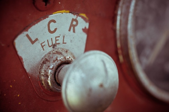
At the average gasoline station, there are three different fuels to choose from. Each is calibrated to engine design. The owner of the automobile knows what fuel should be purchased because he has read the owner’s manual and was instructed when the car was purchased. Knowing which fuel is optimal and choosing that fuel are two different things. We know very well that putting the wrong gasoline into the car is not good for the engine, but oftentimes because of cost, we ignore this advice. We choose a lessor grade fuel. Hence we have millions of cars, some of which may break down because of this failure of choice.

Human beings have evolved from a more primitive species about which we know surprisingly little. However, we are all perfectly aware that the fuel was available throughout our developmental history. From our teeth design we are known as omnivores (an animal that can eat both meat, vegetables and fruit). Very early in our career we were “hunter gatherers”, meaning that we hunted animals for meat and gathered the nuts seeds and vegetation that were available as food. This is still the fuel that was designed for our machinery. Until very recently in our history, there was no food industry to supply us with the variety of processed foods that we have today.

Fuel Storage, Processing, and Transmission: Mechanics 101

Gasoline is stored in a tank: it does not run directly into the engine and it is not consumed until the engine is running.

Similarly, natural food yields a proper combination of protein, fat and carbohydrate, all of which are processed in the body. The primary fuel is glucose, extracted by chemical processes in the body from the food source. This is conveyed by the blood to the liver where it is turned into a more complex, multi-glucose molecule called glycogen for storage, equivalent to a gas tank.

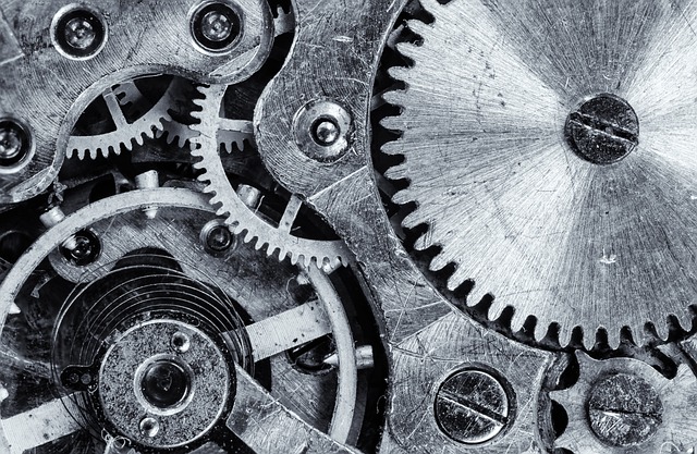
Gasoline is directed from the tank into cylinders that contain pistons. The gasoline is ignited and the explosion drives the pistons connected to the transmission, thus passing the energy to the wheels. The only function required by the car is to move. Note that the engine is producing energy: the transmission is consuming it.

For the body, the process is more complicated but the principles are the same.  All cellular action requires the glycogen to be broken down and released from the liver as glucose that now becomes the fuel source, conveyed by the blood throughout the body. This is an important step because storage of glucose in this manner enables it to be released in proportion to the need for action. It might be compared to fuel injection in an automobile.

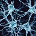
The body consists of 70 to 100 trillion independent cells, all of which require energy to function. Each cell contains microscopic organelles known as mitochondria where energy is generated. This is done by  “burning” glucose that is “ignited” by the action of vitamin B (Glucose + oxygen + vitamin B yields energy). The energy derived from  ignition (oxidation) of glucose  is stored in the form of a chemical compound known as ATP (adenosine triphosphate).  Although an imperfect analogy, ATP can be thought of as somewhat like a battery. Every action, every function of the human body is dependent on this production of energy. ATP is often referred to as the currency of energy transmission

The automobile’s transmission is responsible for passing the energy derived from the engine to the wheels.

In comparison, it is the function of mitochondria to create energy. The next question is how that energy is used. Here there is a striking difference because the body uses that energy in renewing itself as well as dictating function. It uses energy consuming enzymes, chains of which are capable of converting a given substance A to another substance B to C etc. Chains of enzymes carry out the structural details of building new cells as the old ones die off. This is the equivalent of a transmission and energy is consumed.

Energy In Must Match Energy Out

From theses analogies, we can see that all the functions of the human body are detailed in terms of energy production and energy consumption. The production must always keep up with consumption. If the production is inadequate or the consumption is excessive, changes in cellular function follow. If that functional change is not spotted and treated by filling the deficit, cellular damage follows. This must be the basis of true preventive medicine

What about the driver of the car?

Well, until the driverless car becomes a reality, a human being must direct the passage of the car. If we extend the analogy, the driver is the brain that guides the car to its destination.

What about the driver of the body?

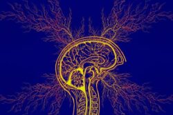
It might be said that the brain is the driver of the body. The body is merely a chassis that enables the brain to move around. It is the personality of every human and nothing happens in the body without the brain. Indeed, the brain can be compared to the conductor of an orchestra. The organs in the body are like banks of instruments, and like instrumentalists, they all know exactly what they have to do but require guidance from the conductor. In order to understand this analogy, the lower part of the brain organizes and computes the functions of all body organs through a nervous system known as autonomic (automatic) and a bunch of glands known as the endocrine system. The autonomic system is a two-way street, used for messages into the brain and out to the organs. The endocrine system produces hormones which act as messengers of the brain. When all of this works appropriately, the “symphony of health” follows. The brain uses a disproportionate amount of energy as compared with the rest of the body. It is therefore not surprising that brain disease is a reflection of a defective energy equation.

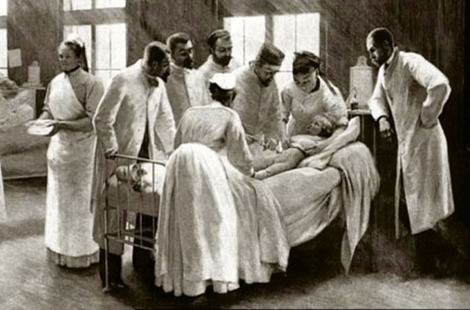
The present approach in medicine is to kill the offending bacteria and viruses that attack us. Alternative medicine, recognizing that the body possesses sophisticated defenses, seeks to assist those defenses by stimulating energy production by the use of nutrients. In other words, alternative medicine looks at correcting the energy equation as the basis for health.

We Need Your Help

More people than ever are reading Hormones Matter, a testament to the need for independent voices in health and medicine. We are not funded and accept limited advertising. Unlike many health sites, we don’t force you to purchase a subscription. We believe health information should be open to all. If you read Hormones Matter, like it, please help support it. Contribute now.

Yes, I would like to support Hormones Matter. 

Photo by Timon Studler on Unsplash.

Vitamins D3 and K2: Power Partners for Mitochondrial Health

6447 views

One of the more common questions regarding vitamin D supplementation is whether or not to supplement with vitamin K also. In this post, I will address those questions as well as look into the roles of these vitamins in mitochondrial function.

If you recall your high school chemistry, mitochondria are the powerhouses of our cells. Their production of cellular energy, or adenosine triphosphate (ATP), depends primarily upon fuel derived from nutrients including vitamins D3 and K2. These two nutrients are co-factors, or partners, synergistically ensuring strong bones and teeth as well as a cardiovascular system void of lingering, plaque-induced calcium.

Why Is Vitamin D3 Important?

Adequate vitamin D3 is essential to our health and quality of life. Every cell in our body contains a vitamin D receptor (VDR). When a VDR is activated by a sufficient intake of vitamin D3, a number of good things happen. Vitamin D3’s mechanisms of action include: anti-microbial, anti-cancer, and anti-inflammatory. Moreover, scientific research suggests that vitamin D3 deficiency is connected to a wide array of serious medical conditions such as cancer, diabetes, mental health disorders as well as multiple sclerosis and other autoimmune diseases.

Due to our modern lifestyles and conventional medical practices, we tend to get little vitamin D3 from its natural source, the ultraviolet B, or UVB, light of the sun. From living, commuting, and working indoors to fretfully slapping sunscreen all over our skin, we appear intent on denying ourselves this essential nutrient.

The re-emergence in the twenty-first century of rickets, a Victorian-era disease causing soft bones primarily in children, calls to action the need for adequate vitamin D3 supplementation. As most diets are severely lacking in vitamin D3, the most practical way of getting adequate amounts of this nutrient is by taking an inexpensive daily, oral D3 supplement. My Facebook support group called “Vitamin D Wellness” provides information about an easy-to-follow protocol designed to increase D3 levels to an optimal value of 100 ng/mL (250 nmol/L).

The Importance of Vitamin K

Before discussing vitamin K2, I would be remiss if I did not mention the first form of vitamin K. Vitamin K1 is literally a vital nutrient. Without vitamin K1 our blood would not clot, and we could bleed to death. The good news is that vitamin K1 (or phylloquinone) is present in all green plants that acquire energy from sunlight. Green leafy vegetables including spinach, kale, collards, broccoli, and brussel sprouts abound with vitamin K1. Better news: vitamin K1 constantly recycles in our bodies so deficiency is rare.

Vitamin K2 (or menaquinone) however differs greatly from K1. There are two forms of vitamin K2: menaquinone-4 (MK-4) found in grass-fed animal protein including meat, egg yolk, butter, some cheeses, and calf’s liver.

A fermented soybean called natto, commonly consumed in Japan, is abundant in the more potent form of vitamin K2 called menaquinone-7 (MK-7). For those who do not want to consume soy, vitamins K2 MK-7 supplements made from chickpeas and formulated with coconut oil are readily available. My Facebook group protocol “Vitamin D Wellness” includes guidance on K2 MK-7 supplementation.

Health benefits of adequate vitamin K2 levels include potential prevention of osteoporosis, arterial plaque, and dental issues. Vitamin K2 moves calcium to the bones and teeth, as well as sweeps calcium from soft tissue lining such as arteries. Specifically, vitamin K2 activates proteins (osteocalcin and matrix gla protein) produced by vitamin D3 that facilitate moving calcium to where it belongs: the bones and teeth.

Low vitamin K2 levels are common. First, unlike vitamin K1, vitamin K2 is not recycled in the body. Second, the vitamin’s natural sources are lacking in most diets. Owing to the preponderance of industrial farming in many parts of the world, many people are low in vitamin K2. When insufficient vitamin K2 is in the blood stream, calcium can linger along arterial pathways potentially causing calcification, the process whereby calcium deposits form plaque accumulating in the cardiovascular system. These calcium deposits can cause heart disease.

The Vitality of Vitamins D3 and K2

Specific research on vitamin D3 and vitamin K2’s association with mitochondrial function remains limited yet fruitful:

Published in a 2013 issue of the Journal of Clinical Endocrinology and Metabolism, a Newcastle-upon-Tyne University, UK study led by Dr. Akash Sinha demonstrated, for the first time, a connection between vitamin D3 intake and mitochondria function in human skeletal muscles. Sinha and his team concluded that muscle function, including muscle fatigue, improved with vitamin D3 supplementation.

The positive effect of vitamin K2 on apoptosis, or natural cell death, in various types of cancer cells has been established. For example, researchers in China discovered the apoptotic influence of vitamin K2 on bladder cancer cells through mitochondrial pathway. Their findings are reported in an August 2016 edition of the journal PLoS One.

In summary, the powerful partnership of vitamins D3 and K2 is essential to mitochondrial health. By enjoying optimal daily intake of D3 and K2, you are more likely to have strong bones, teeth, and muscles as well as a calcium-free cardiovascular system. Oh, and your risk of developing cancer is diminished!

Do You Need to Supplement Vitamin D3 and K2?

Given the paucity of vitamins D3 and K2 in our diets, I think daily supplementation provides the most effective way to increase and maintain these nutrient levels. I find taking a daily dose of 10,000 iu of vitamin D3 and 90-120 mcg of vitamin K2 MK-7 soft gel capsules with or after breakfast works best for me. What an easy way to enjoy the benefits of this powerful partnership of mitochondrial nutrients!

We Need Your Help

More people than ever are reading Hormones Matter, a testament to the need for independent voices in health and medicine. We are not funded and accept limited advertising. Unlike many health sites, we don’t force you to purchase a subscription. We believe health information should be open to all. If you read Hormones Matter, like it, please help support it. Contribute now.

Yes, I would like to support Hormones Matter. 

Semmelweiss Syndrome: Ignoring the Obvious to Save Face

2906 views

Who was Semmelweiss? Please read the post below to find out. We have an urgent message for both patient and physician. It isn’t really new because it started with Hippocrates in 400 BC “when he said ”let food be your medicine and medicine be your food”. He also said that no kind of treatment should ever be used that may do harm. All you have to do is to read the Physicians’ Desk Reference and read about almost any drug. It begins with a short sentence about what it must be used for. Then it very often states that nobody knows why the drug works. What follows is a lengthy discussion of side effects, pure evidence of its toxicity. The potential for harm is implicit. Hippocrates realized that healing was a natural process within the body itself and that any treatment method should assist this process. Today, we are well aware that this healing process requires a huge amount of energy that must be conserved and focused by resting body and mind. Modern  biochemistry provides us  with facts that show the wisdom of Hippocrates when he said “let food be your medicine”!

An Obvious Fallacy in Modern Medicine

Healing is a natural process from within the body that operates best when it is focused. The modern hospital, with its constant clatter and its complete failure to give the patient the rest and tranquility required for energy conservation, works against the healing process. The question that we must ask, is how we should proceed in order to assist healing. The answer must be obvious: help to supply the means by which energy is produced and conserved in the body. Health is a balance between the attack of environment (the enemy) and the constant mobilization of adaptive function that requires energy to maintain functional efficiency (the defense)

Alternative Medicine

This is the philosophy that has given rise to the development of a small group of physicians that use nutrient-based medicine. The point is this: modern medicine attempts always to kill the enemy. Kill the bacteria, the virus, the cancer cell, without killing the patient. Alternative medicine respects this only if it does no harm to the patient. The main approach of these physicians is to anticipate the body’s requirements in all its different ways in attempting to heal itself, the philosophy that was put forward by Hippocrates. These physicians often find that they are excluded from the conventional medical hierarchy, spurned by their former colleagues and despised. When they have a brilliant success with a patient, it is invariably labeled as “spontaneous remission” and we have wondered why this seems to be so inevitable in human affairs.

The Story of Semmelweis

The apocryphal story is that of Ignaz Semmelweiss who was a European physician who practiced before microorganisms had been discovered. He observed that doctors came in from the morgue and delivered their patients without washing their hands or changing clothes. The puerperal fever (childbed fever) rate was excessively high, of course. Semmelweiss concluded that the doctors must be bringing something in on their hands. He divided the ward into two sections and directed that doctors attending patients on one side should wash their hands in chlorinated lime before they performed a delivery, whereas the doctors on the other side should continue in the same old way. It did not need a statistician to see the difference in the incidence of infection. But what happened to Semmelweiss was just as predictable. He had done something new that disagreed with the medical establishment of the day. He was accused of being unscientific by suggesting that an unknown substance on the hands of the doctors was the cause of the problem. He was fired from the hospital and eventually died in a mental institution. He was right and they were wrong. The truly amazing thing is that the medical establishment never bothered to look at the results and times have not changed.

Nutrients Matter: It’s That Simple

People who have to admit their loved ones to hospital as an emergency, knowing and understanding the dramatic effect of nutrient-based treatment, are almost always completely powerless to get the attending physician even to listen to them. A physician of my acquaintance had a patient in hospital with pneumonia due to a resistant bacterial organism. He gave the patient nutritional IVs with water soluble vitamins. The patient recovered. In the next bed was a patient with exactly the same pneumonia who was under the care of another physician. My acquaintance approached what should have been his colleague and suggested that he do the same treatment, using the vitamin therapy. He was told flatly to mind his own business. The patient died. You would think that the second physician would want to discuss the reasoning and the scientific evidence to support the action. Was the belief in his own competence (ego) more important than the life of his patient? This is happening today in the world of medicine. We have come to accept it as the great “Bonanza” of scientific advance. Readers should be aware that the leadership for change will not come from the medical profession. It will come from those most interested in solving those personal health problems that have defied solution, sometimes for years. This website can spread the good news that there are often alternatives to be sought.

We Need Your Help

More people than ever are reading Hormones Matter, a testament to the need for independent voices in health and medicine. We are not funded and accept limited advertising. Unlike many health sites, we don’t force you to purchase a subscription. We believe health information should be open to all. If you read Hormones Matter, like it, please help support it. Contribute now.

Yes, I would like to support Hormones Matter. 

Image: An injection against croup at the Hôpital Trousseau, Paris. Photogravure by Bruun Clement, 1899, after P.A.A. Brouillet, 1893. Wellcome Collection. Public Domain Mark. Source: Wellcome Collection.

A New Model for Medicine

3779 views

What is a Medical Model?

 In the Oxford English dictionary the word model is defined as “design to be followed, style of structure”. Then it follows that there must be a model to distinguish health from disease, that differentiates the two states of being. No disease can be treated without knowing exactly what caused it. Let us go back to Hippocrates, 400 BCE, who said “let food be your medicine and your medicine be your food”. What Hippocrates was saying was essentially that nutrition was the core issue in the maintenance of health. At this time and throughout the Middle Ages there was no model for the cause of disease. Consequently, treatment was extremely primitive and almost purely empirical. In the time of ancient Egypt it was believed that mental illness was caused by the presence of evil spirits in a person’s head. They bored holes in the skull to let the evil spirits out. If you think about that, perhaps it relieved the occasional headache because of increased pressure in the skull caused by a brain tumor. Hence, a few successes might have caused it to be retained as beneficial. During the Middle Ages, the only treatment that seems to have been used is bloodletting. It might have been temporarily useful in people with high blood pressure. A few successes yielded the conclusion that it was beneficial for all disease.

The First Controlled Experiment

Semmelweis was a 19 th century Hungarian physician. In those days, the incidence of puerperal disease (childbed fever) was absurdly high. Semmelweis made the observation that doctors, delivering their patients, entered the delivery room and went directly to their patients without changing their garments or washing. He came to the simple conclusion that the doctors were bringing something in with their hands that caused the problem. The obstetric ward consisted of a number of beds on each side of the room and Semmelweis directed that doctors delivering their patients on one side should wash their hands in chlorinated lime, while doctors on the other side of the room would continue in the old way. Of course, the incidence of childbed fever was so different that it did not need a statistician to document the difference. Semmelweis’s observations conflicted with the established scientific and medical opinions of the time, particularly as he was unable to explain what was on the hands of the doctors. Some doctors were even offended at the suggestion that they should wash their hands. It is truly an amazing vision of human behavior. Innovation carries with it loss of reputation for the innovator, no matter how successful the innovation. Well, of course everyone today knows that it was microorganisms on the hands of the doctors that caused the disease, but they had not yet been discovered. Poor Semmelweis wound up in a lunatic asylum and died in his 40s after a beating by attendants. Today, he is regarded as the first person to introduce antiseptic medicine.

The First Paradigm in Medicine: Microscopic Organisms

Most people are aware that the invention of the microscope, and the work of historical figures like Louis Pasteur, led to the discovery of organisms, that could only be seen with the microscope, caused what we now call infection. We are all familiar with the fact that a tremendous number of diseases are due to infection by bacteria, viruses or fungi. It was a perfectly logical conclusion that the development of treatment should be aimed at killing these organisms. This was the first paradigm in medicine, meaning that it was accepted by all. A glance at history will tell us that the search for medication that would kill these organisms was hard won. It was difficult to find something that would kill the germs without killing the patient and many patients lost their lives as a result of this search. The discovery of penicillin represented a dramatic change in perspective as it gave birth to the antibiotic age. Millions of lives have been saved. However, we are now entering an era where the development of antibiotic resistance is becoming an increasing problem. More and more potentially damaging antibiotics have been synthesized that present their own problems in therapy.

The Second Paradigm in Medicine: Immunity

It has been said that Louis Pasteur made the statement on his deathbed, “I was wrong: it is the defenses of the body that matter”. I believe that this may well become the second paradigm in medicine. So what are we talking about? Everyone recognizes that we have immunity but the average person has only the vaguest idea of what this really means. In fact, body defenses against infection are exquisitely complex and incredibly efficient when the immune system is healthy. The primary mechanism for health maintenance is exactly what Hippocrates said, not only the quantity but the quality of nutrition. By recognizing this, the concept is offered that preventive medicine, the use of nutrients based on a knowledge of the biochemical machinery that give our cells function, is the second paradigm.

Presently, we stimulate our immunity by the use of vaccines. However, each vaccine gives a protection to a specific microorganism, perhaps the best example being the flu. Most of us are aware that there are many strains of the flu virus and it may not be possible to predict the particular strain responsible for the “next epidemic”. Natural immune defense mechanisms recognize most invaders as “enemies”. Those whose adaptive/immune mechanisms cannot respond will succumb to the infection. Assisting the immunity mechanisms by making energy synthesis as efficient as possible and killing the “enemy” with maximum safety to the patient might just be the way of the future.

How the Body Responds to Environmental Stressors  

Each one of us comes with a “blueprint” derived from our parents in the form of genes that carry a code called DNA. This code is unique for each person and provides the structure that makes up a living person. The body is composed of 70 to 100 trillion cells, all of which have to cooperate to produce what we call function. I think of it being like an orchestra where all the organs are made up of cells, each one of which has a specific specialty to provide its contribution. Like instrumentalists in an orchestra, the cells within each body organ have to work together. This requires a conductor, a function that is performed by the subconscious brain. Coordination is administered through an automatic (autonomic) nervous system and a bunch of glands known as the endocrine system that produce messengers called hormones.

Consider what happens when a person is attacked by a pathogenic Streptococcus, for example. The throat becomes sore, the marker of inflammation. Controlled and executed through the brain, it increases local blood supply, bringing white blood cells into the area and is part of a defensive process. Glands in the neck become enlarged and this is also a defensive process, designed to catch and destroy the germs beginning to spread. Body temperature becomes elevated because disease producing bacteria are most virulent at normal body temperature and their efficiency is reduced at a higher body temperature. A standard procedure in medicine for many years has been to reduce the fever and it has always seemed to me to be a disadvantage, based on this explanation. We sweat when the environmental temperature is high and evaporation from the skin results in cooling. When the environmental temperature is low, we shiver and the muscular activity produces heat to maintain body temperature. These are examples of how we are able to adapt to changes in our environment that threaten our well-being. All of this is purely automatic and the only thing to complete the picture is how our food (fuel) is used to create energy. Maximum efficiency of brain metabolism is mandatory. Assist and protect the “conductor”.

How We Create Energy: Enter the Mitochondria

Because any form of burning is the union of oxygen with the fuel, in the body it is termed oxidation. The process is complex and many vitamins and minerals are involved, besides calories. It has long been known that thiamine (vitamin B1) deficiency is the cause of beriberi, the disease that had plagued humanity for thousands of years. Because this deficiency affects every cell in the body, it can degrade the efficiency of virtually any organ. But because different tissues have their own rate of metabolism and the brain and heart are the two tissues that require fast and efficient oxidation, it is the cells in those tissues that are most affected. Therefore, thiamine deficiency has its major effect in the brain and heart, but they are not exclusive.

Glucose is the main fuel, but like any other fuel used to produce energy, it has to be ignited. Thiamine, much like a spark plug in a car, processes this ignition. All simple sugars taken in the diet are broken down to glucose.  But before this happens in the body, dietary sugars have two effects. The first is a signal from the tongue to the pleasure zones of the brain. It is this sweet taste that makes sugar addictive. The second is that this excess of sugar overwhelms the capacity of thiamine to oxidize glucose to create energy. A person may have a perfectly normal thiamine level in the blood that is inadequate to meet the demand. It is the ratio of “empty carbohydrate calories” to the concentration of available thiamine that counts. I have called this “high calorie malnutrition” that seems to be an oxymoron since malnutrition is generally considered to be on the way to starvation. The patients with this form of malnutrition may be obese, remain relatively active, do not look ill and multiple symptoms are regarded by their physicians as “psychologic, or psychosomatic”. There appears to be no reason to seek laboratory evidence of malnutrition and the patient is written off as a “problem patient”. It is hardly surprising that the patient leaves the doctor’s office angry and tells friends that “the doctor told me that it was all in my head”.

The irony is that it IS in the patient’s head, but because of electro-chemical changes in brain metabolism. It has always seemed odd to me that physicians often consider that “psychological issues” are somehow “invented” by patients without thinking that every thought, every action, has a mechanism produced in a chemical “machine” called a brain. Distortions are the result of a combination of cellular energy deficiency (malnutrition), coupled with a potential genetic risk and perhaps a stress factor such as an otherwise mild infection/injury, or an inoculation. Any one of the three factors may dominate the clinical presentation, but in most cases the other two are involved.

A New Model: Genetics, Nutrition, and Stress

Throughout life each of us depends on our ability to survive in an essentially hostile environment. The first thing that it depends upon is our genetic inheritance that I have called “the blueprint”. But we also know that the “engines” of our cells, known as mitochondria, have their own genes in which the DNA is more susceptible to damage than our cellular genes. A new model must consider the fact that any stress requires energy in an adaptive response to any form of environmental attack resulting from a mental or physical problem or infection. The only way that we can protect the structural components of our bodies is by the use of the natural ingredients of nutrition, the ancient teaching of Hippocrates. The new science of epigenetics finds evidence that nutrition and lifestyle can make changes to our genes that might be beneficial or not, according to the circumstances. If a person has become sick from an excess of empty calories and refuses to change, the only way to treat that person would be by increasing the concentration of the missing nutritional ingredient in the form of a supplement. It is of paramount interest that in 1962 a paper was written in a prestigious medical journal. The author had found 696 medical journal manuscripts that reported 250 different diseases that had been treated with supplementary thiamine, with varying degrees of success. This suggests the possibility that health is produced by a combination of genetic influence, how we meet the daily impacts of stress and the quality of our nutrition. Disease results from, either genetic failure (cellular or mitochondrial), failure to meet stress because of energy deficiency, malnutrition, or combinations of the three elements.

We Need Your Help

More people than ever are reading Hormones Matter, a testament to the need for independent voices in health and medicine. We are not funded and accept limited advertising. Unlike many health sites, we don’t force you to purchase a subscription. We believe health information should be open to all. If you read Hormones Matter, like it, please help support it. Contribute now.

Yes, I would like to support Hormones Matter.

Photo by Sebastian Unrau on Unsplash.

Thoughts on Inflammation, Vaccines and Modern Medicine

6561 views

One of the core components of an HPV vaccine adverse reaction inevitably includes some degree of seemingly unexplainable but observable brain inflammation and white matter disintegration. The brain inflammation falls under a number of different names and diagnoses, some are regionally specific, cerebellar anomalies for example, while others demarcate a more diffuse injury including, acute disseminated encephalomyelitis (ADEM), myalgic encephalomyelitis (ME), sometimes known as chronic fatigue, multiple sclerotic (MS) type lesions and, the newest and perhaps more prescient among them, a set of conditions designated as Autoimmune/Inflammatory Syndrome Induced by Adjuvants or ASIA that denote chronic inflammation both centrally and peripherally relative to vaccine adjuvant exposure.

Is the Brain Immune Privileged?

Despite the observance of brain inflammation in many post HPV vaccine victims, many practitioners, and indeed, the FDA and CDC, seem loathe to recognize that an aluminum lipopolysaccharide adjuvanted virus vector might induce a neuro-inflammatory response, leaving patients with little recourse post injury. The difficulties attributing brain inflammation to a vaccine reaction stem from a long held belief that the blood brain barrier is stalwart in its protection against peripheral trespassers.  The brain has long been considered, ‘immune privileged’ having little to no communication with peripheral immune function. Indeed, the perceived impenetrableness of the blood brain barrier is so extensive that brain-body separation might as well be complete, with a brain in bottle and a decapitated body.

Logically, we know this cannot be true. There must be crosstalk between the immune systems of brain/central nervous system and that of the body. How else could we survive if the two modalities were segregated so completely? It turns out, that logic may be prevailing. A decade of research suggests that the long held notion brain immune – privilege is completely and utterly incorrect. Indeed, the immune system not only guides early neurodevelopment (and so mom’s immune function matters) but communicates and affects brain morphological changes chronically. Likewise, signals from the brain continuously influence peripheral immune function.

The immune system appears to influence the nervous system during typical functioning and in disease. Chronic infection or severe illness may disrupt the balance of normal neural–immune cross-talk resulting in permanent structural changes in the brain during development, and/or contributing to pathology later in life. The diversity, promiscuity, and redundancy of “immune” signaling molecules allow for a complex coordination of activities and precise signaling pathways, fundamental to both the immune and nervous systems. 

It should not be surprising then, that nutrient status and toxicant exposures in the periphery, in the body, affect central nervous system function and are capable of inducing brain inflammation and vice versa. And yet, it is; perhaps even more so than any of us realize.

Re -Thinking Brain Inflammation

When one reads through the definitions, research and case reports of ADEM, ME, MS or other instances of brain inflammation, the notion that biochemical lesions in the periphery are linked to observed neuro-inflammatory reactions is far from center stage. Nevertheless, if we can accept the premise what happens in and to the body does not stay in the body, then we can begin to re-frame our approach to brain inflammation. Specifically, we can look at inflammation more globally and ask not only what triggers inflammation, but allows inflammation to persist chronically, regardless of its location. If there is an on-going peripheral inflammatory response, is it not prudent to suspect that a similar response might be occurring within the central nervous system, even if our imaging tools are not yet capable of visualizing the inflammation; even if it is too premature to observe demyelination, neuronal, axonal swelling or other telltale signs of chronic brain inflammation?  I think it is.

Vaccine Adjuvants: A Pathway to Brain Inflammation

With the HPV vaccine, and indeed, any vaccine, the deactivated viral vectors come with a cocktail of additional chemical toxicants and a metal adjuvant to boost the recipient’s immune response, as measured by the increase in post vaccine inflammatory markers. It is believed that without these adjuvants (and data back this up), the recipient’s immune response is insufficiently activated to merit ‘protection’ against the virus. The strength or size of the immune response is then equated with success and protection.

By this equation, an excessive immune response that continues chronically and is eventually labeled ‘autoimmune’ as innate systems begin to fail, is in some way not a failure or side effect, but an example of extreme success; the larger the immune response, the stronger the vaccine. And so, skewed as this observation may seem, within the current vaccine-paradigm there can be no ‘side-effects’, not really. By design, there should be inflammation, even brain inflammation; the more the better. Also by design, metal, lipid soluble, adjuvants cross the blood brain barrier and directly induce brain inflammation. To say vaccines don’t or somehow couldn’t induce brain inflammation is ignorant, if not, utterly negligent, and quite simply, defies logic. Again, for prudence and safety, shouldn’t we assume that an inflammatory reaction in the body might also ignite some concordant reaction in the central nervous system?

Why Aren’t We All Vaccine Injured?

What becomes apparent though, is even with exposure to the most toxic brew of vaccines, not all who receive vaccines are injured, at least observably. (I would argue, however, even those who appear healthy post vaccine, had we the tools to observe brain inflammation more accurately, would show a central inflammatory response, at least acutely, and likely, progressively). So what distinguishes those individuals who seem fine post vaccine, particularly post HPV vaccine, from those who are injured severely and sometimes mortally?

More and more, I think that the fundamental differences between vaccine reactors and non-reactors rest in microbial and mitochondrial health. Indeed, all vaccines, medications, and environmental toxicants damage mitochondria, often via multiple mechanisms, while altering microbial balance. Whether an individual can withstand those mitochondrial insults depends largely upon a balance struck among three variables: 1) heritable mitochondrial dysfunction, genetic and epigenetic; 2) the frequency and severity of toxicant exposures across the lifetime; and 3) nutrient status. Those variables then, through the mitochondria, influence the degree and chronicity of inflammation post vaccine. With the HPV vaccine in particular, the timing of the vaccine, just as puberty approaches and hormone systems come online, may confer additional and unrecognized risks to future reproductive health.

Mitochondria and Microbiota

The mitochondria, as we’ve written about on numerous occasions, control not only cellular energy, but cell life and death. Every cell in the body, including neurons in the brain, require healthy mitochondria to function appropriately. Healthy mitochondria are inextricably tied to nutrient concentrations, which demand not only dietary considerations but balanced gut microbiota. Gut bacteria synthesize essential nutrients from scratch and absorb and metabolize dietary nutrients that feed the mitochondria. Indeed, from an evolutionary perspective, mitochondria evolved from microbiota and formed the symbiotic relationship that regulate organismal health. Disturb gut bacteria and not only do we get an increase in pathogenic infections and chronic inflammation, but also, a consequent decrease in nutrient availability. This too can, by itself, damage mitochondria.

When the mitochondria are damaged, either by lack nutrients and/or toxicant exposure, they trigger cascades of biochemical reactions aimed at conserving energy and saving the cell for as long as reasonably possible. When survival is no longer possible, mitochondrial sequestration, and eventually, death ensue, often via necrosis rather than the more tightly regulated apoptosis. Where the mitochondria die, cells die, tissue dies and organ function becomes impaired. I should note, as steroid hormone production is a key function of mitochondria, hormone dysregulation, ovarian damage and reduced reproductive capacity may be specific marker of mitochondrial damage in young women.

Mitochondria and Inflammation

Mitochondria regulate immune system activation and inflammation and so inflammation is a sign of mitochondrial damage, even brain inflammation. Per a leading researcher in mitochondrial signaling:

The cell danger response (CDR) is the evolutionarily conserved metabolic response that protects cells and hosts from harm. It is triggered by encounters with chemical, physical, or biological threats that exceed the cellular capacity for homeostasis. The resulting metabolic mismatch between available resources and functional capacity produces a cascade of changes in cellular electron flow, oxygen consumption, redox, membrane fluidity, lipid dynamics, bioenergetics, carbon and sulfur resource allocation, protein folding and aggregation, vitamin availability, metal homeostasis, indole, pterin, 1-carbon and polyamine metabolism, and polymer formation.

The first wave of danger signals consists of the release of metabolic intermediates like ATP and ADP, Krebs cycle intermediates, oxygen, and reactive oxygen species (ROS), and is sustained by purinergic signaling.

After the danger has been eliminated or neutralized, a choreographed sequence of anti-inflammatory and regenerative pathways is activated to reverse the CDR and to heal.

When the CDR persists abnormally, whole body metabolism and the gut microbiome are disturbed, the collective performance of multiple organ systems is impaired, behavior is changed, and chronic disease results.

Reducing Inflammation

Instinctively, we think reducing inflammation pharmacologically, by blocking one of the many inflammatory pathways, is the preferred route of treatment. However, this may only add to the mitochondrial damage, further alter the balance of gut microbiota and ensure increased immune activation, while doing nothing to restore mitochondrial and microbial health. In emergent and acute cases, this may be warranted, where an immediate, albeit temporary, reduction in inflammation is required. The risk, however, is that short term gains in reduced inflammation are overridden by additional mitochondrial damage and increased risk of chronic and/or progressive inflammation. The whole process risks becoming a medical game of whack-a-mole; a boon to pharmaceutical sales, but devastating to those who live with the pain of a long-standing inflammatory condition.

In light of the the fact that damaged mitochondria activate inflammatory pathways and that vaccines, medications and environmental toxicants induce mitochondrial damage, perhaps we ought to begin looking at restoring gut microbial health and overall mitochondrial functioning. And as an aside, perhaps we ought to look at persistent inflammation not as an autoinflammatory reaction, but for what is it, an indication of on-going mitochondrial dysfunction.

We Need Your Help

Hormones Matter needs funding now. Our research funding was cut recently and because of our commitment to independent health research and journalism unbiased by commercial interests we allow minimal advertising on the site. That means all funding must come from you, our readers. Don’t let Hormones Matter die.

Yes, I’d like to support Hormones Matter.

Image by Pavlo from Pixabay.

This post was published originally on Hormones Matter on September 22, 2014.

A Question of Responsibility in Health and Disease

2902 views

Self-responsibility is much needed in the quixotic culture that surrounds us today. It should begin to be acquired even in infancy as we learn to navigate life. The difficult job of parenthood, perhaps the most important one of all, has to be undertaken without previous experience or training. In former years the wisdom of grandparents was sought avidly when families tended to remain in the same locality. Geographic separation has caused them to be largely discarded.

This post states that there is no more important example of self-responsibility than in maintenance of health. When we are struck down by disease, we have been taught that it is purely an act of nature: that it has nothing to do with our own actions. It is regarded as bad luck or an inevitable effect of genetic predisposition. We have also been taught that when we get sick, whatever the cause may be, that the wonders of modern medicine will take care of it. We accept a prescription as a birthright, often without seeking why it is being prescribed or how it is expected to cure us. Is that really how we want to live?

Self-Responsibility is Critical to Health

When I emphasize dietary indiscretion as the harbinger of ill health, some readers will say, “oh yes, we’ve heard all that stuff before. It is so boring”, not even bothering to read further. So let us use an analogy that I have used before in posts on this website. You have bought a car and the owner’s manual tells you that the engine uses regular gas. However, a friend has told you that high octane gas increases acceleration and makes the car livelier. You have decided that the feel of the car with high octane gas appeals to you, even though you have also been told that it increases the wear-and-tear on the engine, possibly leading to an eventual breakdown. With that knowledge, you are faced with a choice. If your decision is to continue using a fuel for which the engine has not been designed, it might be referred to as indiscretion, or even lack of self-responsibility. When the forecast of breakdown becomes a reality you might even blame the car maker. Cursing the necessary expenditure, you might expect a skilled mechanic to repair the damage, even forgetting that it may have been your own fault. Could this be compared with dietary indiscretion? Of course, you need to have the knowledge of how and why the “wrong choices” do, in fact, result in health breakdown. If you persist in making those “wrong choices”, are you in fact exercising self-responsibility towards your own health?

Natural Sugars versus Sugary Sweets

However we arrived on the face of the earth, we could not have survived if the fuel had not been available to us. Anthropologists tell us that our ancestors were “hunter gatherers”. The food (fuel) was provided by Mother Nature in the form of nuts, seeds, roots, leaves and fruits. In particular, there was no such thing as sugar in a free state. It was locked up in the fruit and leaves. There are at least 40 or more nutrients in natural food that are mandatory to the maintenance of health and many may not even have been discovered yet. None of them are contained in the highly processed, heavily sweetened substances we call food.

Where did we go wrong? Believe it or not, sugar is the villain. We can now go on the Internet and are told repeatedly that it is more addictive than cocaine and yet 80% of the artificial foods on the shelves of a groceries store contain sugar. In fact, these “foods” would not sell unless they were sweet to the taste. People are so bored with hearing this that it is virtually ignored. Because the characteristic symptoms develop slowly and do not produce abnormal conventional laboratory studies, the connection is almost invariably lost. When symptoms do emerge, they are often mistakenly diagnosed as psychosomatic, for which the standard treatment is a prescription for one of the many tranquilizer pills. Self-indulgence as the cause is never considered by patient or physician.

Of Different Fuels

Let’s try to keep it simple by turning once again to analogy. Gasoline in a car engine has to be ignited. The explosion that occurs represents a union of gasoline with oxygen. The resultant energy has to be captured in a cylinder in order to drive a piston. This connects with a flywheel that transmits the energy to the wheels through a transmission. Our bodies have exactly the same problems but the mechanisms are widely different. Glucose, derived from simple sugars, is the primary fuel of our cells, particularly in the brain. It is “ignited” by uniting it with oxygen and this is done by means of an enzyme. In order to function properly, this enzyme requires the presence of vitamin B1 (thiamine) and magnesium. You could say that thiamine and magnesium “ignite the glucose”, releasing energy in the form of electrons. The energy from electrons synthesizes a kind of energy currency known as ATP. This works a little like a battery. Chemical energy derived from “burning” (oxidizing) glucose must be transduced to electric energy for physical or mental function. If those nutrients are not present, the sugars remain unprocessed, free to evoke the host of modern disease processes that fall under the rubric of Type 2 diabetes.

Returning to our engine analogy, many car owners will remember that they had to use a mechanism called a choke when starting the cold engine. This resulted in a temporary high concentration of gas. Perhaps it will be remembered that if and when the choke was not released or discontinued when the engine had warmed up, the engine would run distinctly badly and black smoke would emerge from the exhaust pipe. The black smoke represents inefficient combustion of the gasoline. Therefore, there should be a much lower ratio of gasoline to oxygen when the engine has warmed.

Cellular Engines Need Fuel

Each of all our cells have “engines” called mitochondria that generate energy. They work constantly, do not have to be started like a car engine and are always warm. They do not need a choke. When we take an excess of calories that do not contain the necessary vitamins and minerals, it is exactly like choking our mitochondria, creating inefficiency of energy production. This is particularly true of sugar that overwhelms the ability of vitamin B1 to “ignite” it. Inefficient combustion (oxidation) gives rise to organic acids that are the equivalent of black smoke in the car exhaust and they can be found in the urine. This inefficiency of energy production affects the part of the brain that is responsible for our ability to adjust ourselves (adapt) to the changes that occur in our environment. We develop functional changes such as “brain fog”, palpitations of the heart, unusual or excessive sweating and “goosebumps” may appear on the skin. We may have a drop in blood pressure, associated with a fainting attack. Because the standard laboratory tests are normal, it is concluded that the symptoms are psychosomatic.

I remember the case of an adolescent whose diet contained a lot of “junk foods”. He climbed a rope in the gymnasium, entailing the consumption of energy. When he came down he passed out and was removed to the nearest hospital. Without knowing that he had vitamin B1 deficiency, they gave him intravenous fluids containing glucose. He had eleven bloodstained bowel movements and died. Giving sugar to somebody who is deficient in vitamin B1 is extremely dangerous and the trouble is that ingestion of sugar leads to vitamin B1 deficiency. There is considerable evidence that dietary indiscretion of this nature, continued over years, may eventually give rise to a brain disease that is given a name. Alzheimer’s, senile dementia, Parkinson’s disease and other well-known scourges may well be the legacy in your later years.

What We Eat and Drink Matters

In light of this discussion, who is responsible for the current health crisis? While it is tempting to blame others, and certainly the food and pharmaceutical industries benefit greatly from our incessant need to indulge, the blame ultimately must reside with each of us. We have abdicated our responsibility to manage our own health. Like the car owner who ‘likes the feel’ he gets from his car with high octane gas, we like the feel we get from when we eat sweets and other junk foods. Ultimately though, without the correct fuel, engines clog and sputter. Whether those engines reside in our vehicles or in our bodies, absent the correct fuel, damage accrues. It is a relatively simple equation, but one that requires a modicum of self-awareness and responsibility. Unfortunately, I am afraid self-responsibility seems to have disappeared from modern concepts of health and disease. I suspect that until it is found and embraced again as core human value, diseases of consumption and indulgence will continue to flourish.

We Need Your Help

More people than ever are reading Hormones Matter, a testament to the need for independent voices in health and medicine. We are not funded and accept limited advertising. Unlike many health sites, we don’t force you to purchase a subscription. We believe health information should be open to all. If you read Hormones Matter, like it, please help support it. Contribute now.

Yes, I would like to support Hormones Matter. 

Image by Tumisu from Pixabay.


More About Eosinophilic Esophagitis

8470 views

Seeing some of the comments following the appearance of my post Eosinophilic Esophagitis May Be a Sugar Sensitive Disease, it seemed that it was necessary to provide a little more explanation for how the conclusions were reached. Hopefully this may produce less misunderstanding.

Compartmentalized Medicine

The present model for disease is being rapidly outdated, so let me first of all review how a diagnosis is made in modern medicine. When a patient pays a visit to a physician, a medical history is recorded. The history begins by the patient describing symptoms, the sensory afflictions experienced since the loss of health began. This is followed by a physical examination when the physician is looking for evidence of malfunction. For example, this may include finding enlargement of a given organ, point tenderness when pain is elicited or a neurological deficit. Family history and the history of previous illnesses are both taken into account. The physician may or may not have a working idea of the nature of the disease process at this stage and a series of laboratory tests are requested. All of this is put together and the physician then has to consider what is generally referred to as a differential diagnosis. Which part of the physical examination, combined with the tests, all point conclusively to a diagnostic category?

This method of making a diagnosis was derived from the Flexner report initiated by Rockefeller in 1910. It was adopted from the German method in which laboratory confirmation was emphasized. This gave rise to the methodology that we now call “scientific medicine”. The symptoms, signs and laboratory reports are then put together and a given disease is named as the most likely fit.

So let us examine for a moment how this confuses us. All sensations are perceived in the brain and symptoms are merely a method by which the brain/body provides a warning that something is wrong. The “wrongness” has to be interpreted. In the present model, each constellation of symptoms, signs and laboratory reports are then given a name. For example, because somebody by the name of Parkinson was the first to describe a given constellation, it is called Parkinson’s disease, even though the underlying cause is completely unknown. Research has been aimed at finding a cure for that disease without giving full recognition to the fact that the constellation of findings overlaps with the constellations exhibited in other brain diseases, each being named separately. Furthermore, if the constellation points to an organ as the seat of a given problem (such as the intestine), the patient is referred to a specialist (a gastroenterologist) whose practice is confined to diseases of that organ (organic disease). An attempt to improve the symptoms by prescribing drugs is the chosen method, without considering the complex connection of the sick organ with the brain. An “anti-inflammatory” drug is prescribed, without asking why or what caused the organ to become sick.

In the case that I wrote about previously, the disease process called eosinophilic esophagitis or EoE, results from ingesting food. The presently accepted cause is “food allergy”.

Understanding Disease Differently: A Connected System

Let me provide an example to illustrate the change in perspective that occurs if the whole person is considered. On one of these posts a mother reported that her daughter had eosinophilic esophagitis, “associated with idiopathic gastroparesis” (partial or complete paralysis of the intestine). The word idiopathic stands for the simple sentence “the cause is unknown”. Evidently, no attempt had been made to connect the two conditions together. Is it likely that two unusual conditions will exist at the same time in one individual? By recognizing that the brain is always involved with body disease and brain disease is always involved with the body, it is possible to provide a solution for a connection between eosinophilic esophagitis and gastroparesis. It depends completely on an understanding of the profound genius of the brain/body interconnection.

The post that led to all of these comments asks the question, is this disease caused by the ingestion of sugar? We know that ingestion of sugar can easily induce thiamine deficiency because we have the ancient model of beriberi where white rice (without its surrounding cusp) ingestion, consumed as a staple, was found to be the cause. (Rice grain is starch and is broken down in the body to glucose. The cusp around the grain contains the vitamins. When the cusp of the rice is removed, as it is in white rice, the vitamins are removed leaving only the starch, which is converted to glucose.)

Digestion: Where Mechanical Meets Chemical

The vagus nerve is the 10th cranial nerve. Its action, initiated in the lower part of the brain, is to send outgoing messages to the spleen, an important organ that is used for controlling inflammation. The vagus nerve uses a neurotransmitter called acetylcholine and it also deploys messages to the esophagus and the entire intestinal tract. The wave pattern in the respective parts of the intestine that is induced by this nerve is called peristalsis. It pushes the contents along while the complex process of digestion occurs. Without going into details, the synthesis of acetylcholine depends on vitamin B complex, dominated by thiamine. Without thiamine, there is less acetylcholine and without this vital neurotransmitter, the control of inflammation and peristalsis in the esophagus, the intestinal tract, or both, are all compromised.

Eosinophilic Esophagitis and Food Allergy

In EoE, food sensitivity, occurring for whatever reason and known as food allergy, is causing inflammation that might occur in either the esophagus or any other part of the intestinal tract. When it occurs in the intestine it is called eosinophilic enteritis. Although the mechanism is the same, the locality differs but the esophagus is more commonly the affected part. The inflammatory response gets out of control because the vagus nerve, lacking acetylcholine to transmit the necessary information, is failing to suppress esophageal inflammation by sending a proper message to the spleen. The association of eosinophilic penetration into the intestinal tissue is part of the inflammation and it is interesting that a similar event has been associated with asthma in bronchial tubes. Asthma was a recurrent problem in the history of my patient.

Like the famous poem:

“for the want of a nail a shoe was lost; for the want of a shoe a horse was lost; for the want of a horse a battle was lost; for the want of a battle a kingdom was lost”.

To paraphrase this in biochemical terms “for the want of thiamine (vitamin B1), action of the citric acid cycle (engine of the cell) was lost; for the want of the citric acid cycle, acetylcholine (neurotransmitter) was lost; for the want of acetylcholine, suppression of inflammation was lost; for the want of acetylcholine, normal peristalsis (wavelike action) in the esophagus and intestinal tract was lost.

The loss of the peristaltic wave in the intestine was given the name “idiopathic gastroparesis”, a clear indication by the diagnostician that “its cause is unknown”. Like the blind men and the elephant the present medical model looks at a segment of the problem and fails to see the big picture. The trouble with this failure to understand the full nature of the problem is because we have divided brain disease from body disease. If it is suspected that the brain is the cause of the problem and all laboratory studies are negative, it is assumed that the symptoms are psychosomatic in nature and have been “imagined by the patient”. When the patient is told that it is “psychological”, it naturally induces anger.

My patient’s symptoms, recurring through infancy to the age of 8 years, were thought to be psychosomatic until endoscopy revealed the esophagitis. The “psychosomatic symptoms” were resulting from thiamine deficiency affecting the brain. His dramatic growth spurt during treatment strongly suggested that the autonomic (automatic) nervous system was at the seat of the complex problem. That conclusion can be supported by the medical literature concerning a well known genetically determined disease called Familial Dysautonomia, a disease whose clinical course results in growth failure. In the case of my patient, the dysautonomia was reversible and the result of thiamine deficiency, hence the growth spurt.

Nobody is looking for evidence of a vitamin deficiency because it has been assumed that that kind of disease is of only historical interest. This idea is so impregnated in the modern medical psyche that we can actually miss such a diagnosis when it is staring us in the face! That was the case here and may be the case in many other instances of eosinophilic esophagitis or enteritis.

We Need Your Help

More people than ever are reading Hormones Matter, a testament to the need for independent voices in health and medicine. We are not funded and accept limited advertising. Unlike many health sites, we don’t force you to purchase a subscription. We believe health information should be open to all. If you read Hormones Matter, like it, please help support it. Contribute now.

Yes, I would like to support Hormones Matter. 

Very high magnification micrograph of eosinophilic esophagitis.

Nephron, CC BY-SA 3.0, via Wikimedia Commons.

What is Fluoroquinolone Toxicity?

17902 views

What happened to me? Why did it feel as if a bomb had exploded within my body and mind?  Why did I go from doing CrossFit to being unable to walk a block? Why did I lose my memory and reading comprehension?  Why was I so anxious and scared?  Why were my tendons so weak and sore?  Why did I suddenly lose my energy, endurance and flexibility?

I knew the short answer to those questions. I had suffered from an adverse reaction to ciprofloxacin, a fluoroquinolone antibiotic, and I had gone through fluoroquinolone toxicity syndrome—a multi-symptom, “mysterious” illness that involves damage to connective tissue (tendons, ligaments, cartilage, fascia, etc.) throughout the body, damage to the nervous systems (central, peripheral and autonomic), and more. Even though I knew why I was sick, I was still left wondering, what does fluoroquinolone toxicity mean?  How do fluoroquinolones damage tendons, muscles, cartilage and nerves?  What, exactly, is fluoroquinolone toxicity?  What happened in my body?

No doctors that I consulted were able to give me any answers to those questions, so I went digging around myself. Here are some hypotheses for the mechanisms by which fluoroquinolones cause nervous system and musculoskeletal damage that manifests as multi-symptom, often chronic, illness.

Mitochondrial Toxicity

Is fluoroquinolone toxicity syndrome a result of mitochondrial damage? This hypothesis has the most evidence to support it. The FDA noted, in their April 27, 2013 Pharmacovigilance Review, “Disabling Peripheral Neuropathy Associated with Systemic Fluoroquinolone Exposure,” that:

“Ciprofloxacin has been found to affect mammalian topoisomerase II, especially in mitochondria. In vitro studies in drug-treated mammalian cells found that nalidixic acid and ciprofloxacin cause a loss of motichondrial DNA (mtDNA), resulting in a decrease of mitochondrial respiration and an arrest in cell growth. Further analysis found protein-linked double-stranded DNA breaks in the mtDNA from ciprofloxacin-treated cells, suggesting that ciprofloxacin was targeting topoisomerase II activity in the mitochondria.”

It is also noted in an article in Science Translational Medicine entitled “Bactericidal Antibiotics Induce Mitochondrial Dysfunction and Oxidative Damage in Mammalian Cells” that fluoroquinolones, and other bactericidal antibiotics, “damage mammalian tissues by triggering mitochondrial release of reactive oxygen species (ROS).” And that “increases in ROS led to DNA, protein, and lipid damage in vitro.”

Mitochondrial damage, and the vicious cycle of damaged mitochondria creating oxidative stress (another name for reactive oxygen species/ROS), which leads to more mitochondrial damage, which leads to more oxidative stress, and so on, and so on, can lead to multi-symptom, chronic illness. It is noted by Doctors Bruce H. Cohen, MD and Deborah R. Gold, MD, in Mitochondrial Cytopathy in Adults:  What we Know So Far, that:

“A problem that has vexed the study of mitochondrial diseases ever since the first reported case (in 1962) is that their manifestations are remarkably diverse. Although the underlying characteristic of all of them is lack of adequate energy to meet cellular needs, they vary considerably from disease to disease and from case to case in their effects on different organ systems, age at onset, and rate of progression, even within families whose members have identical genetic mutations. No symptom is pathognomonic, and no single organ system is universally affected. Although a few syndromes are well-described, any combination of organ dysfunctions may occur.”

Doctors Cohen and Gold go on to say that:

“symptoms (of mitochondrial damage) such as fatigue, muscle pain, shortness of breath, and abdominal pain can easily be mistaken for collagen vascular disease, chronic fatigue syndrome, fibromyalgia, or psychosomatic illness.”

Mitochondrial dysfunction, and ROS overproduction (aka, oxidative stress), are associated with many chronic diseases including Parkinson’s, Alzheimer’s, ALS, autoimmune diseases, cancer, fibromyalgia, chronic fatigue syndrome, autism and more.

Fluoroquinolone toxicity symptoms resemble those of autoimmune diseases, neurodegenerative diseases, and mysterious diseases. On many levels, it makes sense that fluoroquinolone toxicity syndrome is a disease of mitochondrial damage and ensuing oxidative stress. Mitochondrial damage and oxidative stress are almost certainly part of the fluoroquinolone toxicity puzzle.

Recommended reading:

  1. Science Translational Medicine, “Bactericidal Antibiotics Induce Mitochondrial Dysfunction and Oxidative Damage in Mammalian Cells”
  2. Journal of Young Pharmacists, “Oxidative Stress Induced by Fluoroquinolones on Treatment for Complicated Urinary Tract Infections in Indian Patients
  3. Molecular Pharmacology, “Delayed Cytotocicity and Cleavage of Mitochondrial DNA in Ciprofloxacin Treated Mammalian Cells

Neurotransmitter Malfunctions in Fluoroquinolone Toxicity

The central nervous system (CNS) symptoms of fluoroquinolone toxicity include depression, anxiety, psychosis, paranoia, severe insomnia, paraesthesia, tinnitus, hypersensitivity to light and sound, tremors and suicidal ideation and tendencies. Many of the CNS symptoms of fluoroquinolone toxicity can be attributed to the effects of fluoroquinolones on GABA receptors. Fluoroquinolones “are known to non-competitively inhibit the activity of the neurotransmitter, GABA, thus decreasing the activation threshold needed for that neuron to generate an impulse.” (source)  Inhibition of GABA-A receptors, as well as activation of NMDA receptors, can lead to the many severe adverse effects of fluoroquinolones on the central nervous system.

Basically, fluoroquinolones do the same thing to GABA neurotransmitters as a protracted benzodiazepine withdrawal. It is noted in “Benzodiazepine tolerance, dependency, and withdrawal syndromes and interactions with fluoroquinolone antimicrobials” that:

“Chronic use of benzodiazepines causes compensatory adaptions which cause GABA receptors to become less sensitive to GABA. On discontinuation of benzodiazepines, withdrawal symptoms typically develop which may persist for weeks or months. Antagonism of the GABA-A receptor is believed to be responsible for the CNS toxicity of fluoroquinolones affecting 1–4% of patients treated. Fluoroquinolones have also been found to inhibit benzodiazepine receptor binding. The results of this small study seem to confirm that adverse reactions to fluoroquinolones occur more frequently in the benzodiazepine-dependent population than the 1–4% seen in the general public and may be severe.”

Those who have gone through benzodiazepine withdrawal can tell you that all aspects of one’s body are affected. It is possible that neurotransmitter dysfunction is at the root of all fluoroquinolone toxicity symptoms—though I strongly suspect that all of the potential theories that I mention in this post work in tandem.

Recommended reading:

  1. Toxicology Mechanisms and Methods, “Ciprofloxacin-induced neurotoxicity: evaluation of possible underlying mechanisms.
  2. British Journal of Clinical Pharmacology, “Neurotoxic effects associated with antibiotic use: management considerations
  3. Pharmacology Weekly, “What is the mechanism by which the fluoroquinolone antibiotics (e.g., ciprofloxacin, gemifloxacin, levofloxacin, moxifloxacin) can increase a patient’s risk for developing a seizure or worsen epilepsy?”

Magnesium Deficiency

Fluoroquinolones deplete intracellular magnesium. Magnesium is necessary for more than 300 enzymatic reactions, it is vital for the synthesis of vitamins, and magnesium depletion can lead to many symptoms of fluoroquinolone toxicity and other chronic diseases. Many people suffering from fluoroquinolone toxicity are helped by supplementing magnesium (in various forms). Studies have indicated that the binding of quinolones to DNA is mediated by magnesium.

The hypothesis that fluoroquinolones deplete intracellular magnesium is well described in the article, “Fluoroquinolone antibiotics and type 2 diabetes mellitus:”

“Fluoroquinolones are broad-spectrum antibiotics derived from nalidixic acid that inhibit bacterial topoisomerases. Although very effective therapeutically, fluoroquinolones have been linked with serious side effects such as tendinopathy, peripheral neuropathy, retinopathy, renal failure, hypertension, and seizures. These effects can be rationalized as resulting from a drug-induced magnesium deficiency, and according to the hypothesis it is not coincidental that they resemble the complications resulting from type 2 and gestational diabetes. There has, moreover, been a history of dysglycemia associated with certain fluoroquinolone antibiotics. Gatifloxacin was withdrawn from clinical use after reports of drug-induced hyperglycemia and other fluoroquinolones have been reported to interfere with glucose homeostasis.”

“The precise mechanism by which fluoroquinolones might induce intracellular magnesium deficiency is unclear. It may involve the metal-chelating properties of the 3-carboxyquinolone substructure that is common to all fluoroquinolone antibiotics and the fact that the 6-fluoro substituent on the pharmacophore gives rise to sufficient lipophilicity that the drugs can dissolve in and penetrate cell membranes. It has been suggested that intracellular fluoroquinolones may exist almost exclusively as the magnesium complex. Diffusion or active transport of such a complex into the extracellular environment would lead to depletion of intracellular magnesium – a process that may be stoichiometric or catalytic and would be only very slowly reversible, if at all. Thus, the effects of fluoroquinolones on intracellular magnesium levels might be considered to be almost cumulative (and it is noteworthy that the side-effects of fluoroquinolone therapy may manifest or persist many months after treatment). Alternatively, it is perhaps possible that fluoroquinolones could affect magnesium metabolism by disruption of renal reabsorption of this electrolyte.”

Recommended reading:

  1. Medical Hypotheses, “Fluoroquinolone antibiotics and type 2 diabetes mellitus
  2. Natural News, “Magnesium Helps Heal Cipro Damage
  3. Proceedings of the National Academy of Sciences of the United States, Biochemistry, “Quinolone Binding to DNA Mediated by Magnesium Ions

Microbiome Destruction

Fluoroquinolones are powerful antibiotics that wreak havoc on the bacteria in the gut. In addition to indiscriminately killing bacteria in the gut of the person who takes a fluoroquinolone, fluoroquinolones have also been shown to induce large amounts of oxidative stress within the gut.

The importance of the microbiome for all areas of health is just now being explored.  An unhealthy microbiome is associated with Parkinson’s, Alzheimer’s, depression, rheumatoid arthritis, diabetes, Crohn’s, and many other diseases. There are many who think that the root of all disease is in the gut.

Destruction of vital bacteria, and the induction of oxidative stress within the gut, could be responsible for the havoc wreaked on the health and well-being of those who take fluoroquinolones.

Recommended reading:

  1. Antimicrobial Agents and Chemotherapy, “The Fluoroquinolone Levofloxacin Triggers the Transcriptional Activation of Iron Transport Genes That Contribute to Cell Death in Streptococcus pneumonia
  2. National Institutes of Health, “Human Microbiome Project
  3. Scientific American, “Think Twice: How the Gut’s ‘Second Brain’ Influences Mood and Well-Being

Epigenetic Changes

Fluoroquinolones are topoisomerase interrupters. The mechanism for Cipro/ciprofloxacin, and all other fluoroquinolone antibiotics is:

“The bactericidal action of ciprofloxacin results from inhibition of the enzymes topoisomerase II (DNA gyrase) and topoisomerase IV (both Type II topoisomerases), which are required for bacterial DNA replication, transcription, repair, and recombination.”

Topoisomerases are enzymes that are necessary for DNA and RNA transcription.  Topoisomerase interrupting drugs have been found to profoundly affect gene expression.  It may be possible that fluoroquinolones trigger the expression of dormant genes. So, for example, those who have a predisposition toward an autoimmune disease may bring on the autoimmune disease with the fluoroquinolone. Anecdotally, it seems as if any existing weakness a person has is exacerbated by fluoroquinolones.

It is hypothesized in “Epigenetic side-effects of common pharmaceuticals: A potential new field in medicine and pharmacology,” that all adverse reactions to fluoroquinolones are due to epigenetic mechanisms:

“The quinolones are a family of broad-spectrum antibiotics. They inhibit the bacterial DNA gyrase or the topoisomerase IV enzyme, thereby inhibiting DNA replication and transcription. Eukaryotic cells do not contain DNA gyrase or topoisomerase IV, so it has been assumed that quinolones and fluoroquinolones have no effect on human cells, but they have been shown to inhibit eukaryotic DNA polymerase alpha and beta, and terminal deoxynucleotidyl transferase, affect cell cycle progression and function of lymphocytes in vitro, and cause other genotoxic effects. These agents have been associated with a diverse array of side-effects including hypoglycemia, hyperglycemia, dysglycemia, QTc prolongation, torsades des pointes, seizures, phototoxicity, tendon rupture, and pseudomembranous colitis. Cases of persistent neuropathy resulting in paresthesias, hypoaesthesias, dysesthesias, and weakness are quite common. Even more common are ruptures of the shoulder, hand, Achilles, or other tendons that require surgical repair or result in prolonged disability. Interestingly, extensive changes in gene expression were found in articular cartilage of rats receiving the quinolone antibacterial agent ofloxacin, suggesting a potential epigenetic mechanism for the arthropathy caused by these agents. It has also been documented that the incidence of hepatic and dysrhythmic cardiovascular events following use of fluoroquinolones is increased compared to controls, suggesting the possibility of persistent gene expression changes in the liver and heart.”

Fluoroquinolones have been found to deplete mitochondrial DNA, and mitochondria have been found to affect gene expression.

It is possible that fluoroquinolones are profoundly changing gene expression, and that the adverse effects of fluoroquinolones are a result of altered gene expression.  Fluoroquinolones are, after all, topoisomerase interrupters.

Recommended reading:

  1. Medical Hypotheses, “Epigenetic side-effects of common pharmaceuticals: A potential new field in medicine and pharmacology
  2. Mutation Research, “Ciprofloxacin:  Mammalian DNA Topoisomerase Type II Poison In Vivo
  3. Nature, “Topoisomerases facilitate transcription of long genes linked to autism

Thyroid Harm

The harm that fluoroquinolones do to the thyroid is described well in “Fluoroquinolone Antibiotics and Thyroid Problems: Is there a Connection?” on Hormones Matter.  A more in-depth look at fluoroquinolone induced thyroid problems can be found on FluoroquinoloneThyroid.com.

Please don’t interpret the brevity of this section as a reason to discount it as a possible explanation for fluoroquinolone toxicity syndrome. Thyroid dysfunction can wreak havoc on a person’s health and the deleterious effects of fluoroquinolones on the thyroid are a potential explanation for the multi-symptom, chronic illness of fluoroquinolone toxicity. The articles by JMR on both Hormones Matter and fluoroquinolonethyroid.com explore the relationship thoroughly.

All of the source links in “Fluoroquinolone Antibiotics and Thyroid Problems: Is there a Connection?” on Hormones Matter are recommended.

Post-hepatic Syndrome / Liver Damage

Do fluoroquinolones form poisonous acyl glucuronides that lead to post-hepatic syndrome?

I think that it’s a definite possibility, but I also think that I’m not qualified or able to form a coherent and correct explanation of this hypothesis.  Please ask a biochemist to explain the following articles to you:

  1. Drug Metabolism and Disposition, “Acyl Glucuronidation of Fluoroquinolone Antibiotics by the UDP-Glucuronosyltransferase 1A Subfaminly in the Human Liver Microsomes”. 
  2. Current Drug Metabolism, “Editorial [Hot Topic:Acyl Glucuronides: Mechanistic Role in Drug Toxicity? (Guest Editor: Urs A. Boelsterli)]
  3. BMC Public Health, “Adverse effects of the antimalaria drug, mefloquine: due to primary liver damage with secondary thyroid involvement?”  (Note that mefloquine and fluoroquinolones are cousin drugs – they’re both quinine analogues.)

This is a long, and fairly complex, post. Yet I am leaving out many possible explanations for the causes of fluoroquinolone toxicity. Not even touched on are the possibilities that fluoroquinolone toxicity is a lysosomal disorder, autoimmune disease, fluoride overdose, “leaky gut,” histamine response, mast cell activation, serum sickness, and many other possibilities. I suspect that fluoroquinolone toxicity is a combination of all of the things mentioned above, and in this paragraph, and that there are multiple interactions between all of the biological systems. Fluoroquinolone toxicity is a systemic illness and all bodily systems work together.

Though fluoroquinolones can throw a wrench in our biochemistry, the multiple systems that work together to make us sick can also work together to make us well. We have amazing healing mechanisms that are less-understood than the mechanisms that make us sick (and those are pitifully poorly understood).  Our bodies are a complex and wondrous web.  All of our bodily systems work together perfectly most of the time, and they strive to return to health in every moment of life.

Though there are hundreds of articles about the deleterious effects of fluoroquinolones, the question – What is fluoroquinolone toxicity? – remains unanswered.  Any one of the possibilities mentioned above is complex. Put them all together and, well, comprehension becomes close to impossible.  Luckily, comprehension isn’t required for healing.  But it would be very, very, very nice if some efforts toward understanding adverse reactions to fluoroquinolones were being made.

Post script:  The first post I ever wrote on my site about fluoroquinolone toxicity (www.floxiehope.com) was a post like this one, also entitled, “What is Fluoroquinolone Toxicity?”  You can read it HERE.  The 2015 post above is much more thoughtful, well-researched and, in almost every way, it is better. But the 2013 post on Floxie Hope is interesting in its own right.  It went over what it feels like to go through fluoroquinolone toxicity, and it was written from the perspective of a recent victim of fluoroquinolones, not the perspective of someone who has spent hundreds of hours researching fluoroquinolones. The patient perspective, and noting what fluoroquinolone toxicity feels like is valuable, though I think that the connections made in this 2015 post are more accurate.

Information about Fluoroquinolone Toxicity

Information about the author, and adverse reactions to fluoroquinolone antibiotics (Cipro/ciprofloxacin, Levaquin/levofloxacin, Avelox/moxifloxacin and Floxin/ofloxacin) can be found on Lisa Bloomquist’s site, www.floxiehope.com.

Participate in Research

Hormones MatterTM is conducting research on the side effects and adverse events associated with the fluoroquinolone antibiotics, Cipro, Levaquin, Avelox and others: The Fluoroquinolone Antibiotics Side Effects Study. The study is anonymous, takes 20-30 minutes to complete and is open to anyone who has used a fluoroquinolone antibiotic. Please complete the study and help us understand the scope of fluoroquinolone reactions.

Hormones MatterTM conducts other crowdsourced surveys on medication reactions. To take one of our other surveys, click here.

To sign up for our newsletter and receive weekly updates on the latest research news, click here.

What Else Can I Do To Help?

Hormones MatterTM is completely unfunded at this juncture and we rely entirely on crowdsourcing and volunteers to conduct the research and produce quality health education materials for the public. If you’d like help us improve healthcare with better data, get involved. Become an advocate, spread the word about our site, our research and our mission. Suggest a study. Share a study. Join our team. Write for us. Partner with us. Help us grow.

To support Hormones Matter and our research projects – Crowdfund Us.