mitochondrial disease

Medication and Vaccine Adverse Reactions and the Orexin – Hypocretin Neurons

11671 views

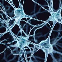
A paper published in Science Translational Medicine, provides preliminary evidence that the H1N1 Flu Vaccine Pandemrix can evoke immune system mediated damage to the orexin – hypocretin neurons and induce narcolepsy in individuals with a particular genetic variant. The orexin – hypocretin neurons were only recently discovered in the mid 1990s, by two separate research groups, hence, the two names for the same molecule. For this paper, we’ll be utilizing the orexin nomenclature.

Initially, the orexin neurons were thought to be involved only in feeding behavior, as damage elicited hypophagia in animals. Soon it was learned that more severe damage to the orexin neurons induced narcolepsy and the orexin system became a key focus in narcolepsy related research. With time, however, it became quite clear that these neurons were involved in regulating a myriad of hormone and neurotransmitter systems and their consequent behaviors. Narcolepsy or rather the ability to sustain wakefulness, is but one of the many functions regulated by the orexin system.

In a previous paper, I touched briefly on the possibility that the orexin neurons might be damaged and have diminished functionality in individuals suffering from post Gardasil side effects. In particular, I suspected these neurons were indicated in post-Gardasil hypersomnia, a derivative of narcolepsy. That may be only the tip of the iceberg. As I soon learned, the hypocretin/orexin neurons are brain energy sensors and may be involved in array of post medication or vaccine adverse reactions. Indeed, they may be central to the ensuing state of sickness behaviors that emanate once an organism becomes overwhelmed.

The Orexin – Hypocretin Basics

Orexin nuclei are located in the lateral hypothalamus, the section of the hypothalamus that is most known for regulating feeding, arousal and motivation. The hypothalamus is the master regulator for all hormone systems and hormone related activity including feeding, sleeping, reproduction, fight, flight, energy usage – basically every aspect of human and animal survival. It sits at the interface between the central nervous system functioning and the endocrine system functioning.

From the lateral hypothalamus, orexin neurons project across the entire brain with its two receptors (OXA and OXB) differentially distributed throughout the central nervous system and even in the body, including in the kidney, adrenals, thyroid, testis, ovaries and small intestine. The orexin neurons also modulate local networks of adjacent neurons within the hypothalamus that in turn influence a myriad of behaviors.

The most densely innervated brain regions include the thalamus, the locus coeruleus, dorsal raphe nucleus, accounting for the hormone’s role in arousal, feeding and energy management. At the most basic level, release of the orexin induces wakefulness. When orexin neurons are turned on and firing appropriately, arousal is maintained. When orexin neurons are turned off, diminished or dysfunctional, melatonin, the sleep promoting hormone, is turned on. The two work in concert to manage wakefulness and sleep.

Orexin receptors are also located in the amygdala, the ventral tegmental area (VTA) and throughout the limbic system, accounting for its role in emotion and the reward system. Orexin directly activates dopamine in the VTA. The VTA is the reward, addiction, and in many ways, the pleasure center of the brain. All drugs of addiction, all pleasurable activities, activate dopamine in the VTA. Through the release of dopamine, here and elsewhere, orexin modulates the motivation to sustain pleasurable activities. When orexin is diminished, not only does dopamine diminish, but the motivation to sustain behaviors decreases and dysphoria increases.

That’s not all. Orexin influences the release of many other neurotransmitters and hormones, several of which are co-located on the orexin neurons themselves. For example, the neuropeptide dynorphin is co-located on orexin neurons. Dynorphin is an endogenous opioid involved in the perception of pain and analgesia. It has dual actions that can both elicit analgesia or pain depending upon dose and length of exposure. Stress activates dynorphin. Dynorphin then inhibits orexin firing by as much as 50%. Illness is a stressor, a vaccine is a stressor, either could activate dynorphin and inhibit orexin. After the initial activation of dynorphin, and the ensuing decrease in orexin, the presence of chronic stressors and chronic pain could begin a continuous feedback loop of diminished arousal, and increasing pain.

Other Neurochemical Connections

  • Consistent with orexin’s role in arousal, orexin neurons contain glutamate vesicles. Glutamate is the brain’s primary excitatory neurotransmitter. Drugs that increase glutamate, also increase orexin. Drugs that block glutamate, via its NMDA receptor, decrease orexin. Common migraine medications block glutamate and thereby may also diminish orexin.
  • Serotonin and norepinephrine decrease hypocretin/orexin firing (suggesting if one is concerned with hypersomnia, anti-depressants might not be a good option).
  • As one might expect, orexin neurons are inhibited by GABAα agonists – sedatives. From a women’s health perspective, consider that cycling hormones would also affect orexin neurons through the GABAα pathway. Progesterone is a GABAα agonist – a sedative, while DHEA and its sulfated partner DHEAS are GABAα antagonists, anxiolytics that block GABAα, reduce sedation, and thereby increase anxiety and wakefulness. There may be a cyclical nature to orexin firing that has yet to be investigated.
  • The hypocretin/orexin neurons also influence galanin, a GI and CNS hormone that seems to inhibit the activity of a variety of other neurons in those regions.

These are but a few of the brain systems that the orexin neurons touch in some way or another. Damage to this system would have serious health consequences by initiating a cascade of biochemical changes within the brain and body. Many of which, we have yet to fully understand.

How Might the Orexin Neurons Become Inhibited?

Quite easily, apparently. In addition to the orexin’s vast interconnected pathways with a myriad of neurotransmitters and neuropeptides, the orexin neurons act as energy and activity sensors with some unique intracellular mechanics that make them especially sensitive to the changing dynamics of the extracellular milieu. Disruptions in ATP, glucose and temperature, elicit reactions in orexin functioning.

Orexin neurons require as much as 5-6X the amount of intracellular ATP to maintain firing, and to maintain a state of wakefulness or arousal. This extreme sensitivity to reduced ATP makes the orexin neurons uniquely positioned to sense and monitor brain energy resources, early, before ATP levels become critical in other areas of the brain. The orexin neurons cease firing when ATP stores become low, thereby allowing the reallocation energy, perhaps to those cells required for survival, breathing and heart rate. As Hans Selye observed many decades ago, one of the first, and indeed, most consistent of the sickness behaviors, no matter the disease, is lethargy, fatigue and sleepiness. Orexin is at the center of this behavior.

Orexin neurons react to extracellular glucose levels, though perhaps not as one might expect. When extracellular glucose levels are high, orexin neurons stop firing via what is called an inward rectifying potassium (K+) channel that is ATP dependent. That means that when extracellular glucose is high, intracellular ATP is allocated to open K+ channels and flood the cell with the inhibitory K+ ions. K+ hyperpolarizes the cell, prohibiting it from firing. This mechanism reminds me of Dr. Peter Attia’s talk about the nature of Type 2 Diabetes and our approach to treatment. He proposes that the body’s metabolic response – the conservation of energy – to Type 2 Diabetes is not something aberrant but is exactly as it should be with a disease state. We’re just not treating the correct disease state.

Another way we can shut down the orexin neurons is via increased temperature. The orexin neurons are very sensitive thermosensors. Increased temperatures shut down orexin firing via the inward K+ flow. Again, this is consistent with sickness behaviors and the reallocation of resources.

Orexin – Hypocretin Neurons in Migraine and Seizures

Diminished orexin has been linked to migraine and seizure activity. With migraines specifically, orexin may contribute to the early warning, hours to days, of impending cortical disruption via changes in feeding and sleep patterns that often precede migraine onset. Orexin may also be linked to the pre-migraine aura mediated by changes in brain electrical activity that prelude the migraine pain itself by minutes, called cortical spreading depression or more appropriately, cortical spreading depolarization – the massive spreading change in ion balance of the neurons. Initially the wave is excitatory, neurons are firing, but that is soon followed by a period of neural silence. Finally, orexin is also connected to the vasodilation of the trigeminal nerve, the nerve responsible for migraine pain. These findings have led some to call orexin a migraine generator.

Diminished cerebral spinal concentrations of oxerin have been found in patients generalized tonic-clonic seizures. Conversely, in rodent studies, injections of orexin elicit seizure activity. Despite the somewhat contradictory findings in seizure activity versus migraine activity, it is likely that the orexin system is involved both disease processes.

Pulling it all Together: Orexins Monitor and Mark Disruptions in Brain Homeostasis

Here’s where it gets really interesting. Although some have argued orexin, particularly diminished orexin functioning, is the cause and culprit of disruptions in brain homeostasis, leading to narcolepsy, excessive sleepiness, migraine, seizures and other diseases, I think this system represents merely a marker of a disease process. I think the orexin system is the stopgap, the final barrier of disrupted cellular energetics, of mitochondrial function. Mitochondrial ATP is the key.

When we consider orexin’s role in migraine, in particular, we see clearly how environmental changes (diet, stress, illness, medication/toxin exposure) can lead to changes in the extracellular milieu where orexins reside. The orexin sensors adjust to these changes, mostly by reducing neural firing in attempt to counteract damages. The reduction in orexin then elicits the premonitory phases of the impending brain disruptions, sleep and hypophagia – the sickness behaviors. If it progresses, the massive waves of electrical disruption ensue, and migraine, perhaps even seizures are evoked. When the extracellular environment become chronically disrupted, so too does the diminishment of orexin activity, thereby initiating a perpetuating loop of dysregulated brain activity. We can hypothesize that similar progressions exist with disease processes marked by aberrant electrical activity, such as epilepsy.

We know that mitochondrial dysfunction is often generated by genetic polymorphisms and can predispose individuals to an array of seemingly unrelated conditions like migraine and fibromyalgia, dysautonomias and cognitive deficits. At the root of the dysfunction is a error of some sort in mitochondrial energy processing – ATP.

What has become increasingly clear, is that the production of cellular energy, can be disrupted environmentally, by diet, illness and exposures, if co-factors necessary for the production ATP like thiamine are diminished. It is via diminished ATP production, that I think some medications and vaccines evoke adverse reactions in some individuals. The orexin system, because it is so exquisitely sensitive to changes in cellular energy, is our warning system; first by subtle changes in neurochemistry, then by changes in arousal and feeding behavior, and finally, by an all-out reallocation of resources – excessive sleeping. If ATP remains deficient chronically, and an individual is so disposed, then the cortical misfiring we see in migraine and seizure ensues, along with autonomic dysregulation and the syndromes associated therewith. It is not the orexin – hypocretin system that is at root of many of these diseases, but rather, the causes are deeper yet and reside with mitochondrial health.

We Need Your Help

More people than ever are reading Hormones Matter, a testament to the need for independent voices in health and medicine. We are not funded and accept limited advertising. Unlike many health sites, we don’t force you to purchase a subscription. We believe health information should be open to all. If you read Hormones Matter, like it, please help support it. Contribute now.

Yes, I would like to support Hormones Matter. 

Image created using Canva AI.

This article was published originally on January 29, 2014.

Cyclic Vomiting Syndrome and Mitochondrial Dysfunction: Research and Treatments

13815 views

Cyclic vomiting syndrome (CVS) is a debilitating disease characterized by episodes of severe nausea and persistent vomiting interspersed with periods of wellness. CVS affects about 2 percent of school-aged children, and also affects adults, although in adults it is often not recognized. Getting a diagnosis can be challenging, and sometimes takes a long time. Episodes of CVS can be extremely debilitating, and are sometimes difficult to treat and require hospitalization.

My daughter has suffered from this disease for 10 years, since she was 2 years old (see her story here). Her episodes were somewhat predictable when she was younger, but have changed and become less predictable, and more difficult to manage with medication, as she gets older. Although we try to avoid triggers such as stress and fatigue, being a pre-teen girl, she likes to have sleepovers with her friends and stay up chatting all night.  Unlike other pre-teen girls, however, she suffers the aftereffects of the sleepovers sometimes by vomiting for 24 hours or more.

What Causes Cyclic Vomiting Syndrome?

Although it has long been thought to be related to migraines, many sources state that the cause of cyclic vomiting syndrome is not known. Mechanisms that may be involved include episodic dysautonomia (malfunction of the autonomic nervous system that can result in a variety of symptoms), mitochondrial DNA mutations that cause deficits in cellular energy production, and heightened stress response that causes vomiting. However, there is mounting evidence for the role of mitochondrial dysfunction in the pathogenesis of this disease, a fact that is not often understood by the average practicing gastroenterologist. The connection to mitochondrial dysfunction has important implications for effective treatment of cyclic vomiting syndrome.

Mitochondrial Dysfunction, Cyclic Vomiting and Other Conditions

Mitochondria are small organelles within the cell responsible for energy production and other critical functions. Because of these crucial functions, Dr. Richard Boles, Director of the Metabolic and Mitochondrial Disorders Clinic at Children’s Hospital Los Angeles, explains that “30 years or so ago, many scientists couldn’t believe that mitochondrial disease could exist, because how does the organism survive?” However, mitochondrial dysfunction plays a role in many diseases, including CVS, and according to Dr. Boles:

“these are partial defects. Mitochondrial dysfunction doesn’t really cause anything, what it does is predisposes towards seemingly everything. It’s one of many risk factors in multifactorial disease. It can predispose towards epilepsy, chronic fatigue, and even autism, but it doesn’t do it alone. It does it in combination with other factors, which is why in a family with a single mutation going through the family, everyone in the family is affected in a different way. Because it predisposes for disease throughout the entire system.”

DNA mutations that affect mitochondrial function can occur in the DNA that is found in the nucleus of the cell (genomic DNA), or they can occur in the DNA that is found within the mitochondria themselves. Mitochondrial DNA is inherited differently than nuclear DNA. Most people are familiar with the inheritance of nuclear DNA, in which we have two copies of every gene, and we inherit one copy from each of our parents. However, mitochondrial DNA is inherited exclusively through the mother; therefore, mutations that affect the mitochondrial DNA can be traced through the maternal lineage of a family.

A possible relationship between cyclic vomiting syndrome and mitochondrial dysfunction was suggested by the finding that in some families, CVS was maternally inherited. Mitochondrial DNA mutations and deletions have been reported in patients with CVS, and disease manifestations of mitochondrial dysfunction have been found in the maternal relatives of patients with CVS. In other words, conditions such as migraines, irritable bowel syndrome, depression, and hypothyroidism, are often found in the maternal relatives of patients with CVS.

Mitochondrial DNA mutations don’t cause CVS directly, in the way that a DNA mutation causes cystic fibrosis, for example. In some patients, mitochondrial dysfunction plays a greater role in the causation of their disease, and in other patients, it may be less of a factor. Dr. Boles explains: “In some cases it’s a clear mitochondrial disorder, they have multiple other manifestations and it drives the disease. However, in most patients, it is one of many factors in disease pathogenesis.” Patients with classical mitochondrial disorders have disease manifestations such as muscle weakness, neurological problems, autism, developmental delays, gastrointestinal disorders, and autonomic dysfunction. Some patients with CVS have these other disease manifestations, and some have only CVS symptoms.

Treatment for Cyclic Vomiting Syndrome and Mitochondrial Dysfunction

As with many diseases, understanding as least some of the cause of CVS has allowed for the development of treatments tailored towards fixing the root cause. Co-enzyme Q10 and L-carnitine are two dietary supplements that have been used to treat a wide variety of conditions.  Both supplements may be able to assist the mitochondria with energy production and thus, help compensate for mitochondrial dysfunction. A retrospective chart review study found that using these two supplements, along with a dietary protocol of fasting avoidance (having three meals and three snacks per day), was able to decrease the occurrence of, or completely resolve, the CVS episodes in some patients. In those patients who didn’t respond to treatment with supplements alone, the addition of amitriptyline or cyproheptadine, two medications that have been used for prevention of CVS episodes, helped to resolve or decrease the episodes. Treatment with the cofactors alone was well tolerated with no side effects, and treatment with cofactors plus amitriptyline or cyproheptadine was tolerated by most patients. Therefore effective treatment for prevention of CVS episodes does exist, although it may not be widely employed by most gastroenterologists.

My daughter is currently trying to treat her CVS with the combination of co-enzyme Q10 and L-carnitine. So far she hasn’t experienced any side-effects, and over the next few months we will see if she experiences a decrease or even a complete cessation of her episodes. My hope for her is that she won’t have to choose between missing out on a fun night with her friends, and being able to be functional for the rest of the weekend. Maybe she can be like every other teenager and go to a sleepover, and just be grumpy the next day, instead of spending the next day vomiting and lying on the bathroom floor in pain.

Dr. Richard Boles, MD:  Dr. Boles completed medical school at UCLA, a pediatric residency at Harbor-UCLA, and a genetics fellowship at Yale. He is board certified in Pediatrics, Clinical Genetics and Clinical Biochemical Genetics. His current positions include Associate Professor of Pediatrics at the Keck School of Medicine at USC, an attending physician in Medical Genetics at Children’s Hospital Los Angeles, and Medical Director of Courtagen Life Sciences. Dr. Boles practices the “bedside to bench to bedside” model of a physician-scientist, combining an active clinical practice in metabolic and mitochondrial disorders with clinical diagnostics (DNA testing) and research. Dr. Boles’ clinical and research focus is on polymorphisms (common genetic changes) in the DNA of genes involved in energy metabolism, and their effects on the development of common functional disorders. Examples include migraine, depression, cyclic vomiting syndrome, complex regional pain syndrome, autism and SIDS. He has 50 published papers on mitochondrial disease.

Postscript: Using this advice, we were able to manage my daughter’s vomiting. Here is the follow-up story.

We Need Your Help

More people than ever are reading Hormones Matter, a testament to the need for independent voices in health and medicine. We are not funded and accept limited advertising. Unlike many health sites, we don’t force you to purchase a subscription. We believe health information should be open to all. If you read Hormones Matter, like it, please help support it. Contribute now.

Yes, I would like to support Hormones Matter.

Image by jcomp on Freepik

This article was first published on January 28, 2014. 

Are Your Mitochondria Stuck in Battleship Mode?

7667 views

As part of my work here, I am regularly confronted with desperately ill individuals who have seen dozens of physicians over the course of many years only to have their health continue to decline. Among the more frustrating aspects of this work is the failure of these physicians to assess and address the most basic aspects of health and healing. Namely, they fail to ask what the body needs to be healthy and whether the patient is getting those things. Of course, since evaluating these aspects health comes down to nutrition and exercise it does not align well with the practice of medicine, and if we are honest with ourselves, it is not something many of us want to deal with either. We want to eat what we want to eat and do what we want to do without regard to our health. Conventional medicine has capitalized on those sentiments, arguing persuasively for decades that a disease process is not real if all it requires to resolve is nutrients. Real illness requires medication, we now believe, having forgotten the very real nutrient deficient scourges like beriberi, Wernicke’s, pellagra, rickets, scurvy, and such.

Like most physicians, we have bought into the corresponding notions that fortification of foodstuffs assures that nutrient requirements are met and that in the land of plenty, where obesity reigns, malnutrition is rare. Neither is true of course, but belief in those myths absolves us of looking more closely at the chemistry of health and disease. For when we look at that chemistry, when we follow each of the altered signal transduction pathways, when look at the various patterns of deranged protein expression, and the myriad of genetic and epigenetic markers, we inevitably land at starving, inefficient mitochondria and the simple truth that they require nutrients to function; nutrient requirements that cannot be met by fortification alone and nutrient requirements, that if not met, lead to disease. When we dig a little deeper, we are also faced with a rather inconvenient truth that not only will pharmaceuticals not recover these deficiencies but they damage the mitochondria further. This does not accord well with the practice of conventional medicine and certainly does not fit into our busy, convenience-based lifestyles.

The Capacity to Survive

Among the more remarkable aspects of human physiology, however, is the capacity to survive all manner of illnesses and insults. We are supremely adept at adapting and surviving. We may not be living healthy, but we live. Mitochondria mediate these survival functions. They are responsible for converting the foods we consume and the oxygen we breathe into cellular energy (ATP).  With that energy, they regulate all aspects of basic survival at the molecular level, including survival itself – cellular respiration – but also things like inflammation, immune function, steroid hormone production, cellular life and death cycles, and whole bunch of other stuff. As one might expect, each of these functions is energy dependent.

Decrements in cellular energy, thus, elicit those survival mechanisms. If they are not resolved appropriately, when the threat persists, and/or when there is limited energy to face the threat, these normal responses lead to all sorts of tissue and organ dysfunction. It is this mismatch between the energy available and the energy required that leads to the persistence of not only the original illness, but because of the chronically activated survival cascades, leads to new and more complicated illnesses. On the one hand, decrements in ATP lead to things like inflammation and immune system activation – the normal, programmed and encoded survival cascades – but on the other hand, the survival cascades themselves lead to decrements in ATP, which in turn leads to more inflammation and immune reactivity. Without resolution, these cascades can easily become ingrained and ultimately lead to death. This suggests that energy availability is the key to health, or more specifically, that insufficient mitochondrial energy, and thus, impaired mitochondrial function leads to illness.

From Power Plant to Battleship: Mitochondrial Healing Cycles and the Necessity of Sufficient ATP

A recent paper suggests this is true. Just last month, one of the leading experts in mitochondrial function, put forth a compelling synthesis of research delineating what he called the mitochondrial healing cycles. Specifically, he demonstrated by what systems level mechanisms mitochondria maintain health or initiate and maintain disease. Dr. Naviaux argues that chronic illness is initiated by the “biological reaction to an injury and not the initial injury or the agent of injury itself.” He uses melanoma as an example, illustrating how it is not caused by the sun per se, but our biological, or more specifically, our metabolic (mitochondrial) response, to the sun. Chronic illness, he argues, becomes chronic only when there is incomplete healing of the original injury and/or when secondary injuries occur before primary injuries have healed. Illness, he suggests, is a multi-hit proposition.

To Naviaux, illness begins and ends in the mitochondria. Mitochondria are responsible for enacting what he terms the ‘cell danger response’ (CDR), the survival mechanisms that I spoke of earlier. There are three phases of the CDR:

  1. The initial inflammatory/immune response: “activation of innate immunity, intruder and toxin detection and removal, damage control, and containment.” He aptly describes this phase as a shift in mitochondrial energetics and function from power plant to battleship. ATP has to be diverted to fight the threat, initiate and maintain the characteristic inflammatory response. The reduction in ATP results in the characteristic fatigue we all experience at the beginning of an illness.
  2. Once the damage from the initial injury is contained, phase 2 begins. This involves replacing the dead and damaged cells as well as walling off any remaining damaged tissue that was not completely cleared in phase 1. Here stem cells are recruited and enter the cell cycle. Mitochondria in stem cells are critical for this phase, supplying the stem cells with ATP as well as key substrates to help with healing process. An interesting aspect of this phase, is that cell – cell communication ceases. There is no metabolic cooperation between cells as they are continuing grow and migrate. It is only when growth is complete and migration ceases that cell-cell communication reemerges.
  3. In phase 3, we get a return to “cell differentiation, tissue remodeling, adaptive immunity, detoxification, metabolic memory, sensory and pain modulation and sleep tuning”. Once the cells have been fully differentiated and re-educated, cell-cell communication reinstates and healing is complete.

The healing cycles are linear, sequential and ATP intensive. Each must be completed before the next can begin, before a secondary injury takes place, and each requires a continuous supply of ATP. Too many hits and/or too little ATP will derail healing. When we consider mitochondrial metabolism as the root cause of persistent disease, it is difficult not to ask what constrains the availability of energy and thus blocks the body’s ability to progress across each healing cycle.

Recovering Mitochondria: The Role of Nutrition

To answer this question, one has to look at how we produce ATP. Absent outright starvation, to get from food to ATP we need a few things: macronutrients and micronutrients or proteins, fats and carbohydrates along with vitamins and minerals. That’s it. Nothing fancy or complicated, just basic nutrition.

When we look at macronutrient consumption, one of the leading problems in western cultures is the high consumption of junk, carbohydrate-based foods. These foods, though they are often fortified with vitamins and minerals, come with far more sugar and other toxicants than the body can handle. Rather than being a net gain in energy, ultimately, become a net loss, both in macro, but especially, in micronutrients. Without sufficient micronutrients, none of the enzyme machinery, whether in the cytosol of the cell or in the mitochondria themselves, can perform the required functions that moves the macronutrient through the factory and produces ATP. Indeed, even the consumption of molecular oxygen requires the presence of vitamins and minerals. Absent those vitamins and minerals, a sort of cellular hypoxia sets in; one that activates inflammatory pathways, and ultimately, the shift in tale tell shift energy production associated with cancer known as the Warburg effect.

Conversely, because of decades-long advertising campaigns, most folks, but women especially, consume insufficient quantities of protein and fat. This skewed consumption of macronutrients places a high demand on the OXPHOS (oxidative phosphorylation) pathway of the mitochondria to produce ATP, while simultaneously not providing sufficient micronutrients to fuel the enzyme machinery to produce this ATP. It also increases the need for detox, while again, failing to provide adequate substrates to do. Moreover, if the diet is high in the staple sweetener high fructose corn syrup, in addition to everything else that becomes dysregulated at the mitochondrial level, the ability to covert fatty acids into energy, can be shut down entirely, conferring a metabolic inflexibility that is common in western cultures.

Along with issues with macronutrient consumption, large percentages of the population are deficient in one or more of the micronutrients required for healthy mitochondria. Individuals with chronic illness are severely deficient. The mitochondria require 22 vitamins and minerals in varying concentrations to convert the food we eat and the air we breathe into cellular energy or ATP (Figure 1.). Absent sufficient concentrations of one or more of those nutrients, mitochondrial function deteriorates and healing will not progress. Survival mode is all that can maintained.

mitochondrial nutrients
Figure 1. Mitochondrial Vitamins and Minerals

With Naviaux’s framework, it becomes clear that healing is an energy intensive process. The only way to boost energy is via good nutrition. Sure there are compounds that can override certain pathways within the mitochondria and, at least temporarily, provide additional energy, but if the core requirements for optimal mitochondrial health are not met, it is only a matter of time before initial benefits become problematic. (I should note that exercise is also critical for healing the mitochondria. Exercise forces mitochondrial biogenesis among other important processes.) The questions that physicians and individuals with chronic illness should be asking are:

  1. What is required for health?
  2. Is this patient or am I getting those things?

Unless and until those aspects of health are addressed, chronic illness will persist because the mitochondria simply do not have the resources to progress through the healing cycles. There are no short cuts here.

We Need Your Help

More people than ever are reading Hormones Matter, a testament to the need for independent voices in health and medicine. We are not funded and accept limited advertising. Unlike many health sites, we don’t force you to purchase a subscription. We believe health information should be open to all. If you read Hormones Matter, like it, please help support it. Contribute now.

Yes, I would like to support Hormones Matter.

Image credit: ImgFlip, Template ID: 24838763

This article was published originally on October 17, 2018. 

Thriving Despite Mitochondrial Disease

3003 views

Mitochondrial Disease with Opthalmoplegia and Hearing Loss

Four years ago, I was diagnosed with mitochondrial disease, the form known as chronic progressive external ophthalmoplegia (CPEO). Mostly, it affects muscles from the neck up but can also affect my heart as well as my overall energy levels. It was a diagnosis I fell into. I had been hit in the eye with a rock around 6 years old and slowly the eye lid muscle started not working as well. I had two surgeries on my eyelid by 21 years old and all looked good until about 40 years old. I noticed my eyelid would not open as well. The eye surgeon performed eyelid surgery on my left eye again.

All my young years, I had extreme balance issues. I would be standing or sitting and just lose my balance.  The pediatrician said it was just bad balance. However, I found most of my balance issues were resolved when I took up running / jogging at 23 years old.

At age 42, I was diagnosed with a bone tumor in my right femur. Thankfully it was benign, but it was the size of a golf ball and I had an 8.5 inch plat right behind my iliotibial band for a year.

At age 43, I found that eating all-natural, no preservative, raw foods enabled me to think so much more clearly than ever in my life. It felt as if a cloud had lifted.

I started natural bodybuilding around 47 years old and found the more I lifted weights along with running, the better my energy levels and cognitive function was.

I found that high sugar or alcohol several limited not only my energy level, but a few days of poor eating limited my cognitive thinking.

In 2015, I found I was seeing double and went to the eye doctor for help. The doctor recognized that I had slight facial paralysis as well, indicating an issue beyond their scope of care and referred me to the Jules Stein Center in UCLA. That doctor advised that I had more issues that needed clarification by UCLA neurologist before he could assist with my eye muscles.

Diagnosing Mitochondrial Disease

The neurologist went through a long screening process from my childhood to present. He had a theory that my condition was related to mitochondrial disease and requested a muscle biopsy to confirm. He cleared me for eye surgery and collected a muscle sample from my strabismus surgery which confirmed the diagnosis. Since then, I have had three more surgeries. My eye muscles will always be moving, so we are hoping to hold off more surgeries because they can only handle so much.

Some of the issues that assisted the doctor in the diagnosis of mitochondrial disease included my height. I am 5’1” and the rest of my family is well over 5’5”, my eyes, and I have had issues swallowing  sometimes, and almost choking. My throat muscles sometimes do not work well when I do not eat natural food. In addition, I have otosclerosis (abnormal remodeling of the bone in the middle ear that causes hearing loss). I was diagnosed with otosclerosis in my early 30s and, even after stapedectomies (a surgery to improve hearing), I was 100% deaf in my left ear and only 60% on my right – this was managed with a hearing aid for over 20 years.

They suspect my disease may be genetically inherited, but I come from a large family and in all of them there were never similar issues. The other option is that my body exposed to some kind of pollutant.  While it could be anything growing up in NYC, I feel certain it had something to do with a marble size mercury ball I swallowed at 5 years old that had we found from a broken thermometer. Although, from the age of two, I had been very ill, by the time I was five years old, before the mercury, I had had scarlet fever, mumps, rubella, constant tonsillitis, and all the way to adulthood, multiple ear infections. In fact, all the way into my late 30s, I caught a cold with fever swollen glands and cough right around the time of my period every single month. It wasn’t until I went all raw for a couple of years, that I stopped getting sick. Now except for September of 2018, I rarely get sick.

Managing Mitochondrial Disease: Diet, Exercise and Avoiding Toxicants

There is no cure. The only way to manage mitochondrial disease is healthy nutrition as well as using vitamins and supplements. Unfortunately there are no doctors in the area that even know what the disease is. There are multiple medications that I hesitate to take because they can damage more mitochondria. I am not interested in a clinical study at this time unless it is with 100% natural ingredients.

In 2018, I realized that in bodybuilding the tanning spray causes mitochondrial issues. Immediately after spray, I lose balance and become exhausted. I started powerlifting instead so I could still compete, as I found competition pushes me to be more dedicated to exercise. However, I stopped eating like a bodybuilder and started adding sugar, alcohol etc. to my diet to gain mass, not realizing how much damage it was doing to my body.

In September 2018, I had an undiagnosed autoimmune response in which my lymph gland close to my right ear swelled so much that I was losing more hearing ability. Though not totally deaf, I went to the ER, multiple ENTs, even the dentist, in case it was TMJ. I had a biopsy of my lymph node as well and it came back negative. On December 28, 2018, I woke up completely deaf. A physician at UNLV School of Medicine came in from his vacation to give me steroid shots in my ear drum in hopes of restoring at least some hearing. This did not work. In February 2019, I received my first cochlear implant and in August 2019, I received the second. Both work extremely well and I can now hear again.

The Loss of Energy with Mitochondrial Disease

My exhaustion levels for most of early 2019 year have been relatively high. Slowly, I am regaining strength and managing my energy levels. I am very careful what I eat and drink because I feel my life is at stake. I started doubling up my L-Carnitine levels in late July and have seen an improved energy level.

Over the next few weeks, I will start experimenting with CoQ10, PQQ and there is a new product I heard about that is MitoQ that I may try after. I just want to be sure it will benefit me individually. I have found that while some supplements work for many with the disease, they are not beneficial to me.

I did try the allithiamine supplement for 60 days and there was no positive or negative affect.

There is definitely a correlation of eating well and adding supplements to be noted.

At this point, my daughter suffers the same issue where she gets sick every time she has her period. Like me, she does not have the time to keep going to the doctor who simply diagnoses it as a cold or upper respiratory infection rather than look at the source.

I have no faith in doctors here, as when I tell them I have mitochondrial disease, they do not even know what it is. Even when I show them the mitochondrial toxin list, they prescribe toxins to me that may hurt me.

The United Mitochondrial Foundation is available for doctors to use as resources for information.

I truly believe the best I can do is monitor what I eat, keep trying supplements and hope that I do not get sick again. If I am unsuccessful, I am certain it will definitely kill me.

We Need Your Help

More people than ever are reading Hormones Matter, a testament to the need for independent voices in health and medicine. We are not funded and accept limited advertising. Unlike many health sites, we don’t force you to purchase a subscription. We believe health information should be open to all. If you read Hormones Matter, like it, please help support it. Contribute now.

Yes, I would like to support Hormones Matter. 

Thiamine Deficiency Amid a Constellation of Mitochondrial and Metabolic Disorders

7961 views

This is a bit of a long story because I can’t tell if I have a genetic problem that only shows up under stress or just plain deficits due to extreme stress. I am a 58 year old woman having some pretty severe health problems that I have recently begun to think might be due to thiamine deficiency, malnutrition and/or possibly some sort of genetic problem.

Medical History

I was hospitalized with pneumonia three times before I was 5 years old, once again in isolation ward at age 7. I also had several childhood illnesses including Rubella, chicken pox, and mumps. At age 5, I was diagnosed with exercise induced asthma and prescribed Quibron. Once I hit puberty, my respiratory problems seemed to resolve themselves as long as I avoided aerobic exercise.

At age 9, I was diagnosed with progressive sensorineural hearing loss (an indicator of mitochondrial disease that I only recently learned about). I had a curved loss with 60% gone in the higher frequencies down to about 40% loss in the low ones. My mother was also diagnosed with hearing loss that had gone unrecognized. My daughter also has a mild hearing loss that was diagnosed as an adult.

Major Life Stressors and Emerging Problems with Nutrient Absorption

From 2000 to 2012, had several major life stressors including the death of my second husband to colon cancer, a dog snapped at me and severed my lower lip, my third husband lost his job and attempted suicide, early retirement, bankruptcy, foreclosure on my home and then moving to a new state. I was diagnosed with post-traumatic stress disorder (PTSD) and received weekly trauma therapy for a year.

During the aftermath of the PTSD trigger event, I began having problems with nutrition. I would eat a meal and get no energy from it. After taking an early retirement from my job and losing my house to foreclosure, we packed up and moved out of state.

I never recovered from the exhaustion and stress of the move. We were nearly homeless one night after our car broke down and I had no more money because we had just paid the deposit to rent a house. I managed to leave all our keys in the car at the repair shop. My first husband wired us money for a hotel for the night.

The problems with digestion continued and my health seemed to go downhill. I was tired all the time and my brain was not functioning well. I tried going for walks but would come home exhausted. To make matters worse, we were scraping by on one third of the income I had before I retired and I had no health insurance. I didn’t know how to go about finding help in those conditions. I’ve always been in the system.

More Digestive Problems

In 2013, my digestive problems got much worse and I had diarrhea almost continuously for three weeks before finally going to the local university emergency room where I was surprised to find my blood pressure was 230/140.  I think they had to give me at least two potassium drips. After they initially got my blood pressure down, an intern came in to give me some sort of eye exam.  He started lecturing me on using the ER for primary care. He then proceeded to interrupt me when I tried to tell him about zonking out (see below). My blood pressure went up 100 pts before he was finished. That experience just increased my reluctance to go to physicians when I am sick.

I was admitted for four days and my major systems checked and seemed fine.  They gave me scripts for Lisinopril and Coreg and referred me to a local clinic.

The NP I saw at the clinic put me on 1000 mg Metformin ER.  She referred me to a GI who diagnosed me with GERD, Pancreatic Exocrine Insufficiency of unknown cause and prescribed Omeprazole and Creon.

The diarrhea died down but I continued to have steatorrhea and my energy did not improve. I had a constellation of general vague symptoms as well as unusual ones. I also continued to have problems eating. The recommendations for GERD are to eat small meals a day. I found that five small meals of no more than about one cup of food a day were all I could tolerate. I also stopped eating vegetables or high fiber foods. Even following those rules there were many days when I had nausea and could not eat very much.

My type 2 diabetes has not been that bad, but my day to day glucose readings were very variable. My A1C was 6.5 2013, 5.8 in 2015, 6.8 in 2016, 7.0 in 2017, and 7.4 in 2018. As I try to get health care professionals to address my problems I keep running into diabetes. As if my diabetes where the cause of everything I could ever experience. I recently ordered a fasting C-peptide test which showed I was in the high normal range. Glipizide did me no favors as it likely increased my insulin resistance.

I know what is normal for my body and what is not and what I have been experiencing is NOT NORMAL!!

Catatonia-like Episodes upon Eating

Starting in 2013, I started having episodes that I call zonking. I would eat something and almost immediately get very sleepy and usually would just fall into a sort of zombie-sleep-like state for 10-30 minutes. Sometimes it seemed sort of catatonic and others is was more like a strong urge to sleep. It wasn’t normal sleep in that there was no dreaming. Sometimes I was in a kind of frozen pose instead of laying my head sideways or getting comfortable. At first it seemed like they were correlated with high-availability carbs, but over time they became an almost daily occurrence with any type of food I ate.  These episodes would sometimes disappear for a few weeks and then reoccur. There was no pattern I could discern nor tie to hormonal cycles.

I have been keeping daily logs from 2013-2016. I reported the zonking episodes to my NP and she prescribed Lactulose which just made the diarrhea worse. She ordered an ultrasound on my gallbladder and liver and no major problems were found.

The zonking episodes were really disabling. I would be feeling fine until I ate something and within a 1/2 hour I would zonk out and feel very groggy afterwards. It didn’t feel safe to drive so my husband had to take me shopping or do the shopping himself when I was too tired. I didn’t drive from 2013 to 2016. At times, I didn’t want to eat at all so I could keep myself functional long enough to do something. I found out the limits of fasting pretty quickly.

Left Temple Headaches, Migraines with Visual Aura, and Cognitive Deficits

I also had left temple headaches starting in 2013 and a few migraines also involving left temple pain but mostly just visual aura. I never had migraines previously. After I fell going up the concrete steps to our front door, I became very cautious about moving when I was in one of these states. I noticed that I became very clumsy with tasks that you normally don’t think about like opening a can or a pill bottle.

I also noticed a significant decline in my ability to comprehend speech when I was in one of these states. This was first mentioned in my logs in 2013. I have always had a problem due to my hearing loss but there seemed to be a magic something taking place in my brain that took care of turning sounds into words. Somehow that process was failing when I was in the dizzy and ditzy state.

My condition was deteriorating in 2016 and I was experiencing more severe brain problems that are what I now call dizzy and ditzy. Dizzy and ditzy feels like being very drunk. I was spending more and more time in bed which was making my diabetes worse. When my brain was functional I was researching and trying to figure out what might be causing my condition.

In August 2016, I came across a description of gluten ataxia. The symptoms seemed similar to what I was experiencing so I tried eating a gluten free diet for a few weeks and started feeling better. After two weeks, I decided I was imagining the improvement and tried eating something with gluten in it. After about two hours I began to experience the dizzy state and the left temple headache.  From that time I have remained gluten free.

It was my intent to get tested for Celiac at my next appointment, but my husband was hospitalized in October 2016 for sepsis due to undiagnosed type-1 diabetes. This was the worst kind of trauma for me because my second  husband died of colon cancer. Knowing that, I applied the techniques I learned during trauma therapy to keep my stress level down. As a result I was able to return to the hospital for his follow-up visits with no increase in blood pressure or other signs of HPA activation. I think it helped me overcome my doctor phobia and I have been increasingly more able to deal with medical situations. My health and energy levels were also improving due to the gluten free diet. I tried various diets and found that I had the best energy levels on a gluten free vegan diet.

Since I did not get in to see my primary until six months had passed gluten free my celiac tests were inconclusive. I found a really good allergist and in January 2017 was diagnosed with 44 food allergies including wheat, barley, oats, eggs, milk, and assorted nuts, fruits, and vegetables and yeast which is everywhere. Six months later, I was diagnosed with multiple environmental allergies. My condition began improving dramatically as I stopped eating the foods that I was allergic to.  However I found that I was still unable to eat solid food and get energy from it. Meanwhile my NP added 10 mg glipizide which caused hypoglycemia when I was feeling well and active.

I didn’t experience any more dizzy and ditzy episodes from October 2016 through March 2018.

Diet and Malnutrition

I did have, and still have, episodes where I become very sleepy and nauseous after eating something. This leads to a feeling of being ill and not enough energy for normal activity. My condition improves if I semi-fast for the remainder of the day and then reduce my intake of protein and fat for a few days. As a result, I became more depleted nutritionally as time went by.

I don’t think I’ve managed to convey to anyone just how my diet has shifted from normal and especially what they seem to expect of diabetics. Many days I was unable to even manage 50 mg of protein and if I didn’t eat enough carbohydrates I had difficulty sleeping. I was on homemade liquid meals from 2017-2018 and only recently was able to transition onto whole foods after stopping Metformin.

I still struggle to explain why I suspect nutritional deficits in the face of a body that is down 10lbs but still 220. I think my body compensates by doing things like lowering my body temperature. It was 96-97 on a regular basis over the past 6 years, yet my thyroid panels are normal except for high TSH which may be due to my Biotin supplementation. In May 2018, I had more thyroid testing done.

  • TSH: 7.5
  • fT3: 3.1
  • fT4: 1.34
  • rT3: 22.2
  • TPO: 12
  • Antibody <1.

Sometimes my body heats up immediately after eating.  I’ve also had iron deficiency anemia where my hands and feet got painfully cold. Sometimes I get chilled and find my body temperature is 95.8 – even in July.

Other Weird Symptoms

Visual Disturbances

I haven’t been able to match up these phenomenon with technical names so I am just describing them as best I can. There is some overlap as I am not sure what is part of which phenomenon.

  • Problem:  After-images occurring too easily
  • Duration:   3-4 years
  • Frequency:  Intermittently – seems to be worse when I am having diarrhea episodes
  • Details:  Can’t find exactly what these are called. In normal vision if you stare at something for a long time then look at a blank surface you can see a phantom image of inverted colors. What has been happening intermittently with me is that I will see these images instantly and they take longer to fade than normal. This is especially bad in the mornings. I made up a test for myself to track how bad my vision was on any day by looking at the GOOGLE logo. On bad days it bounces around in afterglow very strongly; other days not at all or not as strong.  Sometimes if we are out on a sunny day a glance at a bright reflection off metal will create a strong after image that persist for 20-30 seconds or more. I have to be very careful not to let my eyes linger on anything shiny.

Along with the above problem I also experience a kind of sparkle sometimes where if I look out the window, even on a gray day and then back inside there is a brief field of sparkle in my vision.

  • Problem:  Seeing my pulse in my eyes
  • Duration:  6 years
  • Frequency:  Not as much these days, but was especially bad when my blood pressure was uncontrolled.
  • Details:  I would see a grayed pattern around the lower outer edge of my vision that would flash in intensity seemingly in conjunction with my heart rate.

Finally, my left eye seems to flutter and roll.

  • Problem:  Left eye flutter and involuntary eye roll and moving illusions in vertical images.
  • Duration:  9 months in 2015, confirmed by eye doctor.
  • Frequency:  Started very infrequently but has progressed to almost daily.
  • Details:  Irritating flutterly feeling in my left eye. When I looked in the mirror I noticed the left eye was rolling left or attempting to roll left involuntarily. This may be related to another problem I have been having for several years where my vision wobbles. It comes and goes in severity, but if I look at any image that has strong vertical contrast it appears so move. For example a picture of a coliseum or tiger will appear to be moving like the way optical illusions move. The moving illusion also occurs more frequently when I am having bouts of diarrhea.

Migraines With Aura

  • Problem:  Migraines with Aura
  • Duration:  2 years
  • Frequency:  3-4 times a year – sometimes in clusters
  • Details: I get migraines where I see an aura. It starts with a blind spot in the center of my vision, in less severe ones it is more of a sparkle overlay and not as blind. The circle then becomes a semi-circle of the outer edge of each eye that becomes bigger until it moves out of the eye. Then pain comes or not. I can sometimes reduce severity by immediately going into a dark room and lying down. Sometimes I get the headache and sometimes just the visual aura. I mention these here because I have wondered if some of my visual problems are migraine auras that haven’t fully developed.

Allergy Shots and More Symptoms: Ophthalmoplegia, Dizziness, Speech Impairment

In January 2018, I started desensitization shot therapy with my allergist. In February 2018, I had another episode of dizzy and ditzy that was complicated by additional symptoms of hypoglycemia. After consulting with my allergist, he set my shots back a few weeks and set up a standing lab order to see if he could tease out the problem. In March, I had the worst episode of dizzy and ditzy I have experienced to date. It included ophthalmoplegia, dizzy, very sleepy, altered consciousness, and personality changes. I was walking with a sort of limp as if my left leg were injured. I laid down on a bench outside the allergist office until the sleepiness passed. When the people at the allergy clinic got back from lunch they checked my blood pressure which was normal and did a blood draw for labs.

I also had the worst episode of speech impairment ever. I remember thinking it was funny because it sounded like they were speaking so much gobbledy gook. I used mindfulness to notice and try to quantify what it was like. I noticed that my intellect was intact, which distinguishes the condition from brain fog where it’s hard to think at all.  But things like trying to speak or find words was hard and I got confused easily. Oddly, I had no problems reading and comprehending research papers and I could write, except I made more typing errors. I had a quarter sized mark on one of my arms from the previous day’s shots, but my allergist did not consider my brain symptoms to be due to the allergies. My movements and general condition was very much like being very, very drunk.

Up until that point I had associated dizzy and ditzy with gluten exposure. Once I was sure there was no gluten in the shots, I was a lot more worried about my condition. I regretted not going to the ER since that might have helped me get a diagnosis. As it was, the allergy doctor only checked my tryptase levels and blood sugar which was 119. That level of blood sugar is hypoglycemia for me. I start getting symptoms under 130. This second episode was much harder to recover from than the first and I had lingering symptoms for months afterwards.

Medication Reactions

The glipizide combined with 1000 mg Metformin ER for the type 2 diabetes was causing me severe problems, even as my energy levels and inflammation was improving. The repeated episodes where I had to lower my protein and fat intake was really difficult with the medications I was on.

One day I had a different sort of drunk feeling with sweating and distorted vision while out shopping. It got better when I ate something. I got home in one piece and spent the entire afternoon eating every hour until things improved. That was the last time I took glipizide. I started looking for a new primary who understood nutrition.

I found a new primary two hours away who seemed like a good fit but this story is already too long to describe what went wrong. Sometimes a bad thing has to happen before good ones. I found myself without a primary and experiencing more dizzy and ditzy symptoms. The problems started at the request of my new primary PA, I changed my metformin dose from 2 500 mg tablets in the evening to 500 mg morning and 500 mg evening. After a few days I woke up with a 206 fasting glucose. So I doubled down and started taking 1000 mg 2 times a day. That made modest improvement of fasting glucose but also caused steatorrhea and diarrhea to return.

Thiamine Pyrophosphate Order Delayed

I began taking 13 mgs of thiamine pyrophostate (TPP) in 2014.  Recently, I ran out of the TPP sublingual I normally take due to a shipping delay, so I switched to oral 500 mg Thiamine HCL capsule. Over the next two days, I had cramps in my calves, congestion and coughing after my normal light exercise and a body temp of 95.8 in the evening. Then another episode of dizzy and ditzy with mild ophthalmoplegia and dehydration. I was drinking salt water and still couldn’t retain water. I always drink my water with electrolytes and I went through a whole gallon in a day. My blood glucose was 289 in the evening.

I’ve been having problems with burning feet and neuropathy since March 2018, it comes and goes and makes it hard to get to sleep. I first reported mild symptoms of neuropathy in my logs in 2013. TPP and B2 and sometimes calcium supplements help.

Once TPP arrived I started taking it 3 at a time (3x13mg) as well as extra magnesium glycinate and other B vitamins, but my symptoms weren’t improving as much as I expected so I started looking for other causes.

While researching various genes, I came across a paper on thiamine transporters and metformin. I have at least one mutation in one of those transporters but it doesn’t have any established meaning that I can find. I had not considered thiamine deficiency as a possible cause for dizzy and ditzy even though the symptoms seemed similar because I was taking 13 mg of TPP sublingual daily.

I started easing off metformin and my energy and burning feet symptoms improved. A side effect was the metabolism issues that had kept me on a liquid diet improved and I can eat and metabolize solid food again.

Going off metformin caused a slight rise in my fasting glucose so I had a lab test run myself that shows I still have normal insulin production.  My glucose response varies quite a bit but I sometimes get good response when I do daily treadmill with glucose in the 110 range two hours after a meal

One night I had a lot of congestion and couldn’t sleep so I took a dose of 3x TPP and the congestion went away and I was able to sleep. I have had ongoing issues with lung congestion since 2010. I think this may be a return of the lung problems I had as a child. I believe my body compensated with hormones and is now decompensating due to menopause. The fact that the congestion got better after thiamine intake is worrying. But I have a strong placebo response so I have to be careful about creating correlations. The lungs issue has been observed by both my primary and my allergist.  I frequently get very thick mucus that may be due to inadequate fat digestion.

Ran out of Thiamine AGAIN!

I had a dizzy and ditzy episode with temple headache 7/11/2018. I went through three bottles of 60 Sublingual TPP in a few weeks and was unable to order more due to factory back order.  I researched other versions of thiamine. Benfotiamine was having no effect that I could detect. Sulbutiamine helped with some of the brain problems like short term memory. I had concerns that I wouldn’t be able to absorb some of the other forms due to my fat malabsorption.  Fortunately, I came across the blog where Dr. Lonsdale explained what “fat soluble” really means regarding lipothiamine and allithiamine, so I ordered both of those as well as liquid TPP from the UK.

Neither of those had arrived by July 14, when I took my last TPP. I had a return of symptoms over the next few days. I started using a B-complex that had TPP in it, but even with that I had another dizzy and ditzy episode that lasted several days. During that time, I got sleepy every time I ate a meal. My meals are small to start with because of the GERD: one cup lentils with tofu or a tofu taco with avocado. Both of those meals that were normally well tolerated gave me symptoms of sleepiness and fatigue.

I found a comment somewhere about the need for manganese, so I got manganese glycinate supplements and that helped ease the neuropathy until the Lipothiamine and TPP liquid arrived. The first Lipothiamine was like magic, my eyes cleared up in about 15 minutes. They do that sometimes after I eat. The neuropathy symptoms were down by the time I went to bed in the evening. In the morning, my feet were hypersensitive and I had leg cramps due to low magnesium. I still have to take extra magnesium during the day and at night as well as other B-vitamins. I have some mild brain symptoms too but they may be due to the stress of starting with a new primary.

The big difference is in my energy levels, which are sufficient to make it through the day and no burning feet at bedtime. I still have limited ability to metabolize food, I tried to add 1/2 cup brown rice to my daily lunch and it sent me into borderline dizzy and ditzy.  A typical day’s intake for me is 1 slice tofu, 1/2 cup lentils, and tofu taco with avocado, some vegetables, and a big leafy green salad with avocado, beans and nuts.  Sometimes I can have a snack of fruit but more than that I start having problems.  I suspect I have other deficits that may be slowing my recovery.  I am waking up at 1 am and having to eat something to get back to sleep.

I had another episode this past week. I was preparing my allergy records for a trip to ER and was very dizzy and clumsy and a band of pressure in my forehead. I had been taking 2 50 mg enteric tablets 3 times a day of Lipothiamine but was experiencing symptoms of illness after eating and tiredness.  I was only rescued from ER by modifying Lipothiamine dose so that it would be absorbed in my stomach instead of my intestines. I’ve had symptoms of  intestinal malabsorption which haven’t yet been evaluated by a GI.  I have checked several nearby GI practices without finding one who takes a self pay patient.

Genetic Testing

Genetic testing has helped me discover other problems I have involving metabolism. I haven’t cataloged all my mutations but I have a diabetes related set of 3 homozygous SNPs called TCF7L2. The way I understand it is that people with this phenotype have increased signaling mechanisms such that when they have excess fat, the pancreas responds by lowering insulin response to food and the liver increases morning glucose. Losing the weight, reduces the expression and returns things to normal. I have other  genetic mutations which may be impacting me such as MTHFR(++), MTRR(++), and a rare BCHE mutation that impacts my choline levels. It also makes me vulnerable to pesticide exposure. I have to take Trimethylglycine (TMG) and various B vitamins to keep homocysteine in check. If I don’t take enough TMG and CDP choline, I get depressed.

I am seeing a new primary who is a doctor of Internal medicine. The new doctor’s choices for medications are spot on for my genetic type, they both work on the GLP-1 pathway, but they also increase insulin response. I don’t see how that doesn’t cause hypoglycemia, since I have normal insulin production. I have more research to do before making a decision on new medications.

While I can see that the deficiency symptoms I have been experiencing may be a result of poor diet and digestion, the fact that Thiamine HCL did not work and my condition improved so much after stopping metformin leads me to believe I may have a genetic condition. I really need to find a university that is researching metabolic and genetic conditions so I can get a diagnosis.

Medications and Supplements

Most of these supplements were added in 2017 after I started homemade liquid meals.  Prior to this I was taking a sublingual B-complex & Magnesium glycinate.

Current Medications

  • Singular 10 mg x 1
  • CoReg 12.5  x 1
  • Lisinopril 10 mg x 1
  • Cetirizine HCL 10 mg x 1
  • Omeprazole DR 40 mg x 1

Most of these were added when I started liquid diet

  • B-6 Pyridoxal-5-phosphate sublingual 15mg  x 1 (more if homocysteine symptoms)
  • B-1 Thiamine pyrophosphate (TPP) sublingual 13 mg x 1 (the one that is backordered)
  • *TPP liquid 3 x a day or as needed with malabsorption due to intestinal swelling
  • *Lipothiamine tetrahydrofuryl disulfate 100 mg  3 times a day with extra Mg.
  • *Liquid CoQ10 100 mg x 3 times a day
  • B-2 Flavin mononucleotide sublingual 18 mg  x 1
  • B-3  27 mg  as Inositol Hexanicotinate, 25 mg as NAD sublingual 25 mg x 1
  • Liquid B5 + Carnetine
  • MethylCobalmin,  sublingual 1 mg x 1
  • MethlyFolate (S6)-5-MethylTetraHydroFolate glucosamine salt  sublingual 800 mcg x1
  • Biotin 5000 mcg sublingual 1 x
  • Chelated Zinc, zinc glycinate  22 mg x 1
  • Selenium as L-Selenomethionine,  capsule 200 mcg x 1
  • Kelp 325 mcg Iodine,  325 mcg x 1
  • TriChromium Chromium Picolinate, Chelavate, & polynicotinate, 500 mcg x 1
  • Magnesium Glycinate (capsule at home) ~ 800 mg per capsule 50% Mg  x 2
  • Liquid chelated Iron biglycinate  10 mg, Yellow dock  10 mg x 1 (not long term)
  • D-3 5000 iu with K2 1 sublingual  125 mcg D3+ 90 mcd K2
  • Ultra K with K2 softgel, K1-1000 mcg + K2 1400 mcg x 1
  • Algae DHA 200 mg x 1
  • Phosphatidylcholine softgel 1300 mg x 1
  • Source Naturals Vitamin A as palmitate 1 x 10,000 iu
  • Buffered C-1000 complex Calcium Ascorbate 1 g x 2
  • Southwest Botanicals Mucuna Powder (self capsule)  ~ 810 mg x 1
  • Citicholine (self capsule) ~820 mg, 1-2 as needed
  • Nootropics Sulbutiamine 200 mg (thiamine derivative) 1 capsule, 200 mg x 4
  • TrimethylGlycine (Betaine) 1 g x 2 with each meal containing methionine
  • *Taurine 1000 mg
  • Liquid Calcium, Magnesium, D3, Phosphorus as needed for sleep

 *Recent additions

We Need Your Help

More people than ever are reading Hormones Matter, a testament to the need for independent voices in health and medicine. We are not funded and accept limited advertising. Unlike many health sites, we don’t force you to purchase a subscription. We believe health information should be open to all. If you read Hormones Matter, like it, please help support it. Contribute now.

Yes, I would like to support Hormones Matter. 

Share Your Story

If you have a complicated health story to share, send us a note. 

NASA/JPL-Caltech/Univ. of Ariz., Public domain, via Wikimedia Commons.

Could the Way You Walk Indicate Mitochondrial Dysfunction?

5035 views

The answer is an unequivocal, yes. The way you walk, your gait, can be diagnostic of mitochondrial disease and dysfunction. Whether your stride is long or short, rhythmic or arrhythmic, quick or slow, symmetrical or asymmetrical, balanced or unbalanced can indicate mitochondrial dysfunction. Even better, the particular pattern of gait disturbance may distinguish between types of mitochondrial disease, at least cursorily, and indicate whether and where there might be neural involvement. This, according to a study published in the Journal of Neurology.

In a small study of 24 patients with genetically confirmed mitochondrial mutations, researchers were able to discriminate between healthy controls and patients and between the two types of mitochondrial mutations assessed (m.3243A>G and m.8344A>G) based upon gait. Both patient groups were selected because of the known associations between those mitochondrial genotypes and gait disturbances. The questions were whether the pattern of gait disturbance could distinguish between the two groups and whether the gait disturbances could be detected early in the disease process before other symptoms fully emerged. That is, could the way patients walked be diagnostic of incipient mitochondrial disease?

About the Mitochondrial Mutations and Patients Tested

The first mutation m.3243A>G (n=18) is associated a disease called MELAS, which stands for mitochondrial encephalopathy, lactic acidosis, stroke like episodes. It is believed to represent one of the more common classes of mitochondrial mutation. The MELAS mutations are associated with a constellation of additional clinical symptoms, including Chronic Progressive External Ophthalmoplegia (CPEO; weakness in the eye muscles causing eyelid drooping), Maternally Inherited Deafness and Diabetes (MIDD), migraine, bowel problems and short stature.

The second mutation, m.8344A>G is associated with a rare mitochondrial disease called MERRF or myoclonic epilepsy with ragged-red fibers. The cardinal symptoms of MERRF include: muscle twitches (myoclonus), weakness (myopathy), and progressive stiffness (spasticity). However, like with MELAS and other mitochondrial diseases, the clinical presentation of symptoms is diverse with the myoclonic seizures developing in only 1 in 5 MERRF individuals. The remainder of patients present with a variety of symptoms including, generalized seizures, ataxia, cognitive decline, hearing loss, eyelid drooping, multiple lipomas (fatty growths or lumps between the skin and muscle), cardiomyopathy, neuropathy, exercise intolerance, increased creatine kinase levels. Individuals with the MERRF mutation may also have increased muscle wasting, respiratory impairment, diabetes, muscle pain, tremor and migraine.

Testing Gait: Walking, Balance, Energy and Strength Disturbances

For this study, researchers looked at five variables associated with gait disturbances:

  • Pace (step velocity and step length)
  • Rhythm (step time)
  • Variability (step length and step time variability)
  • Asymmetry (step time asymmetry)
  • Postural stability (step width, step width variability and step length asymmetry)

The gait testing involved walking on a sensor embedded mat which then calculated the above parameters. Additionally, the researchers assessed:

  • Mutation load with urinary epithelial testing
  • Energy expenditure (a body-worn multi-sensor)
  • Exercise capacity (peak oxygen consumption, heart rate response)
  • Muscle strength (hip flexor ad extensor strength)
  • MRI when available

Results

Compared to the healthy controls, individuals with mitochondrial disease demonstrated significantly reduced gait speed; they walked much more slowly. They also took smaller steps and had increase step time, width and length variability. Individuals with the MERRF mutations were noticeably worse and more globally impaired than those with the MELAS mutations and individuals bearing higher mutation loads and a longer disease trajectory performed most poorly. Universally reduced energy expenditure, exercise capacity and hip flexion and extension strength was observed across both patient groups compared to controls.

One of the more interesting and perhaps unanticipated findings was the association between aspects of gait and cerebellar atrophy. As might be expected, disturbances is balance and symmetry were correlated with cerebellar atrophy. What was interesting is that subtle changes in step width and length variability were observable in individuals with low mutation loads and who otherwise presented with fewer clinical symptoms, suggesting step variability may among the first signs of cerebellar involvement, before full blown ataxia is observed. If this bears out in additional research, walking may become an easy mechanism to test for mitochondrial dysfunction.

Connecting a Few Dots: Medication and Vaccine Induced Mitochondrial Dysfunction

Across many of the patient groups we work with at Hormones Matter, ataxia is a common symptom post medication and vaccine reaction and among individuals with thyroid disease (here and here). Often the ataxia presents with an array of other symptoms associated with mitochondrial disease, seizures, migraines, tremors, GI dysmotility, muscle weakness, neuropathy, to name a few. Since genotyping has not been conducted with these patients, it is not clear whether the medication or vaccine simply unmasked and expedited a latent mitochondrial mutation, triggered a functional mitochondrial deficit with symptoms corresponding with those manifested by more traditional genetic mutations, or some combination of both. Whatever the cause, however, it is becoming increasingly clear that many of the adverse reactions share phenotypes and follow trajectories similar to those associated with mitochondrial disease. Cerebellar involvement, being key among them. Based upon the research cited above, gait disturbances ought to be considered more closely and viewed as a marker of mitochondrial disease or dysfunction, particularly when the constellation of other mitochondrial associated symptoms presents concurrently.

We Need Your Help

More people than ever are reading Hormones Matter, a testament to the need for independent voices in health and medicine. We are not funded and accept limited advertising. Unlike many health sites, we don’t force you to purchase a subscription. We believe health information should be open to all. If you read Hormones Matter, like it, please help support it. Contribute now.

Yes, I would like to support Hormones Matter. 

Image from Pixabay.

This article was posted originally October 13, 2014. 

Thiamine Deficiency and Aberrant Fat Metabolism: Clues to Adverse Reactions

18302 views

Over the last several months, the writers and researchers at Hormones Matter have posted a number of articles about mitochondrial dysfunction and thiamine deficiency.  Thiamin, or thiamine as the internet search engines prefer, is critical to mitochondrial function. We’ve learned that thiamine deficiency can emerge gradually due to dietary inadequacies or more suddenly as a result of a medication, environmental or surgical insult. Regardless of the cause, deficits in thiamine evoke devastating health issues that can be treated easily if identified. More often than not, however, thiamine deficiency is not assessed and symptoms are left to escalate, mitochondrial damage increases, and patient suffering continues. Because thiamine deficiency is rarely considered in the modern scientific era, mild symptoms are ascribed to other causes such as “an allergy” or “it’s all in the patient’s head”. If, however, the cause is not revealed, the same old dietary habits will continue and can be guaranteed to produce much more severe and difficult to treat chronic disease.

Although there are a myriad reasons why mitochondria are damaged, medication or vaccine reactions paired with latent nutritional deficiencies seem to be common. Predicting who and how the mitochondrial dysfunction might appear, however, is more complicated. Quite often, athletes and individuals considered healthy are hit harder by a stress factor such as a vaccine than those whom we might not regard as particularly healthy. There are several potential reasons for this, some of which have been outlined previously. In this post, I would like to add one more reason why highly active, high performing individuals might be hit harder and more quickly than their less active counterparts with vaccine or medication reactions that induce thiamine deficiency.

Mitochondria are the Engines of the Cell

To use an analogy, the usefulness of a car obviously depends upon its engine. Mitochondria are the “engines” of each cell within our bodies, all 70 to 100 trillion cells that make up an adult body. They are known as organelles and are so small that their structure can only be seen with an electron microscope. But we can take this analogy further by comparing each cell to a different car model. A high powered car uses more gasoline than a low powered one and there are many models of each type of car. So some cells in the body require more energy than others, depending on the special function of the cell.  The most energy consuming cells are in the brain, the nervous system and the heart, followed by the gastrointestinal system and muscles. That is why those organs and tissues are most affected in the disease known as beriberi, the thiamine deficiency disease that we have discussed previously in other posts. The function of other organs is affected by the deficiency because of the changes in the control mechanisms originating in the brain through the autonomic (automatic) nervous system.

It has been pointed out that this disease in its early stages affects the autonomic nervous system by causing POTS. Beriberi and POTS, both being examples of dysautonomia (abnormal activity of the autonomic nervous system), can only be distinguished by finding evidence of thiamine or other nutrient deficiency as a cause. Thiamine is but one factor whose deficiency causes loss of cellular energy, resulting in defective brain metabolism and dysautonomia.  Although the relationship with vaccination is conjectural, some individuals with post Gardasil POTS were found to be thiamine deficient and had some relief of symptoms by taking supplementary fat soluble thiamine, an important derivative that occurs in garlic and has been synthesized. Not all of these thiamine deficient individuals have benefited to the same degree, suggesting that other deficiencies might also be involved. This post is to provide some information about more recent knowledge concerning the action of thiamine and the incredible, far-reaching effects of its deficiency, particularly in the brain. Experimental work in animals has shown that thiamine deficiency will damage mitochondria, a devastating effect for an acquired rather than a genetic cause. Far too much research has been devoted to genetic cause without sufficient attention to the way genes are influenced by diet and lifestyle.

The Importance of Enzymes to Mitochondrial Function

Before I provide this new information, let me remind the reader that enzymes, like cogwheels in a man-made machine, enable bodily function to occur. The importance of thiamine is that it is a cofactor to many of the enzymes that preside over energy metabolism. Without its cofactor an enzyme becomes inefficient. Perhaps it might be compared with missing teeth in a cog wheel. With missing teeth the cog wheel may still function but not nearly as well as it would with all of its component parts.

In previous posts we have discussed how thiamine deficiency can be caused by an excess of sugar in the diet. I have likened this to a “choked engine” in a car where an excess of gasoline, relative to insufficient oxygen concentration in the mixture, makes ignition of the gasoline extremely inefficient. Bad diet, one that is rich in sugary, carbohydrate laden foods may be one of the more common contributors to latent thiamine deficiencies. Excessive intake of processed fats and the concomitant changes to mitochondrial function and energy metabolism may be another important contributor.

Thiamine and Fat Metabolism

All the enzymes affected by thiamine deficiency have a vital part to play in obtaining cellular energy from food by the process of oxidation. Most of them have been known for many years but in the nineties a new enzyme was discovered. It has a very fancy name that has been simplified by calling it HACL1.  Only in recent years has it been found that HACL1 is dependent on thiamine as its cofactor. Although not reported, it may mean that it is also dependent on magnesium. This is exceedingly important because it introduces the fact that thiamine is involved in fat as well as carbohydrate metabolism, something brand new, even to biochemists.

Here I must digress again to describe another type of organelle called a peroxisome that occurs in our cells.  Like mitochondria, they are infinitesimally small. Their job is to break down fatty acids and they have a double purpose. One purpose is to synthesize very important substances that construct and maintain cells and their function: they are particularly important in the brain. The other purpose is what might be called fuel preparation. As the fatty acids, consisting of long carbon chains, are broken down, the resulting smaller fragments can be used by mitochondria as fuel to produce energy.  Failure to break down these fatty acids can result in the accumulation of natural components that may be toxic in the brain and nervous system or simply result in lack of one type of fuel. That is why feeding medium chain triglycerides by administration of coconut oil has been reported useful to treat early Alzheimer disease. They can be oxidized in mitochondria.

The Important Use of Fatty Acids in Mitochondrial Health

Here, I want to use another analogy. Imagine a lake that admits water to a river through a sluice gate that has to be opened and closed by a farmer who regulates the supply of water. If the gate is open the river will supply water to the surrounding fields. If however the gate is closed, the river will begin to dry up and the crops in the fields will suffer. Perhaps the farmer half closed the gate during a rainy period and has forgotten to open it when a dry period follows. High temperatures in the dry period results in insufficient water to meet the growth needs of the crops.

In this analogy, the lake represents food, the sluice gate is the HACL1 enzyme and the farmer who controls the gate represents thiamine. The water in the river represents the flow of fatty acids to the tissues for the double purpose of cellular construction and fuel for oxidation. The half open gate represents a minor thiamine deficiency, more or less sufficient for everyday life but not enough when there is greater demand. A high temperature that increases the water needs for crops represents Gardasil and many other medications as a stress factor, placing a greater demand on essential metabolic action.  The analogy also implicates the nature of the crops, some of which require more water than others. The crops, of course, represent body tissues and organs.

If we consider high performing individuals, whether academically or athletically, like high performance cars or crops that demand more nutrients, we can see how a previously unrecognized minor deficiency might trigger clinical disease by the stressful demands of a vaccine or medication. Some pharmaceuticals can attack thiamine directly, like Gardasil and the fluoroquinolones, while others attack different pathways within the mitochondria.

No matter the pathway, high performing individuals, with high energy needs not covered by diet, may be hit harder when a medication attacks mitochondrial energy.

The Outcome of Defective Fatty Acid Metabolism

Returning back to the HACL1 enzyme, we now know that HACL1 is the first thiamine dependent enzyme to be discovered in peroxisomes. It is research news of the highest importance, affecting us all. Its action is to oxidize a diet related fatty acid called phytanic acid and fatty acids with long carbon chains that cannot be used for fuel until they are broken down. Phytanic acid is obtained through consumption of dairy products, ruminant animal fats and some fish. People who consume meat have higher plasma phytanic acid concentrations than vegans. If the action of HACL1 is impaired because of thiamine deficiency the concentration of phytanic acid will be increased. The river in the analogy actually represents a series of enzymatic reactions that may be thought of as down-stream effects, whereas thiamine deficiency, being up-stream, affects all down-stream phenomena. One of the reasons thiamine deficiency is such an important contributor to illness is because its effects are broad.

These enzymatic reactions, known technically as alpha oxidation, involve four separate stages. It has been known for some time that if another enzyme at stage two is missing because of a gene defect, the result will be damage to the neurological system known as Refsum’s disease. Symptoms include cerebellar ataxia (also reported after Gardasil vaccination), scaly skin eruptions, difficulty in hearing, cataracts and night blindness. Other genetic mutations in alpha oxidation, resulting in various biochemical effects, result in a whole variety of different diseases. This places thiamine deficiency as a potential cause for all the down-stream effects resulting from defective alpha oxidation, for it has been shown in mice that this vitally important chemistry is totally dependent on presence of thiamine. Since its complete absence would be lethal, we have to assume that it is mild to moderate deficiency, equivalent to a partial closure of the sluice gate in the analogy.

Sources of Phytanic Acid: How Diet Affects Thiamine

In ruminant animals, our source of beef, the gut fermentation of consumed plant materials liberates phytol, a constituent of chlorophyll, which is then converted to phytanic acid and stored in fat. The major source of phytol in our diet is, however, milk and dairy products.  It raises several important questions. If thiamine deficiency is capable of causing an increase in phytanic acid in blood and urine, it might be a means of depicting such a deficiency in a patient with confusing symptoms. It might also explain why some individuals who have been shown to have thiamine deficiency by means of an abnormal transketolase test have symptoms that are not traditionally accepted as those of such a deficiency, perhaps because of loss of efficiency in HACL1.

If an excess of sugar in the diet gives rise to a secondary (relative) thiamine deficiency, we are provided with an excellent view of the extraordinary danger of empty simple carbohydrate and fat calories, perhaps explaining much widespread illness in Western civilization. Interestingly, it would also suggest that something as benign as milk could give rise to abnormal brain action in the presence of thiamine deficiency, because of phytanic acid accumulation. Our problems with dairy products may go well beyond lactose intolerance and immune dysregulation.

In sum, the discovery of HCAL1 enzyme and its dependence upon thiamine suggests one more mechanism by which thiamine deficiency affects mitochondrial functioning. As emerging evidence indicates a myriad of environmental and pharmaceutical insults impair mitochondrial functioning, thiamine deficiency ought to be considered of prime importance. Deficits in thiamine evoke devastating health issues that can be treated easily if identified.  If, however, thiamine deficiency is not identified and the same old dietary habits continue, the latent thiamine deficiency can be guaranteed to produce a much more severe and difficult to treat chronic disease. Moreover, individuals with thiamine deficiency who do not respond sufficiently to thiamine replacement might also have aberrant fatty acid metabolism. This too should be investigated and dietary changes adopted.