thiamine

Threats to Thiamine Sufficiency in the 21st Century

15.3K views

In the first paper, Thiamine Deficiency in Modern Medical Practice , I provided an overview of why health practitioners should consider thiamine in general practice. In this paper, I would like to delve more deeply into how one becomes deficient in the 21st century.

Thiamine and Its RDA

Thiamine, or vitamin B1, is an essential and rate limiting nutrient required for metabolic health. Like the other B vitamins, it is water-soluble. Unlike some other B vitamins, it has a very short half-life (1-12 hours), and a limited reserve of about 30 milligrams. Absent regular consumption, deficiency arises quickly, manifesting symptoms that range from general fatigue, mood lability, anorexia, and nausea to cardiac irregularities, neuromuscular and neurocognitive deficits. In developed countries, where food enrichment and fortification programs have added thiamine to grain and other products, thiamine deficiency syndromes are considered to be rare and largely confined to specific populations and circumstances where thiamine ingestion, absorption, metabolism, or excretion are impaired such as poverty-based malnutrition, alcoholism, severe gut dysbiosis and/or hyperemesis.

The recommended daily allowance (RDA) put forth by health institutions considers 1.1-1.2mg of thiamine sufficient for most adults to stave off deficiency. This requirement is met easily with any modern diet, even a poor one, suggesting that the suspected low incidence of deficiency is accurate. And yet, across multiple studies that have measured thiamine status in different patient populations, none of whom can be considered malnourished by RDA standards, or alcoholic, the rate of deficiency is found to be between 20-98%; a discordance that suggests both institutional designations of thiamine sufficiency and deficiency are underestimated.

Insofar as thiamine is absolutely requisite for the conversion of food into cellular energy, e.g. ATP, and sufficient ATP is fundamental to metabolic health, something that has become an increasingly rare phenomenon in the Western world, it is possible that our understanding of thiamine sufficiency and deficiency is mismatched to the demands of modern living. If this is the case, then insufficient thiamine may be a key factor in many of the disease processes that plague modern medicine. Indeed, thiamine insufficiency and frank deficiency has been observed with obesity, diabetes, heart disease, gastrointestinal dysbiosis and dysmotility syndromes, post gastric bypass surgery, in cancer, Alzheimer’s, Parkinson’s, and psychiatric patients. Combined, these patient populations represent a far larger percentage of the population than recognized within the current paradigm. From this perspective, it is conceivable that the older designations of sufficiency and deficiency no longer apply and that for the 21st century patient, thiamine stability is a much more fragile endeavor than recognized.

Micronutrients and Cellular Energy

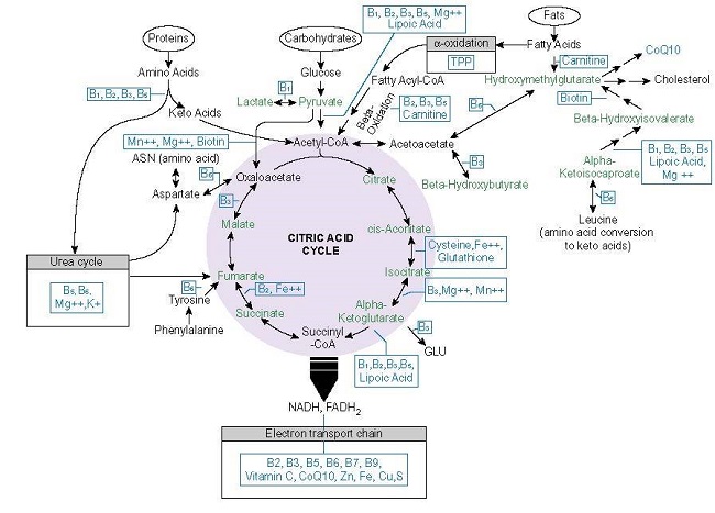
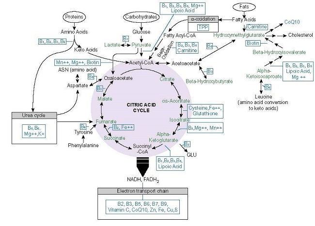
The most fundamental process to health and survival involves the conversion of consumed nutrients into ATP. Absent adequate ATP, health is impossible. Energy metabolism requires a ready supply of macronutrients (carbohydrate, protein, and fats) and at least 22 micronutrients or vitamins and minerals (see Figure 1.).

In developed countries, macronutrients are readily available, often in excess. Micronutrient intake, however, is inconsistent. A review article from the University of Oregon report found that a large percentage of the population had inadequate micronutrient status (4-65% depending upon the nutrient) despite excessive caloric intake. Moreover, much of the supposed nutrient sufficiency came from enriched or fortified foods. In other words, absent food enrichment or fortification, most children, adolescents, and adults had insufficient micronutrient intake. Inasmuch as most fortified foods come with a high caloric content, which effectively demands a higher micronutrient content to metabolize it; this presents a problem.
mitochondrial nutrients
Figure 1. Mitochondrial Nutrients, from: Thiamine Deficiency Disease, Dysautonomia, and High Calorie Malnutrition

Thiamine Dependent Enzymes

From the graphic above, note how many times thiamine (vitamin B1 or TPP) appears. Thiamine is required for the transketolase (TKT), pyruvate dehydrogenase complex of enzymes (PDC), branched chain keto acid dehydrogenase (BCKAD), 2-Hydroxyacyl-CoA lyase (HACL), alpha-ketoglutarate dehydrogenase ([a-KDGH] – also called 2-oxoglutarate dehydrogenase complex [OGDC]) and for lactate recycling as a cofactor for the lactate dehydrogenase complex (LDH). Beyond its coenzyme role, thiamine allosterically regulates the expression and activity other mitochondrial proteins including:

  • Succinate thiokinase/succinyl-CoA synthetase: together with a-KDGH catalyzes succinyl-CoA to succinate.
  • Succinate dehydrogenase: oxidizes succinate to fumarate, uses the electrons generated to catalyze reduction of ubiquinone to ubiquinol for complex II (TCA>ETC linkage)
  • Malate dehydrogenase (MDH): interconversion of malate and oxaloacetate with cofactor NAD+ or NADP+.
  • Pyridoxal kinase: converts dietary vitamin B6 into the active cofactor form pyridoxal 5′-phosphate (PLP) creating a functional deficiency.

With low or absent thiamine, each of these enzymes downregulates from 10% to almost 30% resulting in a reduction of ATP from 38 to ~13 units (in culture).

Thiamine Is Fundamental

Among the 22 micronutrients needed to convert macronutrient ATP, thiamine, along with its cofactor, magnesium, sit at the entry points to this process. That means that thiamine availability controls the rates of carbohydrate, protein, and fat metabolism and their subsequent conversion into ATP. Insufficient thiamine, even marginally so, impedes this process resulting in not only reduced ATP, but also, impaired cellular respiration, and increased oxidative stress and advanced glycation end products (AGEs); the very cascades linked to the preponderance of modern diseases dominating the healthcare landscape.

  • Cellular respiration, the ability to use molecular oxygen, requires ATP, which requires thiamine. Insufficient thiamine causes cell level hypoxia and upregulates the expression of hypoxia inducible factors (HIFs). HIFs are responsible for oxygen homeostasis, regulating at least 100 other proteins including those involved in angiogenesis, erythropoiesis and iron metabolism, glucose metabolism, growth factors, and apoptosis. HIF stabilization is implicated in a range of illnesses from autoimmune disease, to heart disease and cancer.
  • Reactive oxygen species (ROS) are a natural byproduct of ATP production and serve as useful mitochondrial signaling agents. Elevated ROS, relative to antioxidant capacity, however, creates oxidative stress, damaging cellular lipids, proteins and DNA. Antioxidant capacity is reduced with thiamine deficiency while ROS are increased.
  • AGEs, the toxic byproducts of hyperglycemia and oxidative stress, are modulated by thiamine. With sufficient thiamine, AGE precursors are shunted towards energy metabolism via the transketolase and the pentose phosphate pathway rather than accumulating in tissue as reactive carbonyl intermediates common with metabolic disease.

Each of these play a role in the pathophysiology of diabetes, cardiovascular and neurodegenerative diseases. This makes thiamine status, by way of its role in ATP production, cell respiration, ROS management, and AGE metabolism, a critical variable determining health or disease.

Given its position and role in these processes, it is not difficult to imagine how insufficient thiamine intake might derange and diminish energy metabolism and how that, in turn, might impact metabolic health both locally at the cell, tissue and organ level, and systemically. What is difficult to imagine, however, given the miniscule RDA requirement for a little over a single milligram of thiamine, is how anyone in the developed world where food scarcity is rare, where thiamine is readily available in both whole foods and in fortified foods, becomes thiamine deficient. And yet, a growing body of research suggests that is exactly what is happening. Recall from above, that depending upon the population studied, insufficient thiamine to frank deficiency has been found in 20-98% of the patients tested.

Modern Challenges to Thiamine Sufficiency From Consumption to Utilization

As an essential nutrient, thiamine must be consumed from foods, absorbed, activated and transported to where it is needed, and then utilized by its cognate enzymes. At each of these steps there are challenges that diminish thiamine availability, effectively increasing thiamine need well beyond the current RDA values. In fact, many of the products and amenities that make modern living what it is, imperil thiamine status and do so at multiple junctions. The additive effects of these challenges leaves many vulnerable to deficiency.

Dietary Sources of Thiamine

The highest concentrations of thiamine in natural and non-manufactured foods come from pork, fish (salmon, trout, tuna, catfish), many nuts and seeds (macadamia, pistachios, sunflower seeds, flax seed), beans (navy, black, black-eyed peas, lentils), peas, tofu, brown rice, whole wheat, acorn squash, asparagus, and many other foods. A diet rich in organic, whole foods is generally sufficient to meet the daily requirements for the thiamine and other vitamins and minerals. Likewise, though less ideal, a diet of processed foods that has been enriched or fortified with thiamine, will meet the RDA for thiamine quite easily, perhaps even exceed it. Indeed, one serving of breakfast cereal is sufficient to reach the RDA for thiamine.

Despite the ready availability of thiamine in both whole and processed foods, the data suggest that many people find it difficult to maintain thiamine status. This is due to the interactions between the endogenous chemistry of thiamine metabolism and the chemistry of exogenous variables affecting thiamine stability. The most common factors affecting thiamine status, include high calorie, high toxicant load diets, alcohol and/or tobacco use, caffeine products, and pharmaceutical and chemical exposures.

Dietary Impediments to Thiamine Sufficiency

While fortification provides access to thiamine, highly processed foods carry a high calorie and toxicant count making them metabolically deleterious despite any potential gains from vitamin enrichment or fortification. High carbohydrate, highly processed foods diminish thiamine status by multiple mechanisms.

Other common dietary contributors to insufficient thiamine.

Although food scarcity is not as prevalent in developed countries compared to undeveloped regions, poverty still impacts nutrient status. This owes largely to the fact that highly processed foods, high calorie foods are less expensive than whole foods and thus, there is an over-reliance on carbohydrate consumption to meet caloric requirements. Here, obesity and metabolic dysfunction co-occur with micro-nutrient and sometimes macronutrient, e.g. protein, deficiency.

Pharmaceutical and Environmental Threats to Thiamine Status

After high calorie malnutrition and other dietary habits that limit thiamine availability, the next most common threat to thiamine sufficiency is the use of pharmaceuticals. This variable cannot be stressed enough. Pharmaceutical chemicals deplete thiamine and other nutrients, directly or indirectly by a number of mechanisms.  Some of this is by design, such as with antibiotics that target folate and thiamine, some of it represents off-target effects, such as the blockade of thiamine transporters by metformin and the other 146 drugs tested for this action, an increase in demand in order to withstand other mitochondrial damage. Regardless of the intended purpose, however, pharmaceuticals represent chemical stressors to thiamine and nutrient stability. As such, their regular use necessitates a concerted approach to maintain nutrient status. Some of the most commonly used medications are the biggest offenders:

In addition to the ingestion of pharmaceutical chemicals, environmental chemical exposures damage mitochondrial functioning, even at low, and what are considered, non-toxic exposures. These exposures are pervasive, often unavoidable, and tend to accrue over time, with additive and synergistic effects to other stressors. Consider the totality of a patient’s toxic load when addressing the risk of nutrient insufficiency.

Absorption and Metabolism

Assuming sufficient thiamine is ingested from diet and is not blocked or otherwise degraded by food, pharmaceutical or environmental chemicals, it then has to be absorbed in the intestines before it can be activated and transported to organs and tissues for use. Epithelial injury, microbial dysbiosis, and genetic variation, all of which are common, limit the effectiveness of this phase. Epithelial injury and microbial dysbiosis slow passive absorption, while genetic, epigenetic, and environmental variables, slow or block active transport.

At low concentrations, thiamine is absorbed in the small intestine by active transport, while higher concentrations are absorbed by passive diffusion. Active transport is mediated by two primary thiamine transporters, ThTR1 and ThTR2, and a number of additional transporters that fall under the solute carrier family of genes:

  • SLC19A1: folate transporter, but also, transports thiamine mono- and di- phospho derivatives.
  • SLC19A2 (ThTr1): systemic thiamine transport, main transporter in pancreatic islet tissue and hematopoietic cells; most abundant, from highest to lowest in the intestine, skeletal muscle, nervous system, eye, placenta, liver, and kidney.
  • SLC19A3 (ThTr2): primary intestinal thiamine transporter, also located in adipose tissue, breast tissue, liver, lymphocytes, spleen, gallbladder, placenta, pancreas, and brain.
  • SLC22A1 (OCT1): organic cation transporter 1, primary hepatic thiamine transporter; competitively inhibited with transport of metformin, xenobiotics, and other drugs.
  • SLC25A19 (MTPP-1): mitochondrial thiamine pyrophosphate carrier.
  • SLC35F3: endoplasmic reticulum and Golgi thiamine transporter, implicated in hypertension.
  • SLC44A4 (hTPPT/TPPT-1): absorption of microbiota-generated thiamine pyrophosphate in the large intestine.

Although conventional wisdom suggests that only homozygous mutations affect the performance of these proteins, in reality, there is a gradation of abnormalities that challenge thiamine uptake, particularly when environmental or pharmaceutical variables block or otherwise limit the functioning of the same protein. In some cases, genetic difficulties can be compensated for providing nutrient support at supraphyisiological doses, among the better known examples:

  • Thiamine responsive megaloblastic anemia (mutations in SLC19A2/ThTr1); megaloblastic anemia, progressive sensorineural hearing loss, and diabetes mellitus.
  • Biotin-thiamine responsive basal ganglia disease (mutations in SLC19A3/ThTr 2) presents in infancy or childhood with recurrent subacute encephalopathy, confusion, seizures, ataxia, dystonia, supranuclear facial palsy, external ophthalmoplegia, and/or dysphagia or Leigh-like syndrome with infantile spasms. When presenting in adulthood, acute onset seizures, ataxia, nystagmus, diplopia, and ophthalmoplegia.
  • Thiamine responsive Leigh Syndrome (mutations in in the SLC19A3/ThTr2).
  • Thiamine metabolism dysfunction syndrome-4 (mutations SLC25A19/MTPP-1); episodic encephalopathy and febrile illness, transient neurologic dysfunction, and a slowly progressive axonal polyneuropathy.
  • Thiamine Pyrophosphokinase 1 (TPL1) defects cause problems in the activation of free thiamine to thiamine pyrophosphate, rendering much of the thiamine consumed unusable. TPK1 defects have been identified as condition called thiamine metabolism dysfunction syndrome 5 or Leigh-like syndrome because of the similarity in symptoms. More recently, TPK1 defects have been found associated with Huntington’s disease. High dose thiamine appears to overcome the defect in some cases.

Thiamine Activation/Deactivation

Before it can be used, free thiamine has to be phosphorylated into its active form thiamine pyrophosphate (TPP), also called thiamine diphosphate (ThDP/TDP). This is done by the enzyme thiamine pyrophosphokinase (thiamine diphosphokinase), which is magnesium dependent and requires ATP. Magnesium deficiency is common in developed countries. TPP accounts for almost 90% of circulating thiamine.

Additional thiamine metabolites include thiamine monophosphate (TMP) and thiamine triphosphate (TTP) along with the recently discovered adenosine thiamine triphosphate (AThTP) and adenosine thiamine diphosphate (AThDP). AThTP and AThDP are produced by E.coli during periods of nutrient starvation and have been found in most mammalian tissue. This likely represents a salvage pathway common in many pathogenic microbes.

Microbial Thiamine Synthesis

It is important to note, that although the consumption of dietary thiamine provides the main sources of this nutrient systemically, a smaller, but notable (2.3%), percentage of thiamine and other B vitamins is produced endogenously by various commensal bacterial populations in both the small and large intestines. At least 10 species of bacteria synthesize thiamine that is absorbed and utilized by the colonocytes. Endogenous thiamine synthesis is reduced by diets high in simple carbohydrates but increased with complex carbohydrates. Antibiotics and other medications inhibit endogenous synthesis of B vitamins directly by design as in the case trimethoprim and sulfamethoxazole and indirectly via additional that disrupt thiamine availability. Additionally, a number of pathogenic microbes produce enzymes that degrade bacterially produced thiamine suggesting the balance of gut biota is influenced by and influences nutrient availability.

In the large intestine, bacterially synthesized TPP is absorbed directly into the colon via a population of TTP transporters (TPPT-1) in the apical membrane and then transported directly into the mitochondria via the MTPP-1 for ATP production. The reduction of colonocyte thiamine and thus ATP, would force a shift towards the more pathogenic microbial populations that thrive in nutrient deficient environments and dysregulate bowel motility. This local thiamine deficiency may be a contributing factor in large bowel microbial virulence and the dysmotility syndromes so common in modern medical practice.

Enzyme Activation

The final step in attaining thiamine sufficiency is utilization. Returning to Figure 1., the key enzymes involved in this process include: TKT, PDC, HACL, BCKAD, a-KGDH and LDH.  This is an addition to the enzymes involved in the phosphorylation of free thiamine and the remaining enzymes in the Krebs cycle whose gene expression depends upon thiamine status. As with the variances and mutations in the transporters, supraphyisiological doses of thiamine may compensate for decrements in enzyme function. This has been observed in thiamine responsive PDC deficiency, characterized by excessive lactic acid; and in maple syrup urine disease, where mutations in the thiamine dependent BCKAD enzyme responsible for amino acid metabolism is impaired; also in Leigh-like syndrome, where mutations in TPK1 enzyme, which converts free thiamine to active TPP, is affected.

Is the Thiamine RDA Sufficient?

Both the chemistry and the data suggest that the current RDA of just a single milligram of thiamine is insufficient to meet the challenges presented by modern diets and chemical exposures. Owing to its role in energy metabolism, thiamine insufficiency may underlie many of the disease processes associated with metabolic dysfunction, where cellular hypoxia, increased ROS and AGEs are present. These disease processes develop long before, and sometimes absent, frank deficiency suggesting there may be gradations of insufficiency relative to the individual’s metabolic needs. Whether thiamine is a causative variable in these disease processes or simply a consequence of a complicated history of negative interactions between genetics, diet, and exposures is unclear. What is clear, however, is that thiamine insufficiency is likely far more prevalent than recognized and given its role in energy metabolism, ought to be addressed more consistently in clinical care.

We Need Your Help

More people than ever are reading Hormones Matter, a testament to the need for independent voices in health and medicine. We are not funded and accept limited advertising. Unlike many health sites, we don’t force you to purchase a subscription. We believe health information should be open to all. If you read Hormones Matter, like it, please help support it. Contribute now.

Yes, I would like to support Hormones Matter. 

Thiamine Deficiency in Modern Medical Practice

11.8K views

Modern medical practices are plagued with patients who present with chronic, complex, and seemingly treatment resistant illness; illnesses that defy most laboratory testing and abound current diagnostic categories. Some data suggest that 25-75% of symptoms experienced by these patients fall under the umbrella of ‘medically unexplained symptoms’.

What if some of those symptoms were not only explainable but treatable and the expression of these illnesses manifested not from some complicated new disease but from a simple but forgotten nutrient deficiency? What if this nutrient was uniquely critical to mitochondrial competence such that its insufficiency would derail energetic capacity, affect cellular function broadly and diversely, and produce many of the symptoms currently ascribed as medically unexplained? Wouldn’t this be worthy of investigation in your patient population?

That nutrient is thiamine or vitamin B1 and it is essential to mitochondrial energetics – the conversion of food into adenosine triphosphate (ATP). This process is the backbone of all health, and absent sufficient thiamine, it grinds to a halt producing many of the diseases processes vexing modern medicine.

Thiamine is a critical and rate-limiting cofactor to five key enzymes involved in this process, including those at the entry points for the glucose, fatty acid, and amino acid pathways. It has a very short half-life (1-12 hours), limited storage capacity, and is susceptible to depletion and degradation by a number of products that epitomize modern life.

When thiamine is insufficient to overcome these variables, oxidative metabolism falters and the ability to generate molecular energy declines. Over time, aerobic respiration turns anaerobic, oxidative stress increases, and cellular, tissue, and organ function dependent upon steady state energetics deteriorates.

Anaerobic glycolysis, the telltale sign of everything from general metabolic dysfunction to cancer, is, at its root, an adaptive response to insufficient micronutrients like thiamine. Replenish thiamine, recover mitochondrial capacity, and aerobic metabolism and health improve.

Critically Ill Versus Walking Sick: Gradations of Insufficient Thiamine

Conventionally, thiamine deficiency syndromes have been described relative to overt, and often later stage illness in the hospital setting. The most common designations include: Wernicke’s encephalopathy marked by nystagmus, ataxia, and cognitive deficits; wet beriberi or high output cardiac failure with edema and dry beriberi, central and peripheral nervous system and cardiovascular disturbances without edema. More recently, sensorimotor polyneuropathy or neuritic beriberi, gastrointestinal dysmotility syndromes, and the dysautonomias have been included in the spectrum, but recognition is lagging.

These designations give the false illusion of a disease process that happens acutely and one that can be categorized by the afflicted organ system. Neither is accurate. While overt thiamine deficiency is certainly a medical emergency and may sometimes develop acutely, the vast majority of cases represent a culmination of years, if not decades, of insufficient thiamine intake relative to need. Until fulminant, these disease processes are marked by low mortality, but high, chronic, and polysymptomatic morbidity. This suggests ample opportunity to treat and prevent more serious illness, improve the patient’s quality of life, and possibly even regain health. Even in overt and emergent cases, where symptomology is obvious, resolution is possible with thiamine repletion.

Thiamine Depleting Factors

Thiamine deficiency is most commonly associated with food insecurity and chronic alcoholism; a narrow view that risks missing early signals of accruing disease across patient populations. Contributors to this deficiency are far more prevalent in first world countries with westernized food production than is recognized. Among the key dietary contributors to insufficient thiamine:

  • Alcohol
  • Tobacco
  • High carbohydrate, highly processed foods
  • Coffee, tea, energy drinks

Additionally, the regular use of common medications and/or exposures to environmental chemicals independently and synergistically provoke thiamine deficiency. Every medication and environmental chemical depletes thiamine directly or indirectly by a number of mechanisms including blocking thiamine uptake, increasing its degradation, preventing synthesis in gut microbiota, increasing excretion and/or by inducing mitochondrial damage by other means that then necessitates a higher thiamine intake to compensate. Some of the most commonly used medications are the biggest offenders:

Sadly, poor dietary habits trigger thiamine insufficiency independently, leading to the prescription of many of these medications, which then further derail thiamine status and mitochondrial capacity. It is an illness spiral that can only be resolved by addressing diet and mitochondrial nutrients like thiamine.

Genetic Contributors to Thiamine Deficiency

While thiamine deficiency diseases are predominantly attributable to diet and lifestyle variables, a number of common genetic polymorphisms in the solute carriers responsible for thiamine uptake, and in enzyme activity involved in thiamine metabolism, increase the demand for thiamine intake. In these cases, disease expression, particularly later in life, represents a latent genetic vulnerability triggered by environmental or lifestyle stressors. Many medication and vaccine reactions fall into this category.

Prevalence Across Patient Groups

Inasmuch as thiamine status is not regularly evaluated in clinical care, it is difficult to know how pervasive thiamine deficiency is within the general population. Moreover, there are no universally accepted cutoffs demarking the progression from suboptimal to frank deficiency. Of the data that do exist, it is likely far more common than recognized across a broad swathe of patient populations.

Strikingly, diabetes confers one of the largest risks for thiamine deficiency across patient populations. This is largely do to metabolic derangements (to be discussed in a subsequent post) initiated by the hyperglycemia. These include the increased excretion of thiamine, and interestingly, the endogenous production of the anti-thiamine molecule oxythiamine.

Thiamine Testing

Laboratory assessment of thiamine status varies in sensitivity and specificity, with some tests carrying a high false negative rate (standard serum and plasma), particularly when thiamine status is marginal and with recent intake of thiamine. The two most sensitive tests are whole blood HPLC and the erythrocyte transketolase activity/thiamine pyrophosphate effect combination, neither of which is readily available. Urinary organic acid tests, while indirect, may provide useful patterns for determining the need for thiamine and other mitochondrial nutrients.

How To Recognize Thiamine Insufficiency

In light of the difficulties associated with laboratory testing, clinical acumen is required. Given its role in energy metabolism, lack of energy, in multiple manifestations, is a cardinal indicator of insufficiency.

  • Chronic fatigue, muscle weakness, or pain
  • Hypersomnia or anorexia
  • Dysautonomic reactions – exaggerated, ill-timed, or inadequate autonomic responses to stressors, most notably in the brain, heart and/or GI system

Office observations to support thiamine insufficiency:

  • Subtle changes in gait, stability, muscle tone, speech, decrements cognitive or affective acuity or stability
  • Asymmetrical pulse pressure, postural hyper- or hypotension, general tachycardia (early stage), bradycardia (later stage)

Standard labs pointing to problems with energy metabolism:

How to Treat

While clinical practice guidelines exist for overt thiamine deficiency in hospital, which include the use of IV thiamine and additional nutrients at a range of doses dependent upon severity, there are no established guidelines for out-patient thiamine deficiency or insufficiency syndromes. This is partly due to its lack of recognition and partly due to the fact that individual need for thiamine, other mitochondrial co-factors, and response to repletion, varies considerably.

There are no known toxicities to high doses, however, there can be negative reactions in the initial phases of thiamine repletion for a subset of patients. These reactions can occur at any dose. In some cases, the reaction involves the specific formulation of thiamine. In other cases, electrolyte disturbances and/or other micronutrient deficiencies unmasked by thiamine are at fault. To mitigate these reactions, thiamine should always be given with magnesium (~50% of the population consumes less than the RDA and magnesium is required to activate thiamine), a clean, lower dose multi-vitamin and a potassium rich diet. It should be noted that additional calcium may also be needed (here, here), especially when dietary calcium has been low for an extended period. Hypophosphatemia may develop as well in patients with recent or extended GI illnesses and/or have a history of low protein consumption and sodium disturbances are also common.

Consider Thiamine

Thiamine is a safe, non-toxic, essential nutrient that has become increasingly difficult to maintain in the face of modern dietary practices and chemical exposures. Thiamine sufficiency is fundamental to energy metabolism, mitochondrial capacity, and thus, health. Consider thiamine in your practice.

We Need Your Help

More people than ever are reading Hormones Matter, a testament to the need for independent voices in health and medicine. We are not funded and accept limited advertising. Unlike many health sites, we don’t force you to purchase a subscription. We believe health information should be open to all. If you read Hormones Matter, like it, please help support it. Contribute now.

Yes, I would like to support Hormones Matter. 

Healing From Lupron and Endometriosis With Thiamine

11.1K views

I was diagnosed with stage 4 endometriosis in 1996. In 2017, I was ready for a hysterectomy. I had two children and was finished with childbirth. I was having a lot of pain on my left side where my ovary was located. My Veterans Administration GYN refused to do a hysterectomy without first giving me Lupron shots assuming that my pain was due to the endometriosis. I was trying to hold down a very demanding government job and missing a lot of work from the pain. I had two laparoscopic surgeries in 1996 and 2001, respectively. Both were to excise the endometriosis. At the time, I was required to take Lupron in order to have a hysterectomy, I was 46 years old. I was denied a hysterectomy after my son was born in 2000 because I was considered too young at 30 years old to have a hysterectomy.

Endometriosis in the Colon and Lupron

After the injections of Lupron, a colonoscopy confirmed a diverticula pocket in that spot that was painful and others on my large intestine. The laparoscopy and excision in 1996 confirmed that my endometriosis extended to my large intestine. The colonoscopy found that I have so many pockets of diverticulosis, a resection surgery was not possible. Basically, if I were to become septic due to an endometriosis/diverticulosis flare, they would need to remove all of my large intestine. My options were very limited. My GYN wouldn’t perform a hysterectomy and laparoscopy under the assumption that the pain I was having was due to endometriosis. He convinced me to start the shots to see if they would help the pain because he assumed the pain was due to endo. I didn’t research the Lupron injections much prior to receiving them. I fully trusted my GYN. He mentioned hot flashes and suppression of symptoms with estradiol.

Immediately I noticed a difference. I don’t take prescription drugs of any kind unless I am really sick. I had nothing for any preexisting conditions. I could not tolerate the injections and function at work. I had severe hot flashes every few seconds 24/7 for three months even with add back estradiol. Worse, the estradiol made my migraines flare and so I was a hot mess. After stopping estradiol, my migraines continued to flare and still do without supplementation. I was also having diverticulosis flares every month sometimes twice a month. I had terrible gas and severe IBS symptoms. My work leave, FMLA and advanced sick leave were dwindling from all the visits to the various doctors. Within three months of my last Lupron injection, I was forced to retire or be fired for not being able to work. I never fully recovered from the Lupron.

Finally, a Hysterectomy

My GYN finally agreed to the hysterectomy in 2018 where they found my left ovary and left fallopian tube in one mass of adhesion scarring with my large intestine. The GYN removed the left polycystic ovary, left and right fallopian tubes along with my uterus, which had fibroids, and cervix leaving me with just my right ovary. Prior to the hysterectomy, I began noticing some numbness and cramping or burning in my feet at work that was much worse at night. I had the same kind of cramping and burning in my lower back too. I would later learn that these are symptoms of thiamine deficiency. Trying to keep it together at work with all of this was a nightmare.

Around this time, I also began having severe nausea and pain in my stomach. The GI doctors did an upper GI scope to confirm duodenal ulcers. The digestive issues, especially the diverticulosis should disqualify anyone from having Lupron as Lupron causes major digestive upset according to the FDA fact sheet. My digestive tract was inflamed from mouth to anus post Lupron. I had an inflamed esophagus and ulcers, diverticulosis flares, IBS with constipation and diarrhea and hemorrhoids that I couldn’t heal with meds. The low FODMAP diet helped though.

No More Pharmaceuticals

In 2019, I finally stopped taking all pharmaceuticals. No pharmaceutical made me feel better. Every medication I took for GI issues and neuropathy made me worse. I only took one for one or two weeks at a time to log all my side effects from each so I could have them added to my growing list of allergic reactions. I did have some sensitivity issues with prescription drugs prior to Lupron, just not as bad. I have the MC1R redhead gene. Redheads are more sensitive to pharmaceuticals and have more adverse reactions. I struggle with topical solutions as well. I couldn’t use estradiol patches because I’m allergic to the adhesive. Thankfully, my primary care physician also has endometriosis and suggested herbal supplements and remedies. All of this ,surprisingly, is from the veteran’s hospital. I was ordered by her to stop working. This was a final attempt to heal my ulcers, as they would eventually kill me if I could not find relief.

How I Healed Myself With Thiamine and Diet

I decided to try high dose thiamine after researching it via Drs. Lonsdale and Marrs and Elliot Overton. I started with 100mg daily for 6 months. Then 500mg for 3 months and currently 1000mg (500mg 2x daily). The thiamine works as well as the acupuncture with EMS. I also take Alpha Lipoic Acid and Dandelion root daily. The increases in thiamine are proving to be a significant factor in recovery. If I miss one day of supplements I’m sick for several days so I’m convinced that it is working.

To help myself heal, I no longer work a 9 to 5 job. I follow a low FODMAP diet with modification for diverticulosis and supplement with elderberry or dandelion for inflammation and immunity, turmeric, prebiotic + probiotics, magnesium for bone loss, palpations, anxiety, alpha lipoic acid for neuropathy, high dose thiamine for neuropathy, fatigue anxiety and brain fog, b vitamins and D3+K2 for b1 uptake regulation and delta 8 CBD for fibromyalgia pain and fatigue. I have regular chiropractor adjustments of my neck and lower back. Acupuncture and light therapy on my feet helped with the burning and cramping.

Where I Am Now

Currently, I have no endometriosis pain, only some lingering PMDD. I have no ovarian cysts and the migraines are not as frequent. Now only a couple a month versus weekly. I still have some burning and cramping in my legs and feet, but it is tolerable. Before thiamine, I was bedridden. The back and neck pain I had previously has improved with thiamine along with physical therapy/yoga and regular chiropractic care. I no longer experience diverticulosis flares with the new diet and supplements for inflammation like dandelion root, turmeric, and elderberry. I switch out the dandelion and elderberry because they work about the same. Depends on what is on sale.

I am able to stand for longer periods of time. My anxiety is significantly reduced, my palpations are gone, I can remember things, and my ADHD flare ups are minimal. In 2022, I only had two mild diverticulosis flares. Prior to the diet changes and supplements, I was having them once a month. I went from being bedridden completely to cooking (I still need to sit some), cleaning with short breaks, gardening with a sit on garden cart, and walking about a half mile every few days. I still have numbness in both feet. I am hopeful that lowering my A1C will resolve this. It may be permanent. Only time will tell. I’m going to the VA this week for a checkup and requesting more PT to see if it will help. They did an EMP on both legs with normal results. That was pretty painful but I felt nothing in my 3 little toes on both feet. Overall, I am doing much better with the higher dose thiamine and have much more energy.

We Need Your Help

More people than ever are reading Hormones Matter, a testament to the need for independent voices in health and medicine. We are not funded and accept limited advertising. Unlike many health sites, we don’t force you to purchase a subscription. We believe health information should be open to all. If you read Hormones Matter, like it, please help support it. Contribute now.

Yes, I would like to support Hormones Matter. 
Photo by Erol Ahmed on Unsplash.

This story was published originally on February 28, 2023.

Mitochondria Need Nutrients

27.9K views

One of the more common questions I get asked is which nutrients do the mitochondria need to function well? This is really two questions. The first involves which nutrients are involved in the enzymatic processes that allow the mitochondria to convert food to ATP and to manage all of the other tasks that they are responsible for like inflammation, immune function, and steroidogenesis. The second question applies specifically to the individual. It is a question of what he/she needs to be healthy. The answers to both are entirely different. While it is true that there are a set of nutrient co-factors involved in the mitochondrial machinery and these are necessary for mitochondrial function for everyone, which ones and how much of each an individual may need to support his or her health varies significantly. Moreover, although there are baseline minimum nutrient requirements that tell us where insufficiency diseases are likely to develop, what determines an individual’s health or disease is entirely dependent upon genetics, exposures, diet and lifestyle, and even day to day stress. Here, there is no one-size-fits all prescription for nutrient replacement and supplementation or even diet and exercise. This frustrates folks to no end and I think it is one of the reasons both patients and physicians are so reticent to look toward nutrient supplementation seriously as a therapeutic option.

Both the current model of medicine, and to a large degree, the way we approach nutritional therapies, relies very heavily on the silver bullet approach to health. If we’re honest with ourselves, so too do we. It is so much simpler to believe that if we just take X drug or vitamin in Y dose, all of our health issues will disappear and they will disappear at set rate that is linear and predictable. Unfortunately, this is not how the body works. While there is an internal chemistry that requires certain nutrients to function appropriately, that chemistry varies ever so slightly by genetics and is endlessly modified by life itself. There is no one-size-fit-all. There are no magic supplements. There is just your chemistry and your needs.

Since I have written repeatedly on the mitochondria and the reasons why nutrients are required for health, this post will not tackle those topics. Articles on those topics can be found on Hormones Matter with any number of search terms. This information can also be found in the book, Thiamine Deficiency Disease, Dysautonomia, and High Calorie Malnutrition, that I co-authored with Dr. Lonsdale. Here, since many have requested it, I just would like to present a graphic illustrating mitochondrial nutrient requirements. This is from Chapter 3 of our book. Use this as template to understanding your health.

Figure 1. Nutrient requirements for healthy mitochondria.

mitochondrial nutrientsA few things should be pointed out. First, while these nutrients are required by everyone for proper mitochondrial functioning, not everyone needs to supplement with each one, or even sometimes any of them, although that is becoming increasingly rare with modern dietary patterns. Secondly, notice how many times and where vitamin B1 (thiamine) appears in this chart. It is at the entry points of the entire system and at various junctures throughout. This suggests that among all of the nutrients required for healthy mitochondria, thiamine is particularly important. Unfortunately, it is the one nutrient that is so often ignored or missed in testing. Indeed, that is why we wrote the book. Thirdly, notice how many vitamins are required to process the food we eat into ATP. Contrary to popular opinion, we need more than simply empty calories. For the foods we eat to be converted into ATP, there are multitude of vitamins and minerals required that may or may not be included in sufficient density with the macronutrients we consume daily. Finally, not discussed in this chart, but discussed in great detail in the book, synthetic chemicals, whether in form of pharmaceuticals, industrial, environmental, or food production, damage the mitochondria. Some deplete nutrients directly, while others damage aspects of mitochondrial functioning that necessitate increased nutrient density for the enzyme machinery to work. Of course, underlying all of this, are the genetic variables that each of us brings to the table. These influence how well or poorly we metabolize any of these nutrients from the get-go. All of this combines to make nutrient therapies complicated.

What is not complicated, however, is that we need nutrients to function and so, no matter what else we do to improve health, if we do not address nutrient concentrations, we can never be well. Mitochondrial functioning demands nutrients, and thus, health demands the same. Nutrient deficiencies are not something we can override with a pharmaceutical. That being said, addressing nutrient deficiencies holds great promise for those seeking health. If you or someone you love experiences chronic and complicated illnesses that have been treatment refractory, consider healing the mitochondria by tackling nutrient deficiencies. You might be surprised at well this works.

We Need Your Help

More people than ever are reading Hormones Matter, a testament to the need for independent voices in health and medicine. We are not funded and accept limited advertising. Unlike many health sites, we don’t force you to purchase a subscription. We believe health information should be open to all. If you read Hormones Matter, like it, please help support it. Contribute now.

Yes, I would like to support Hormones Matter. 
Image credit: Free AI created illustration from Magnific

This article was originally published on November 11, 2019.

Treating Intractable Insomnia and Cerebellar Ataxia With Thiamine

12K views

Twenty years ago, I attempted suicide after years of alcohol and drug abuse. I almost succeeded, but by some miracle I survived. I suffered a cerebellar stroke and as result was left with severe cerebellar damage. The doctors said that I had lost almost 75% of my cerebellum. I couldn’t walk or talk, I could not swallow and was barely functioning, if you could call it that. At some point, I decided I would live and forced myself to improve. I worked the physical therapy and eventually began lifting weights. I wrote about my journey here. Despite my gains in strength and balance though, since the stroke I had suffered from severe and intractable insomnia. Although I was under the care of multiple doctors, none could offer any help beyond increasing this or that medication, none of which worked. Insomnia is common in individuals with cerebellar injury. Then I learned about thiamine.

Persistent Insomnia Treated with Thiamine

Around the time my initial article was published, I began supplementing my diet with thiamine. This vitamin may have been mentioned in passing by some of my physicians, but it was never prescribed or even really emphasized to any great degree. Neither was it ever touted by the medical community during my initial stroke recovery in 2003. I am fairly sure much of the reluctance to use thiamine could be attributed to the pervasive fear of it not benefitting my health.

Being entrenched in the world of mental health and alcoholism for about 25 years (sober 9), I am well aware of Wernicke-Korsakoff’s syndrome. I had heard about massive doses of thiamine being administered to others in medical detoxification facilities. I also have loved ones grappling with Parkinson’s Diseases. I quickly began to see how many of our neurological symptoms were similar, and that thiamine was slyly mentioned to help all of us.

In early 2021, it was strongly suggested that I gradually increase to a large dose of thiamine (not by doctors, mind you, but by others from this website). At the time, I was lucky if I slept 3 hours per night, which of course, exacerbated my ataxia symptoms. I was on so many medications that it raised red flags, but I was never warned about the negative neurological effects. And worse yet, nothing was working.

Once my initial dose of thiamine was entrenched in my system (50 mg of Thiamax), I began sleeping with greater ease. At first, this was approximately an hour, but I discovered that I also slept more soundly and had acquired greater rest during the night. As time progressed, my sleep patterns became increasingly regular.

Better Functioning With Improved Sleep

For several years, I have been making progress in all areas of my life. By August of 2022, thanks to the thiamine, I was off of all prescription medications. I was sleeping through the entire night, soundly. I was also able to complete difficult feats at the gym without falling asleep on a mat in the stretching area (usually resulting in me being taken home, so I didn’t sleep at the gym.) This is one of the reasons that I didn’t train publicly until recently. With my sleep problems, I wasn’t sure if I’d be overcome with an insatiable need to sleep mid-workout.

In addition to the improvements in sleep and the elimination of medication after thiamine came on board, I was able to lift a barbell by myself, while standing. This is no small feat for someone with ataxia. Remember, I was told I would not walk again, or function in any semblance perceived as normal. They said that all my neurological systems had crossed the threshold believed salvageable. In September of 2022, I competed in my first powerlifting meet, where I deadlifted 182 pounds and bench pressed 78 pounds. I am currently deadlifting 225 pounds (January 2023). While I am still ataxic and I still have struggles relative to my injury, I have improved so much since beginning the thiamine.

thiamine for insomnia and cerebellar ataxia
Me lifting 225lbs at the gym.

As I write this, right before the New Year of 2023, I am overcome with the amount of remarkable progress that has been made in my life since August. To deny that would fact would be denial of any amount of truth or reality in existence. I fail to see how anyone can deny the differences in my life since using regular thiamine.

We Need Your Help

More people than ever are reading Hormones Matter, a testament to the need for independent voices in health and medicine. We are not funded and accept limited advertising. Unlike many health sites, we don’t force you to purchase a subscription. We believe health information should be open to all. If you read Hormones Matter, and like it, please help support it. Contribute now.

Yes, I would like to support Hormones Matter. 

Photo by Jake Charles on Unsplash.

This story was published originally on January 17, 2023.

Medication and Vaccine Adverse Reactions and the Orexin – Hypocretin Neurons

20K views

A paper published in Science Translational Medicine, provides preliminary evidence that the H1N1 Flu Vaccine Pandemrix can evoke immune system mediated damage to the orexin – hypocretin neurons and induce narcolepsy in individuals with a particular genetic variant. The orexin – hypocretin neurons were only recently discovered in the mid 1990s, by two separate research groups, hence the two names for the same molecule. For this paper, we’ll be utilizing the orexin nomenclature.

Initially, the orexin neurons were thought to be involved only in feeding behavior, as damage elicited hypophagia in animals. Soon it was learned that more severe damage to the orexin neurons induced narcolepsy and the orexin system became a key focus in narcolepsy related research. With time, however, it became quite clear that these neurons were involved in regulating a myriad of hormone and neurotransmitter systems and their consequent behaviors. Narcolepsy or rather the ability to sustain wakefulness, is but one of the many functions regulated by the orexin system.

In a previous paper, I touched briefly on the possibility that the orexin neurons might be damaged and have diminished functionality in individuals suffering from post Gardasil side effects. In particular, I suspected these neurons were indicated in post-Gardasil hypersomnia, a derivative of narcolepsy. That may be only the tip of the iceberg. As I soon learned, the hypocretin/orexin neurons are brain energy sensors and may be involved in array of post medication or vaccine adverse reactions. Indeed, they may be central to the ensuing state of sickness behaviors that emanate once an organism becomes overwhelmed.

The Orexin – Hypocretin Basics

Orexin nuclei are located in the lateral hypothalamus, the section of the hypothalamus that is most known for regulating feeding, arousal and motivation. The hypothalamus is the master regulator for all hormone systems and hormone related activity including feeding, sleeping, reproduction, fight, flight, energy usage – basically every aspect of human and animal survival. It sits at the interface between the central nervous system functioning and the endocrine system functioning.

From the lateral hypothalamus, orexin neurons project across the entire brain with its two receptors (OXA and OXB) differentially distributed throughout the central nervous system and even in the body, including in the kidney, adrenals, thyroid, testis, ovaries and small intestine. The orexin neurons also modulate local networks of adjacent neurons within the hypothalamus that in turn influence a myriad of behaviors.

The most densely innervated brain regions include the thalamus, the locus coeruleus, dorsal raphe nucleus, accounting for the hormone’s role in arousal, feeding and energy management. At the most basic level, release of the orexin induces wakefulness. When orexin neurons are turned on and firing appropriately, arousal is maintained. When orexin neurons are turned off, diminished or dysfunctional, melatonin, the sleep promoting hormone, is turned on. The two work in concert to manage wakefulness and sleep.

Orexin receptors are also located in the amygdala, the ventral tegmental area (VTA) and throughout the limbic system, accounting for its role in emotion and the reward system. Orexin directly activates dopamine in the VTA. The VTA is the reward, addiction, and in many ways, the pleasure center of the brain. All drugs of addiction, all pleasurable activities, activate dopamine in the VTA. Through the release of dopamine, here and elsewhere, orexin modulates the motivation to sustain pleasurable activities. When orexin is diminished, not only does dopamine diminish, but the motivation to sustain behaviors decreases and dysphoria increases.

That’s not all. Orexin influences the release of many other neurotransmitters and hormones, several of which are co-located on the orexin neurons themselves. For example, the neuropeptide dynorphin is co-located on orexin neurons. Dynorphin is an endogenous opioid involved in the perception of pain and analgesia. It has dual actions that can both elicit analgesia or pain depending upon dose and length of exposure. Stress activates dynorphin. Dynorphin then inhibits orexin firing by as much as 50%. Illness is a stressor, a vaccine is a stressor, either could activate dynorphin and inhibit orexin. After the initial activation of dynorphin, and the ensuing decrease in orexin, the presence of chronic stressors and chronic pain could begin a continuous feedback loop of diminished arousal, and increasing pain.

Other Neurochemical Connections

  • Consistent with orexin’s role in arousal, orexin neurons contain glutamate vesicles. Glutamate is the brain’s primary excitatory neurotransmitter. Drugs that increase glutamate, also increase orexin. Drugs that block glutamate, via its NMDA receptor, decrease orexin. Common migraine medications block glutamate and thereby may also diminish orexin.
  • Serotonin and norepinephrine decrease hypocretin/orexin firing (suggesting if one is concerned with hypersomnia, anti-depressants might not be a good option).
  • As one might expect, orexin neurons are inhibited by GABAα agonists – sedatives. From a women’s health perspective, consider that cycling hormones would also affect orexin neurons through the GABAα pathway. Progesterone is a GABAα agonist – a sedative, while DHEA and its sulfated partner DHEAS are GABAα antagonists, anxiolytics that block GABAα, reduce sedation, and thereby increase anxiety and wakefulness. There may be a cyclical nature to orexin firing that has yet to be investigated.
  • The hypocretin/orexin neurons also influence galanin, a GI and CNS hormone that seems to inhibit the activity of a variety of other neurons in those regions.

These are but a few of the brain systems that the orexin neurons touch in some way or another. Damage to this system would have serious health consequences by initiating a cascade of biochemical changes within the brain and body. Many of which, we have yet to fully understand.

How Might the Orexin Neurons Become Inhibited?

Quite easily, apparently. In addition to the orexin’s vast interconnected pathways with a myriad of neurotransmitters and neuropeptides, the orexin neurons act as energy and activity sensors with some unique intracellular mechanics that make them especially sensitive to the changing dynamics of the extracellular milieu. Disruptions in ATP, glucose and temperature, elicit reactions in orexin functioning.

Orexin neurons require as much as 5-6X the amount of intracellular ATP to maintain firing, and to maintain a state of wakefulness or arousal. This extreme sensitivity to reduced ATP makes the orexin neurons uniquely positioned to sense and monitor brain energy resources, early, before ATP levels become critical in other areas of the brain. The orexin neurons cease firing when ATP stores become low, thereby allowing the reallocation energy, perhaps to those cells required for survival, breathing and heart rate. As Hans Selye observed many decades ago, one of the first, and indeed, most consistent of the sickness behaviors, no matter the disease, is lethargy, fatigue and sleepiness. Orexin is at the center of this behavior.

Orexin neurons react to extracellular glucose levels, though perhaps not as one might expect. When extracellular glucose levels are high, orexin neurons stop firing via what is called an inward rectifying potassium (K+) channel that is ATP dependent. That means that when extracellular glucose is high, intracellular ATP is allocated to open K+ channels and flood the cell with the inhibitory K+ ions. K+ hyperpolarizes the cell, prohibiting it from firing. This mechanism reminds me of Dr. Peter Attia’s talk about the nature of Type 2 Diabetes and our approach to treatment. He proposes that the body’s metabolic response – the conservation of energy – to Type 2 Diabetes is not something aberrant but is exactly as it should be with a disease state. We’re just not treating the correct disease state.

Another way we can shut down the orexin neurons is via increased temperature. The orexin neurons are very sensitive thermosensors. Increased temperatures shut down orexin firing via the inward K+ flow. Again, this is consistent with sickness behaviors and the reallocation of resources.

Orexin – Hypocretin Neurons in Migraine and Seizures

Diminished orexin has been linked to migraine and seizure activity. With migraines specifically, orexin may contribute to the early warning, hours to days, of impending cortical disruption via changes in feeding and sleep patterns that often precede migraine onset. Orexin may also be linked to the pre-migraine aura mediated by changes in brain electrical activity that prelude the migraine pain itself by minutes, called cortical spreading depression or more appropriately, cortical spreading depolarization – the massive spreading change in ion balance of the neurons. Initially the wave is excitatory, neurons are firing, but that is soon followed by a period of neural silence. Finally, orexin is also connected to the vasodilation of the trigeminal nerve, the nerve responsible for migraine pain. These findings have led some to call orexin a migraine generator.

Diminished cerebral spinal concentrations of oxerin have been found in patients generalized tonic-clonic seizures. Conversely, in rodent studies, injections of orexin elicit seizure activity. Despite the somewhat contradictory findings in seizure activity versus migraine activity, it is likely that the orexin system is involved both disease processes.

Pulling it all Together: Orexins Monitor and Mark Disruptions in Brain Homeostasis

Here’s where it gets really interesting. Although some have argued orexin, particularly diminished orexin functioning, is the cause and culprit of disruptions in brain homeostasis, leading to narcolepsy, excessive sleepiness, migraine, seizures and other diseases, I think this system represents merely a marker of a disease process. I think the orexin system is the stopgap, the final barrier of disrupted cellular energetics, of mitochondrial function. Mitochondrial ATP is the key.

When we consider orexin’s role in migraine, in particular, we see clearly how environmental changes (diet, stress, illness, medication/toxin exposure) can lead to changes in the extracellular milieu where orexins reside. The orexin sensors adjust to these changes, mostly by reducing neural firing in attempt to counteract damages. The reduction in orexin then elicits the premonitory phases of the impending brain disruptions, sleep and hypophagia – the sickness behaviors. If it progresses, the massive waves of electrical disruption ensue, and migraine, perhaps even seizures are evoked. When the extracellular environment become chronically disrupted, so too does the diminishment of orexin activity, thereby initiating a perpetuating loop of dysregulated brain activity. We can hypothesize that similar progressions exist with disease processes marked by aberrant electrical activity, such as epilepsy.

We know that mitochondrial dysfunction is often generated by genetic polymorphisms and can predispose individuals to an array of seemingly unrelated conditions like migraine and fibromyalgia, dysautonomias and cognitive deficits. At the root of the dysfunction is a error of some sort in mitochondrial energy processing – ATP.

What has become increasingly clear, is that the production of cellular energy, can be disrupted environmentally, by diet, illness and exposures, if co-factors necessary for the production ATP like thiamine are diminished. It is via diminished ATP production, that I think some medications and vaccines evoke adverse reactions in some individuals. The orexin system, because it is so exquisitely sensitive to changes in cellular energy, is our warning system; first by subtle changes in neurochemistry, then by changes in arousal and feeding behavior, and finally, by an all-out reallocation of resources – excessive sleeping. If ATP remains deficient chronically, and an individual is so disposed, then the cortical misfiring we see in migraine and seizure ensues, along with autonomic dysregulation and the syndromes associated therewith. It is not the orexin – hypocretin system that is at root of many of these diseases, but rather, the causes are deeper yet and reside with mitochondrial health.

We Need Your Help

More people than ever are reading Hormones Matter, a testament to the need for independent voices in health and medicine. We are not funded and accept limited advertising. Unlike many health sites, we don’t force you to purchase a subscription. We believe health information should be open to all. If you read Hormones Matter, like it, please help support it. Contribute now.

Yes, I would like to support Hormones Matter. 

Image created using Canva AI.

This article was published originally on January 29, 2014.

Lab Mice Get More Thiamine Than We Do

2.1K views

How much of our understanding of molecular mechanisms related to mitochondrial energy metabolism is wrong because the chow fed to the mice and rats used for this research is heavily supplemented with vitamins and minerals at quantities exceeding that of an equivalent human diet? I have been pondering this question for a few years now, but this morning the magnitude of the problem became clear when the pathway I had been studying was viewed positively when (hyper) activated in rodent research, but one that was clearly not so in humans. This is a pathway for which drugs had been designed to activate based upon the patterns identified in said research; research where the animals are fed a steady diet containing markedly higher concentrations of vitamins and minerals than humans will ever get, unless supplementing, and where the animals are sheltered from the bevy chemical stressors that deplete those same nutrients. So, on top of all of the other challenges associated with extrapolating data from animal research to humans, deriving data about the ‘normal’ behavior of molecular pathways that are incredibly sensitive to nutrient status and chemical exposures from well fed and environmentally isolated animals is extraordinarily problematic.

An Eye Opening Experience

As someone not trained in animal research, I never considered chow composition as influencing variable on all research. Sure, if the research question involved diet or nutrients specifically, the composition of the chow would obviously affect outcomes, but that there were would such stark differences in relative micronutrient content compared to humans is something I had not contemplated. A few years ago, however, I worked briefly with a foundation sponsoring some research into the various effects of thiamine supplementation on health. One series of investigations looked into genetic and metabolomic changes, along with variety of cognitive and behavioral measures, relative to a diet high in soybean oil, with and without thiamine supplementation. Since the foundation disbanded and the funding was cut, except for a few posters and abstracts, the results were never published. It was disappointing, because the results were remarkably enlightening – despite the issues with the chow.

That said, what struck me early on was the high intake of thiamine even in the control mice. In this particular set of experiments, the control mice were getting ~2.5mg/kg of bodyweight, while the mice in the two experimental ‘high thiamine’ groups were getting 51 and 102 mg/kg of bodyweight. This is an insane amount thiamine, even in the control group. By comparison, the average human eating a lot of processed and fortified foods, which is never a good idea, may get up to 4mg per day total and a large majority Americans get less than recommended daily allowance (RDA) of 1.1-1.2 mg/d. If he/she weighs 70kg that works out to between .02mg/kg and .06mg/kg daily – a minute fraction of what a lab mouse is given.

Why such a high intake of thiamine? It was explained to me that mice need more thiamine because they have a higher metabolism than humans.

Okay. Sure. But…

This never sat right with me, but not having the experience lab animals, I let drop and moved on. And then I began studying some molecular pathways that just happened to be influenced by thiamine and it occurred to me that conclusions drawn may not accurately reflect the true behavior of those pathways because of the chow fed to these animals. I began digging, and oh, what a mess this is. Not only do most of the published studies not list the vitamin concentrations in the particular chow used and make the assumption that all them contain equivalent amounts, they do not, but some may misinterpret the metrics. Most chow labels report nutrient doses in mg/kg of diet consumed by the animal. I suspect, in some studies they are using, or at least reporting, mg/kg of the animal’s body weight. Is the animal really consuming 5mg/kg of body weight or 5mg/kg of food? It is not always clear. Alternatively, other studies, including the one I reviewed for the foundation, scale the doses between the species using a multiple of 12. Apparently, this is to account for differences in body surface area. It is based upon the presumed rate of which organisms convert food into energy (heat dissipation) and it relies heavily on measures of body mass relative to caloric intake such that while larger animals consume more calories, their ‘metabolism’ by unit of mass is lower. This is a linear metric developed to estimate drug toxicity and may not accord with nutrient needs. Importantly, kcal burn and body mass are poor indicators of metabolic energy (here, here, here).

Reviewing methods sections from random studies that I have reported on in the past, there is no way to tell how much thiamine or other nutrients the animals actually consumed, unless the study was specifically designed to assess something related to thiamine. Even then, however, most studies assessing thiamine deficiency experimentally either use diets that include no thiamine and/or induce deficiency by administering anti-thiamine molecules like oxythiamine or pyrithiamine. That said, whatever the actual amount, even at the lowest possible intakes to prevent deficiency, the animals were consuming far more than a human would consume, and this is what troubles me most. Do lab rodents really require more thiamine than humans or have we simply underestimated human requirements? I believe it is the latter, but let us explore the basis for this argument before drawing conclusions.

Thiamine Requirements For Mice

According to the literature, the daily thiamine requirement for mice used experimentally ranges from ~2ug of thiamine per day up to 6ug per day. This does not sound like much unless we consider that mice weigh only about 25g. This means that a lab mouse gets at least somewhere between .08 -.48 mg/kg. In a 70kg human, this equals 5.6 – 33 mg daily. The human RDA or DRI (daily reference intake) is only 1.1-1.2 for women and men respectively, per day, and accumulating research shows that many people do not even get that much. Nevertheless, by these metrics, the lab mouse is getting 5 -~30x the amount of what is considered necessary for the average human, and as was illustrated previously, if other metrics are used, these animals get even more thiamine.

This begs the question, how are we calculating the metabolic needs of these animals such that they require more nutrients than we do? It turns out that, like anything, the answer depends upon what assumptions we make and how we factor those assumptions into the math we create to estimate those values. As I mentioned previously, the research I reviewed was using a drug toxicity metric that included surface area differences to calculate dosages between humans and mice. This, apparently, is a common metric. Others include different allometric variables like bodyweight and/or caloric intake to calculate metabolism. All of these are linear calculations where the change in one variable is assumed to perfectly and linearly correspond to the other. Biology is not linear, and at some point researchers recognized this and began multiplying the exponents of the body surface area. Needless to say, multiplying by different exponents changes the result dramatically. Whether it changed its accuracy, however, is questionable.

Just How Much Does Nutrient Intake Vary?

Per a review of such things, rodents get between .2 -.26ug of thiamine daily with three different chow formulas. The human equivalent dose (HED) for these values ranges anywhere from 2.8 mg/d to 69 mg/d depending upon which variables are included in the calculations.

For example, if the calculation considers kcal required per day to maintain function (a 25g mouse requires 15kcal daily, while a 70 kg human requires 2000 kcal per day), the human equivalent for daily thiamine intake would be from 2.8-3.3 mg – double and triple the current RDA/DRI. In contrast, if the calculation uses body weight as the key variable, the human equivalent would be 56-69mg per day, or more, as bodyweight increases. This is ~50x more than current RDA/DRI values.

Conversely, if we run the math backwards, the 1.2 mg per day thought to be acceptable male humans weighing 70kg equals only .017mg/kg or .0004mg per day total – clearly below the intake of the chow fed lab mouse (likely below that of the wild mouse), but also, well below what is required prevent deficiency in the mouse. Although I am focusing on thiamine, because that is what I know best, the issues with scaling dosages between mice and humans carries the same problems for each of the micronutrients.

extrapolations of nutrients from mice to humans overestimate the nutrient intake for a human, while scaling for humans to mice would underestimate nutrient intakes for a mouse and theoretically result in nutrient deficiencies.

I will ask the question again: are we really overestimating nutrient doses when scaling from mice to humans or have we fundamentally underestimated the nutrient requirements of humans all along?

The Development of Human Nutrient Requirements

The nutrient estimates of humans to which all animal research is compared is equally flawed but for entirely different reasons than those of animal estimates. Not only was metabolism not considered as variable in human dosing, and is thus not scalable in either men or women of difference sizes or from humans to mice or any other species, but our assumptions regarding the accuracy allometric models of metabolism are likely incorrect as well.

Nutrient requirements for humans were developed using food surveys of different populations. From the average caloric intake of an average man (70kg), thiamine and micronutrient consumption more generally, were estimated. Adequacy was determined purely upon the absences of observable symptoms of deficiency. A number just above the point at which deficiency symptoms were observable became the recommended daily dose. Doses for women and for pregnancy were largely guestimates based upon presumed differences between male and female size, activity levels, and calorie consumption.

There was never a mg/kg dosing strategy developed for micronutrients for humans, so there is no ability to scale relative to bodyweight, caloric intake, or surface area, as there is with lab animals. Neither were there ever any attempts to calculate the actual metabolic rates relative to micronutrient needs, then or since. The human micronutrient standards were developed based entirely on observational data and the absence of deficiency symptoms. Like many accepted medical truths, there was no math, just observation, some contention, but ultimately, consensus, and eventually, entrenchment – even when data suggested otherwise. Notably, there have been no changes to the RDA/DRI for thiamine for over 80 years.

That said, how do we really know if the thiamine requirements for humans are less than that of rodents? Could the recommended dose in humans, which causes deficiency in mice, also be causing deficiency in humans but the complexity of our biology and variability of our environment, paired with the longevity differences between the species, mask that deficiency – sometimes indefinitely? Possibly.

A Question of Metabolism

And then there is this: how do we really now that mice have a higher metabolic rate than humans? We don’t, and that is the other problem with scaling micronutrient needs between rodents and humans. We simply have no logical basis upon which to make these calculations. Certainly bodyweight, surface area and caloric intake differences are important, but those variables do not, in any way, address the fundamental question about metabolism, which is energy used. Energy is the basis for metabolism. Energy usage should be in the calculations we use to estimate nutrient requirements and it is not.

In study published in 2024, researchers did something almost heretical – they added energy variables to metabolic calculations and standardized for body mass, fat free mass, and environmental temperature to more appropriately estimate the energetic needs of rodents, versus other animals, versus humans. In doing so, they were able to calculate expenditures with a common metric that could compared across species: megajoules per day (MJ/d). From there, they calculated total energetic expenditure (TEE), resting energetic expenditure (REE) and active energetic expenditure (AEE).

By these estimates, humans have higher metabolic demands than rodents (and many other animals including apes). Per their research, the TEE of humans globally was ~27% greater than that of rodents. In the US, where the population is largely sedentary and overweight, TEE for humans was still 18% higher than that of rodents. While the AEE are largely the same between rodents and humans (~1% difference), the differences in resting metabolic needs are huge. Humans require 40-45% more energy at rest (US and global, respectively) than rodents.

Per the authors of the study, humans evolved to be an ‘energetically extravagant species’ due in part to our larger brains, which consume an awful lot of energy, even when we do not use them. None of the previous studies considered this variable (or several others) when calculating the presumed metabolic differences between the species.

If humans do indeed have higher metabolic needs than rodents, then shouldn’t we also need comparably higher micronutrient intakes to sustain optimal energetic capacity?

Comparing Well Nourished Mice to Malnourished Humans

To the original question that sent me down this rabbit hole, how do we know what we think we know about the activity of different nutrient- sensitive and nutrient – dependent proteins, either in isolation or within a pathway, when we study those proteins in animals that are very well nourished and the species to which we are extrapolating those findings is most decidedly not? And a question that I did not address, how do we know what we think we know when the study animals are never exposed to the bevy of nutrient depleting chemicals that the human is exposed to? We do not, and that is the problem.

This means that all of the conclusions I (and others) have drawn based upon animal research, where the animals receive far and above the nutrients that a human might, are likely skewed, if not entirely incorrect. That is a sobering realization, particularly when one considers all of the other problems associated with extrapolating data from lab animals to humans. Moreover, this is yet another reason why humans need more thiamine and other nutrients than are currently recommended by governmental entities.

We Need Your Help

More people than ever are reading Hormones Matter, a testament to the need for independent voices in health and medicine. We are not funded and accept limited advertising. Unlike many health sites, we don’t force you to purchase a subscription. We believe health information should be open to all. If you read Hormones Matter, like it, please help support it. Contribute now.

Yes, I would like to support Hormones Matter. 

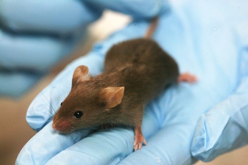
Image: Rama, CC BY-SA 2.0 FR, via Wikimedia Commons

Poor Nutrition Stress: The Enemy of Health

12.9K views

In previous posts, I have indicated that stress can initiate or exacerbate disease and medication or vaccine adverse reactions. Read that statement, you might think I am attributing the onset of serious disease and adverse reactions to a psychosocial cause. That is not the case. Stress comes in a myriad of forms, some external, some internal, and although much of what we call stress relates to psychosocial responses to perceived threats, I think stress encapsulates so much more. At its most fundamental level, stress represents a physical state where the body is performing less than optimally. Let me explain.

What is Stress?

I define the word “stress” as a physical or mental force that is acting upon you. An example of mental or psychosocial stress might be an insult from a person, meaning that the stress comes from a source outside the body. On the other hand, it might be the realization that a deadline has to be met, a mental source from within. Any form of injury is an obvious source of physical stress. Physical action such as shoveling snow is another form of stress, demanding energy consumption imposed by the individual who wishes to get rid of the snow. Being infected with a virus or by bacteria is a form of stress that demands a defensive reaction. In each of these instances, the body reacts to the inflicting stressor. Sometimes, when the resources are available, it reacts efficiently. Other times, when the resources are not available or when additional factors intercede, the body’s response to the stress is ill-adapted.

Your Body is Your Fortress, Your Immune System the Soldiers

Perhaps an analogy might help to provide an explanation for the remarks that follow. I imagine the body as being like an old fashioned fortress. The people living within it go into action when the fortress is attacked by an enemy from outside. It would be of little use if the defense soldiers went to the eastern battlements if the attack came from the west and so there had to be a central figure that would coordinate the defensive reaction. The nature of the attack would be spotted by a guard on duty and the central figure informed by messenger.

The body represents the fortress and the lower part of the brain represents the central figure that coordinates the defense. The cells in the blood known as white cells can be thought of as soldiers, armed with the necessary weapons to meet the nature of the enemy. Suppose, for example, a person’s finger is stuck by a splinter carrying a disease bearing germ. The pain, felt in the brain, recognizes its source and interprets it as a signal that an attack has occurred. White cells in the area can be regarded as the “militia under local command” and a “beachhead” is formed to wall off the attack. The white cells sacrifice themselves and as they die, they form what we call pus. If the beachhead is broken and the germs manage to get into the bloodstream, it is then called septicemia and the brain/body goes into a full defensive reaction where high fever is the most obvious result. Such an illness is an attack/defense battle.

The symptoms that develop from such an infection represent the evidence for this defense, feeling ill, pain and developing a fever are excellent examples. Micro-organisms are most efficient at 37° C, the normal body temperature. The rise in body temperature, initiated by the brain, makes the microorganisms less efficient and may kill some of them. One therefore has to question the time honored method of reducing the fever, during illness, as being an example of good treatment. While reducing fever improves the symptoms caused by the infection, it also reduces the efficiency of the immune battle raging within.

The outcome against the stressor is death or recovery; although it is possible sometimes to end up in a kind of stalemate, represented by prolonged symptoms of ill health. Chronic illness may be viewed as the immune system’s inability to eradicate fully the stressor.

Poor Nutrition and Stress

As I have emphasized in previous posts, the autonomic (automatic) nervous and endocrine systems are used to carry the messages between the body and the brain that enable the defense to be coordinated. This demands a colossal amount of cellular energy, no matter the nature of the stress. That energy to fight stress comes from oxidation of the fuel that is provided from nutrition. Of course, the greater the stress the greater the energy demand, but in the end the equation is quite simple. If the energy required to meet the stress is greater than the energy that is supplied, there must be a variable degree of collapse within the defensive system. That collapse presents as intractable symptoms, where the body is unable provide the energy needed to sustain health. This is the secret of the autonomic dysfunction in the vitamin B1 deficiency disease, beriberi. It is also the secret behind the initiation of POTS because both conditions are examples of defective oxidation. You can read more details regarding thiamine deficiency, beriberi, POTS and other health issues from previous posts on this website

High Energy Demands Equal High Nutritional Demands

Nutrient density of diet might appear to be perfectly adequate for a given individual, but inadequate to meet the self-initiated energy demands of a superior brain/body combination in a highly active individual such as an actively engaged student or athlete. Our genetic characteristics, the quality of nutrition and the nature of life stresses each represent a factor that all combine together to give us a profile for understanding health and its potential breakdown.

Epigenetics and Mitochondria: The Stress of Our Parents

Epigenetics, the science of how our genes are influenced by diet and lifestyle, is relatively new. Epigenetics considers the possibility that genes can be activated and deactivated by nutrition and lifestyle. Stress can come in many forms, from psychosocial trauma, poor nutrition, environmental and medical toxin exposures, to infections. Stress impacts how our genes behave. Even though one may inherit a hard-coded genetic mutation from a parent, that mutation may not be activated unless exposed to a particular type of stress. Similarly, an individual who may have no obvious illness-causing genetic abnormalities but stress, in the form of nutritional depletion, exposures or trauma, can turn on or turn off a set of genes that induce illness. What is remarkable about epigenetics is the transgenerational nature of the stressors. The memories of stressors affecting our parents and even our grandparents can affect our health by activating or deactivating gene programs.

We also have to consider the state of our mitochondria, the “engines” in each of our cells that produce the energy for cellular function (to learn more about mitochondria and health, see previous posts on this website). Mitochondria have their own genes that are inherited only from the mother. Damage to the DNA that makes up these genes sometimes explains the similarity of symptoms that affect a given mother and any or all of her children. For example, although this damage may be inherited, we also have scientific evidence that thiamine deficiency, known to be the result of poor diet, can damage mitochondria. A bad gene might be the solitary cause of a given disease, but even where this is known as the cause, the symptoms of the disease are sometimes delayed for many years, suggesting that other variables must play a part. A minor change in cellular genetic DNA might be alright to meet the demands of normal living, but impose a risk factor that could be impacted by prolonged stress or poor nutrition, and disease emerges.

Nutrition is the Only Factor that We can Control

The imposition of stress on any given individual is variable, most of which is accidental and out of our control. Therefore, if we represent these three factors, genetics, stress and nutrition as three interlocking circles, all of which overlap at the center of such a figure, there is actually only one circle over which we have control and that is nutrition. We now know from the science of epigenetics that nutritional inadequacy can affect our genes. By examining the mechanism by which we defend ourselves against stress, we can also see the effect of poor nutrition.

Poor Nutrition Equals a Poor Stress Response

Using these three variables, perhaps we can begin to understand several unanswered questions. Why does a vaccination negatively affect a relatively small percentage of the total population vaccinated? Or why do some medications negatively impact only some individuals? It might be because of a genetic risk factor or because of a collapse of the coordinated stress response related to quality of nutrition or a combination of both. Why does a vaccination tend to “pick off” the higher quality students and athletes? Again, the same kind of answer; high quality machinery demands high quality fuel. Since the limbic system of the brain has a high energy demand and represents the computer that coordinates a stress response we can understand the appearance of beriberi or POTS and cerebellar ataxia, all examples of a deviant response to stress. Nutrition, therefore, should not be looked at as supplement to good health, but as the foundation of health. When disease or medication and vaccine reactions emerge, efforts to identify and then restore nutritional deficiencies must be the first line of immune system health. Without critical nutrients, the body simply cannot mount a successful stress response and the battlefield will expand and eventually fall.

We Need Your Help

More people than ever are reading Hormones Matter, a testament to the need for independent voices in health and medicine. We are not funded and accept limited advertising. Unlike many health sites, we don’t force you to purchase a subscription. We believe health information should be open to all. If you read Hormones Matter, like it, please help support it. Contribute now.

Yes, I would like to support Hormones Matter. 

Image by Pedro Figueras from Pixabay.

This article was published originally on May 6, 2014. 

Rest in peace Derrick Lonsdale, May 2025.

1 2 3 13